
FIGURE 1.
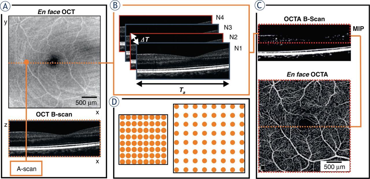
FIGURE 2.
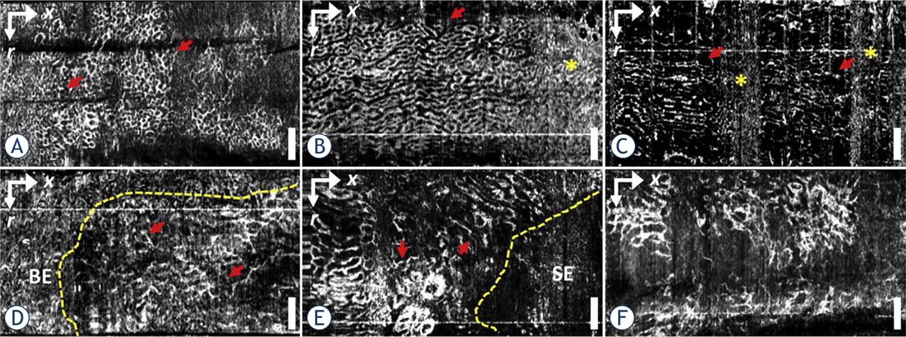
FIGURE 3.
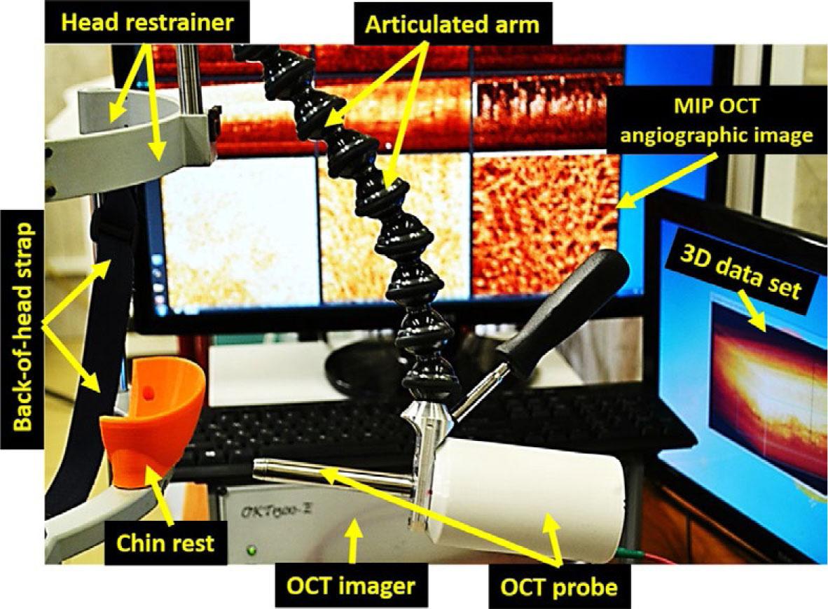
FIGURE 4.
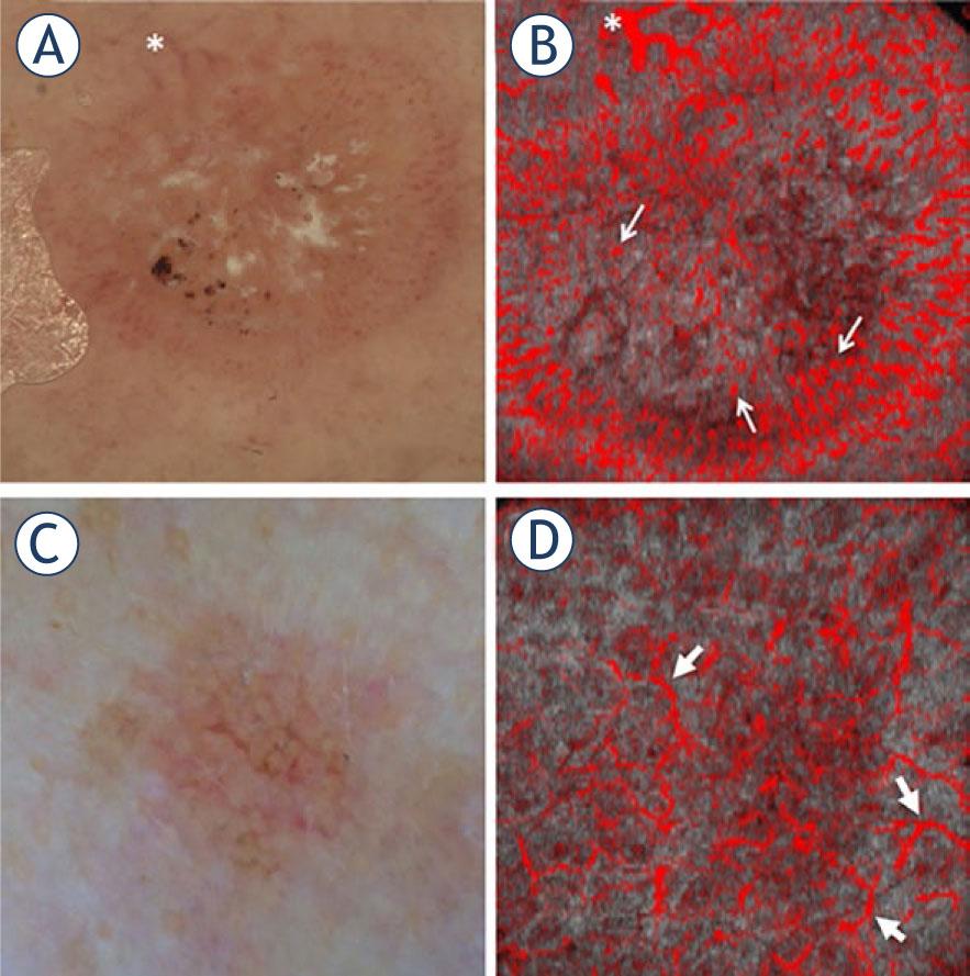
FIGURE 5.
Included articles reporting the use of optical coherence tomography angiography (OCTA) to quantify microvascular changes in nonocular clinical applications in oncology
| Reference | Year of publication | Number of patients | Oncologic setting |
|---|---|---|---|
| GI tract | |||
| Tsai et al.15 | 2014 | 1 | Nondysplastic Barrett's esophagus |
| Lee et al.16 | 2017 | 52 | Nondysplastic Barrett's esophagus surveillance or endoscopic eradication therapies for low-grade/high-grade dysplasia |
| Head and neck | |||
| Maslennikova et al.17 | 2017 | 25 | Radiotherapy of oropharyngeal and nasopharyngeal cancer |
| Skin | |||
| De Carvalho et al.18 | 2016 | 1 | Naevus to melanoma transition |
| Themstrup et al.19 | 2017 | 47 | Actinic keratosis, Bowen's disease and squamous cell carcinoma |
| Themstrup et al.20 | 2018 | 81 | Basal cell carcinoma |
| Meiburger et al.21 | 2019 | 7 | Basal cell carcinoma |
| Gubarkova et al.22 | 2019 | 27 | Basal cell carcinoma |
| De Carvalho et al.23 | 2018 | 127 | Melanoma |
| Welzel et al.24 | 2021 | 159 | Melanoma |
| Perwein et al.25 | 2023 | 130 | Nevi |