
TABLE 1.
Patients, tumor and treatment characteristics
| Characteristics: | n (%) |
|---|---|
| Sex | |
| Male | 12 (71) |
| Female | 5 (29.4) |
| Age at initial diagnosis, years | |
| Median (range) | 69 (43–90) |
| Dimension, cm (diagnostic MRI) | |
| Median (range) | 12.8 (6–30) |
| Pathology | |
| Undifferentiated pleomorphic sarcoma (UPS) | 8 (47) |
| Myxoid liposarcoma (MLS) | 6 (35) |
| Dedifferentiated liposarcoma (DLS) | 2 (12) |
| Pleomorphic rhabdomyosarcoma (PRS) | 1 (6) |
| Grade | |
| 1 | 6 (35) |
| 2 | 7 (41) |
| 3 | 4 (24) |
| RT schedule (total dose, dose fraction) | |
| 50.4 Gy, 1.8Gy | 5 (29) |
| 50 Gy, 2Gy | 10 (59) |
| 45 Gy, 3 Gy | 1 (6) |
| 70 Gy, 2 Gy* | 1 (6) |
| GTV on CTs1 (ml) | |
| Median (range) | 381 (84–2908) |
| PTV on CTs1 (ml) | |
| Median (range) | 1373 (587–5793) |
| D95%PTV on CTs1 (%) | |
| Median (range) | 97.1 (96–99.1) |
| Interval between CTs1 and MVCT 1 (days) | |
| Median (range) | 13 (9–17) |
| Neoadjuvant CT** | |
| No CT | 10 (59) |
| Adriamycin ifosfamide ((doxorubicin 20 mg/m2 and ifosfamide 2500 mg/m2 day 1, 2 and 3 for 4 cycles (21-day cycle)) | 6 (35) |
| Adriamycin (doxorubicin 75 mg/m2 day 1 for 4 cycles (21-day cycle)) | 1 (6) |
CTs1 = CT simulation scan; D95%PTV = dose received by 95% of the PTV volume; MRI = Magnetic Resonance Imaging; MVCT1 = first fraction of RT
GTV = gross tumor volume; MVCT = megavoltage computed tomography;PTV = planned target volume; RT = radiotherapy
TABLE 2.
Gross tumor volume (GTV) and planned target volume (PTV) volumes variations, the largest GTV and D95% PTV changes during the course of treatment
 | Pathology | Vol GTV CTs1 (ml) | Vol PTV CTs1 (ml) | Vol GTV last MVCT | Vol PTV last MVCT | Largest GTV vol change % (week) | Largest D95%PTV change % (week) |
| UPS | 674 | 1373 | 560 | 1150 | +36.4 (2) | −12 (2)* | |
| PRS | 239 | 1037 | 238 | 966 | +33.7 (2) | −3.1 (1) | |
| UPS | 381 | 1432 | 486 | 1706 | +32.3 (5) | −20.1 (6)* | |
| UPS | 2546 | 5292 | 3243 | 6263 | +27.4 (6) | + 1 (4) | |
| DLS | 2908 | 5793 | 3444 | 6628 | +18.9 (4) | −4.6 (3) | |
| UPS | 1356 | 3534 | 1319 | 3446 | +11 (1) | −2.1 (1) | |
| UPS | 234 | 999 | 246 | 1086 | +7.9 (2) | −14.1 (4)* | |
| UPS | 140 | 587 | 144 | 598 | −7.2 (5) | −3.4 (2) | |
| UPS | 826 | 2073 | 781 | 2031 | −9.2 (1) | −7.6 (4) | |
| DLS | 531 | 1679 | 461 | 1509 | −13.2 (6) | −1.3 (3) | |
| MLS | 258 | 952 | 184 | 805 | −28.9 (6) | −1.3 (2) | |
| MLS | 415 | 1514 | 233 | 1098 | −43.8 (6) | + 1.6 (6) | |
| MLS | 192 | 816 | 104 | 630 | −46.2 (6) | −2 (6) | |
| MLS | 228 | 629 | 122 | 398 | −46.4 (6) | +0.4 (4) | |
| MLS | 1062 | 3084 | 568 | 2158 | −46.5 (6) | + 1.14 (5) | |
| UPS | 208 | 961 | 96 | 602 | −52 (6) | −0.9 (1) | |
| MLS | 84 | 606 | 44 | 548 | −52 (5) | −7 (3) |
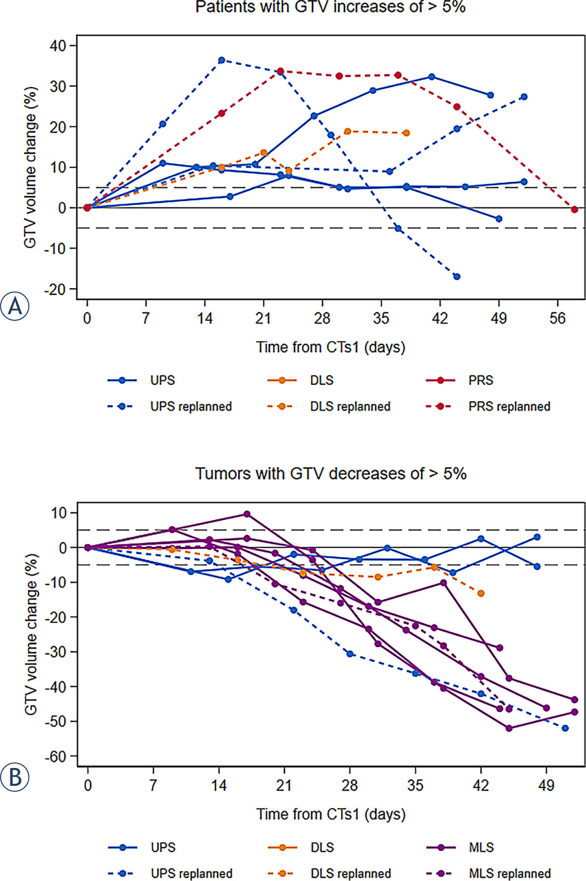
FIGURE 1.
Percentage of maximal GTV changes during treatment: patients with significant gross tumor volume (GTV) increase (A), patients with significant GTV decrease (B)*. Day 0 corresponds to CTs1. Each color represents a different histology subtype; replanned patients appear as dotted line.
DLS = dedifferentiated liposarcomas; MLS = myxoid liposarcomas; PRS = pleomorphic rhabdomyosarcoma; UPS = undifferentiated pleomorphic sarcomas
* Only the largest variation is shown for patients who presented with both an increase and a decrease in tumor size during the course of treatment.
TABLE 3.
Correlations between GTV volume variations and dosimetric and clinical characteristics
| Maximal GTV change | |||
|---|---|---|---|
| Decrease (n=10) | Increase (n=7) | ||
| GTV largest axial axis* | Decrease | 7 (70%) | 0 |
| Stable | 3 (30%) | 3 (43%) | |
| Increase | 0 | 4 (57%) | |
| GTV largest sagittal axis* | Decrease | 8 (80%) | 0 |
| Stable | 2 (20%) | 3 (43%) | |
| Increase | 0 | 4 (57%) | |
| Estimated GTV volume | Decrease | 9 (90%) | 0 |
| Stable | 1 (10%) | 0 | |
| Increase | 0 | 7 (100%) | |
| PTV volume | Decrease | 8 (80%) | 0 |
| Stable | 2 (20%) | 0 | |
| Increase | 0 | 7 (100%) | |
| D95% PTV | Decrease | 0 | 3 (43%) |
| Stable | 10 (100%) | 4 (57%) | |
| Surgical complications** | Yes | 3 (33%) | 4 (80%) |
| No | 6 (67%) | 1 (20%) | |
| Histology subtypes | UPS | 3 (30%) | 5 (71%) |
| DLS | 1 (10%) | 1 (14%) | |
| MLS | 6 (60%) | 0 | |
| PRS | 0 | 1 (14%) | |
| Histology grade | 1 | 6 (60%) | 0 |
| 2 | 2 (20%) | 5 (71%) | |
| 3 | 2 (20%) | 2 (29%) | |
| Neoadjuvant CT | Yes | 3 (30%) | 4 (57%) |
| No | 7(70%) | 3 (43%) | |
* Axial and sagittal linear tumor dimensions were recovered from the same slice for each MVCT, the slice that contained the largest tumor axial and sagittal axis on the CTs1.
** Data available for 14 patients: one patient had not been operated; the two others had not been operated when the analysis was performed.
GTV = gross tumor volume; MVCT = megavoltage computed tomography; PTV = planned target volume
TABLE 4.
Dosimetric and clinical data of patients with significant reductions in PTV coverage
| Dosimetric data at first MVCT showing a variation of volume | Concomitant volume changes | Clinical dimension change* | D95% PTV on last MVCT | ||||||
|---|---|---|---|---|---|---|---|---|---|
| D95% PTV on CTs1 | Fraction n° | Variation of D95 coverage (%) | New D95% PTV | D95% GTV | GTV (%) | PTV (%) | Estimated GTV(%) | ||
| 99%* | 6* | −11.8* | 87.22%* | 72.96%* | +36.4* | +22.8* | +27.6* | + 1.3 cm (ax)* | 98.71%* |
| 96.1% | 6 | −10.4 | 86.49% | 98.03% | +10.7 | +6.2 | +12.2 | + 1 cm (sag) | 77.07% |
| 98.2% | 1 | −5.9 | 92.39% | 98.91% | +2.8 | +3.3 | +2 | No | 94.29% |
Significant reductions in coverage are indicated in bold. The patient who had a plan adaptation is marked with a star (*).
* The “clinical dimension change” corresponds to the largest visible variation, all slices combined.
GTV = gross tumor volume; MVCT = megavoltage computed tomography; PTV = planned target volume
TABLE 5.
Dosimetric and clinical data of patients with a plan adaptation
| Dosimetric data just before plan adaptation | Evolution of D95%PTV | ||||||
|---|---|---|---|---|---|---|---|
| Clinical Reason* | Fr n° | % GTV vol change | %PTV vol change | OAR constraints | At CTs1 | Before re-planning | At CTs2 |
| Ax. increase (+ 1.3 cm) | 13 | + 33 | + 22 | Better | 99.1% | 88% | 97.5% |
| Ax. increase (+ 0.9 cm) | 4* | + 9 | + 9 | Better | 98.1% | 93.5% | 96% |
| Sag. increase (+ 1.2 cm) | 1 | + 23 | + 10 | Better | 96.8% | 93.7% | 95.9% |
| Sag. increase (+ 1 cm) | 11 | + 17 | + 12 | NA | 96.9% | 97.6% | 97.6% |
| Ax. decrease (− 1.8 cm) | 13 | − 36 | − 21 | Bone Dm + 1.8% | 96.9% | 97.2% | 98.7% |
| Sag. decrease (− 2 cm) | 17 | − 28 | − 17 | Bone Dm + 1% | 96.4% | 97.5% | 96.5% |
| Weight loss | 21** | − 13 | − 10 | Bone Dm + 30.7% | 96.4% | 97.1% | 96.9% |
Ax = axial; Bone Dm = average dose to the bone; CTs1 = CT computed tomography simulation scan; CTs2 = new Fr n° = fraction number; GTV = gross tumor volume; NA = result not available; OAR = organ at risk; PTV = planned target volume; Sag = sagittal