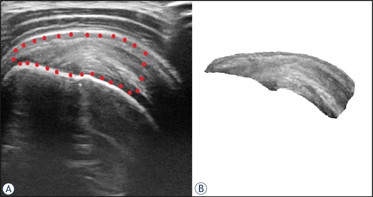
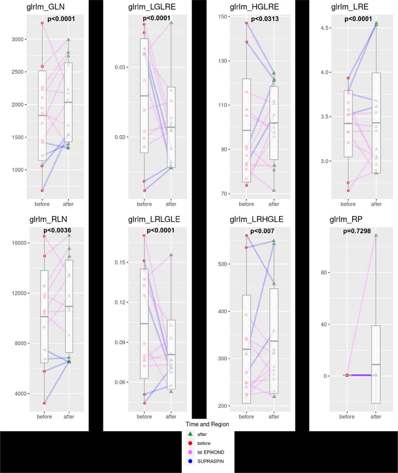
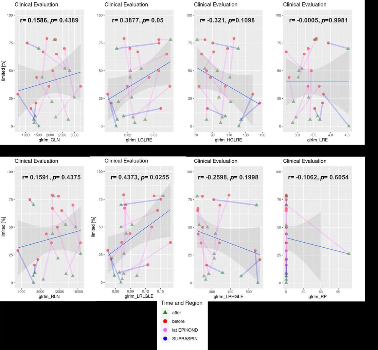
Have a personal or library account? Click to login
Assessment of short-term effect of platelet-rich plasma treatment of tendinosis using texture analysis of ultrasound images Cover

Assessment of short-term effect of platelet-rich plasma treatment of tendinosis using texture analysis of ultrasound images

Open Access
|Nov 2023

Figures & Tables

FIGURE 1.

US image of the representative supraspinatus tendinosis in longitudinal plane (A). Region of interest was selected from the footprint of the tendon to the myotendinous junction of supraspinatus muscle (encircled with red dots) and cropped for the purpose of texture analysis (B).

FIGURE 2.

Box plots of the amplitude using gray level run length matrix method (GLRLM) features before and after platelet rich plasma (PRP) treatment (supraspinatus tendons encoded with blue and common extensor tendons encoded with pink).

FIGURE 3.

Clinical evaluation dependency of average texture feature per subject divided by tissue region and time. Tissues are coded with blue and pink lines, for supraspinatus and common extensor, while the time of measurement is indicated by a red circle for the first measurement before treatment and a green triangle for the measurement after treatment.
DOI: https://doi.org/10.2478/raon-2023-0054 | Journal eISSN: 1581-3207 | Journal ISSN: 1318-2099
Language: English
Page range: 465 - 472
Submitted on: Jun 14, 2023
Accepted on: Aug 6, 2023
Published on: Nov 30, 2023
Published by: Association of Radiology and Oncology
In partnership with: Paradigm Publishing Services
Publication frequency: 4 issues per year

© 2023 Karlo Pintaric, Vladka Salapura, Ziga Snoj, Andrej Vovk, Mojca Bozic Mijovski, Jernej Vidmar, published by Association of Radiology and Oncology
This work is licensed under the Creative Commons Attribution 4.0 License.