
FIGURE 1.
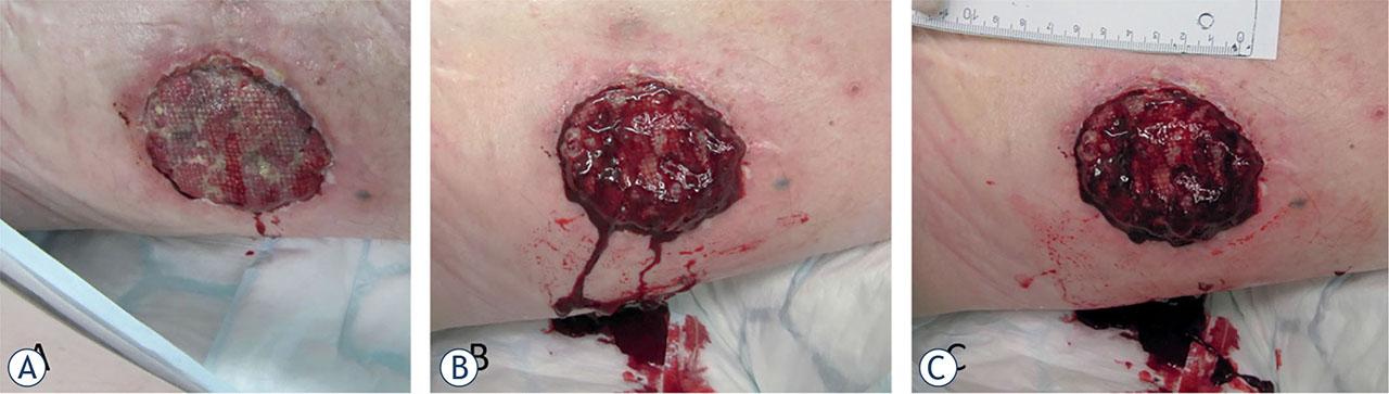
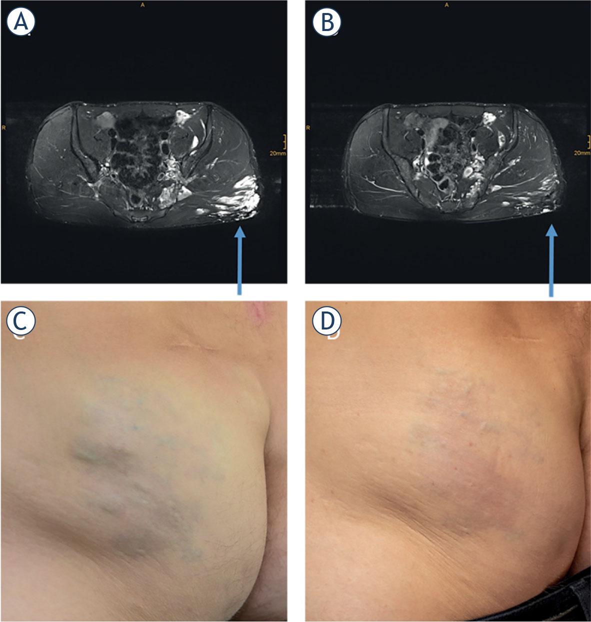
FIGURE 2.
Clinical studies and case reports using bleomycin electrosclerotherapy40,41,42,43,44,45
| Reference | Type of malformation | N of pts | Bleomycin dose and concentration | Electrodes used | N of pulse applications | Response | Comment | |
|---|---|---|---|---|---|---|---|---|
| McMorrow et al., Br J Oral and Maxillofacial Surg 201744 | Venous malformation | 1 | Reduced dose: 1/3 of the standard dose | Not reported | Not reported | Considerable improvement after 6 unsuccessful sessions with bleomycin | Case report with poor respiratory function | |
| Horbach et al., Dermatologic Surgery 202045 | Hypertrophic capillary malformations | 5 pts. (out of 20 planned) | 0.25 mg or units/cm3 | Plate & needle | Not reported | 7–8 weeks | Randomized controlled pilot trial | |
| Dalmady et al., Pediatrics 202043 | Lymphatic malformation | 1 | 0.5 mg/kg (5.4 mg) | Needle | 1st session: 68 applications | 63% growth-corrected volume decrease. | Case report | |
| Wohlgemuth et al., Journal of Vascular Surgery 202140 | Venous malformations | 17 pts. (20 lesions) | Calculated based on the size of the lesion. | Needle & finger | Not reported | 3-month post-therapy Changes in volume MRI: Volume reduction,%: | ||
| > 90% | 9 lesions | Retrospective observational case study | ||||||
| > 70% in < 90% | 6 lesions | |||||||
| > 50% in < 70% | 2 lesions | |||||||
| < 35% | 2 lesions | |||||||
| No response | 1 lesion | |||||||
| Kostusiak et al., Dermatologic Surgery 202242 | Various vascular malformations | 30 pts. | Calculated based on the size of the lesion. | Needle & finger | Not reported | 17 Complete Response | Prospective observational case study | |
| Krt et al., Front Oncol 202241 | Arteriovenous malformation | 1 | 750 IU BLM intralesional | Plate | 15 | CR 18 months after BEST | Case report | |
International Society for the Study of Vascular Anomalies (ISSVA) classification for vascular anomalies 2018
| VASCULAR TUMORS | VASCULAR MALFORMATIONS | |||
|---|---|---|---|---|
| Benign | Locally aggressive | Malignant | Simple | Combined |
| Infantile hemangioma | Kaposiform hemangioendothelioma | Capillary malformation (CM) | CVM, CLM | |
| Congenital hemangioma | Retiform hemangioendothelioma | Lymphatic malformation (LM) | LVM. CLVM | |
| Tufted hemangioma | PILA. Dabska tumor | Epithelioid hemangioendothelioma | Venous malformation (VM) | CAVM |
| Spindle-cell hemangioma | Composite hemangioendothelioma | Angiosarcoma | Arteriovenous malformation (AVM) | |
| Epithelioid hemangioma | Kaposi sarcoma | Arteriovenous fistula | CLAVM | |
| Pyogenic granuloma | ||||