
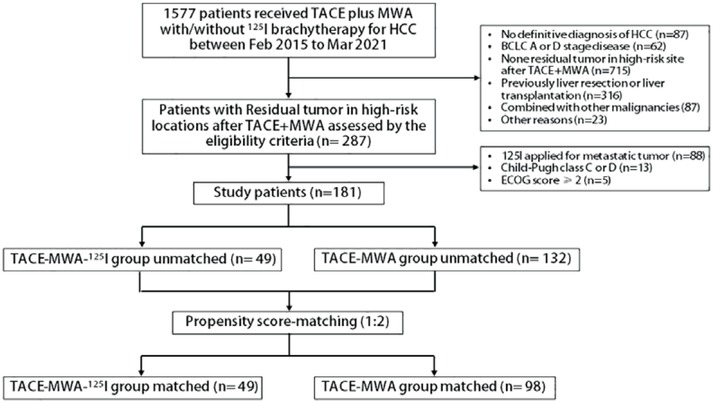
Figure 1
Patient flow diagram.
TACE = transarterial chemoembolization; MWA = microwave ablation
Table 1
Baseline patient characteristics before and after propensity score-matched
| Parameter | Total (N = 181) | Total cohort | PSM cohort | ||||
|---|---|---|---|---|---|---|---|
| Group A | Group B | P | Group A | Group B | P | ||
| N = 49 | N = 132 | value | N = 49 | N = 98 | value | ||
| Sex | 0.37 | 0.751 | |||||
| Male | 162 (89.5%) | 46 (93.9%) | 116 (87.9%) | 46 (93.9%) | 89 (90.8%) | ||
| Female | 19 (10.5%) | 3 (6.12%) | 16 (12.1%) | 3 (6.12%) | 9 (9.18%) | ||
| Age (y) | 56.0 [47.0;64.0] | 56.0 [47.0;68.0] | 55.5 [48.0;62.0] | 0.425 | 56.0 [47.0;68.0] | 55.0 [48.0;62.0] | 0.468 |
| BCLC | 0.041* | 1 | |||||
| B | 72 (39.8%) | 13 (26.5%) | 59 (44.7%) | 13 (26.5%) | 26 (26.5%) | ||
| C | 109 (60.2%) | 36 (73.5%) | 73 (55.3%) | 36 (73.5%) | 72 (73.5%) | ||
| Maximum median (IQR), tumor mm diameter, | 29.0 [18.0;49.0] | 36.0 [22.0;53.0] | 26.5 [18.0;46.0] | 0.039* | 36.0 [22.0;53.0] | 31.0 [18.0;57.8] | 0.579 |
| Tumor number | 0.927 | 0.66 | |||||
| 1-2 | 60 (33.1%) | 17 (34.7%) | 43 (32.6%) | 17 (34.7%) | 29 (29.6%) | ||
| ≥ 3 | 121 (66.9%) | 32 (65.3%) | 89 (67.4%) | 32 (65.3%) | 69 (70.4%) | ||
| PVTT | 0.202 | 1 | |||||
| None | 108 (59.7%) | 25 (51.0%) | 83 (62.9%) | 25 (51.0%) | 49 (50.0%) | ||
| Yes | 73 (40.3%) | 24 (49.0%) | 49 (37.1%) | 24 (49.0%) | 49 (50.0%) | ||
| Distant metastasis | 0.171 | 0.953 | |||||
| None | 109 (60.2%) | 25 (51.0%) | 84 (63.6%) | 25 (51.0%) | 52 (53.1%) | ||
| Yes | 72 (39.8%) | 24 (49.0%) | 48 (36.4%) | 24 (49.0%) | 46 (46.9%) | ||
| High risk location | 0.045* | 0.76 | |||||
| 1 | 98 (54.2%) | 33 (67.3%) | 65 (49.2%) | 33 (67.3%) | 62 (63.3%) | ||
| 2 | 83 (45.8%) | 16 (32.7%) | 67 (50.8%) | 16 (32.7%) | 36 (36.7%) | ||
| TACE sessions | 2.00 [1.00;3.00] | 2.00 [1.00;2.00] | 2.00 [1.00;3.00] | 0.442 | 2.00 [1.00;2.00] | 1.00 [1.00;3.00] | 0.464 |
| MWA sessions | 1.00 [0.00;2.00] | 1.00 [0.00;2.00] | 1.00 [0.00;2.00] | 0.162 | 1.00 [0.00;2.00] | 1.00 [0.00;2.00] | 0.31 |
| Cause of liver disease: | 0.09 | 0.181 | |||||
| Continued | |||||||
| HCV/HBV | 169 (93.4%) | 43 (87.8%) | 126 (95.5%) | 43 (87.8%) | 93 (94.9%) | ||
| Other | 12 (6.6%) | 6 (12.2%) | 6 (4.55%) | 6 (12.2%) | 5 (5.10%) | ||
| ECOG score | 0.189 | 0.669 | |||||
| 0 | 126 (69.6%) | 30 (61.2%) | 96 (72.7%) | 30 (61.2%) | 65 (66.3%) | ||
| 1 | 55 (30.4%) | 19 (38.8%) | 36 (27.3%) | 19 (38.8%) | 33 (33.7%) | ||
| Child-pugh score | 0.563 | 0.386 | |||||
| A | 82 (45.3%) | 46 (93.9%) | 36 (27.3%) | 46 (93.9%) | 86 (87.8%) | ||
| B | 99 (54.7%) | 3 (6.12%) | 96 (72.7%) | 3 (6.12%) | 12 (12.2%) | ||
| AFP, ng/mL | 0.811 | 0.576 | |||||
| < 400 | 125 (69.1%) | 35 (71.4%) | 90 (68.2%) | 35 (71.4%) | 64 (65.3%) | ||
| ≥ 400 | 56 (30.9%) | 14 (28.6%) | 42 (31.8%) | 14 (28.6%) | 34 (34.7%) | ||
| PT (s) | 12.2 [11.5;13.1] | 12.0 [11.5;12.8] | 12.3 [11.6;13.1] | 0.167 | 12.0 [11.5;12.8] | 12.2 [11.5;13.1] | 0.331 |
| ALB (g/L) | 40.35 [36.2;43.9] | 40.8 [36.7;44.1] | 40.2 [36.0;43.9] | 0.329 | 40.8 [36.7;44.1] | 40.3 [36.1;44.1] | 0.518 |
| TBIL (mg/dL) | 13.55 [10.1;19.32] | 12.3 [8.73;18.2] | 14.6 [10.4;20.4] | 0.167 | 12.3 [8.73;18.2] | 14.6 [10.1;20.2] | 0.227 |
[i] AFP = alpha fetoprotein; ALB = albumin; BCLC = Barcelona Clinic Liver Cancer; ECOG = Eastern Cooperative Oncology Group; IQR = interquartile range; MWA = microwave ablation; PSM = propensity score-matched; PVTT = portal vein tumor thrombosis; PT = prothrombin time; TACE = transcatheter arterial chemoembolization; TBIL = total bilirubin
SI conversion factors: To convert albumin to grams per liter, multiply by 10.0; to convert bilirubin to micromoles per liter, multiply by 17.104; and to convert α-fetoprotein to micrograms per liter, multiply by 1.0.
*p value ≤ 0.05 was considered to indicate statistical significance.
Table 2
Intrahepatic and high-risk locations tumor responses in the two groups after propensity score matching (PSM)
| Tumor response | Intrahepatic Tumor | Tumor in high-risk locations | ||||
|---|---|---|---|---|---|---|
| Group A (n = 49) | Group B (n = 98) | P value | Group A (n = 49) | Group B (n = 98) | P value | |
| Complete response (CR) | 4 | 6 | 5 | 9 | ||
| Partial response (PR) | 21 | 23 | 28 | 29 | ||
| Stable disease (SD) | 17 | 41 | 13 | 40 | ||
| Progressive disease (PD) | 7 | 28 | 3 | 20 | ||
| Objective response rate (ORR) (%) | 51.0% | 29.6% | 0.011†* | 67.3% | 38.8% | < 0.001†* |
| Disease control rate (DCR) (%) | 85.7% | 71.4% | 0.055† | 93.9% | 79.6% | 0.025†* |
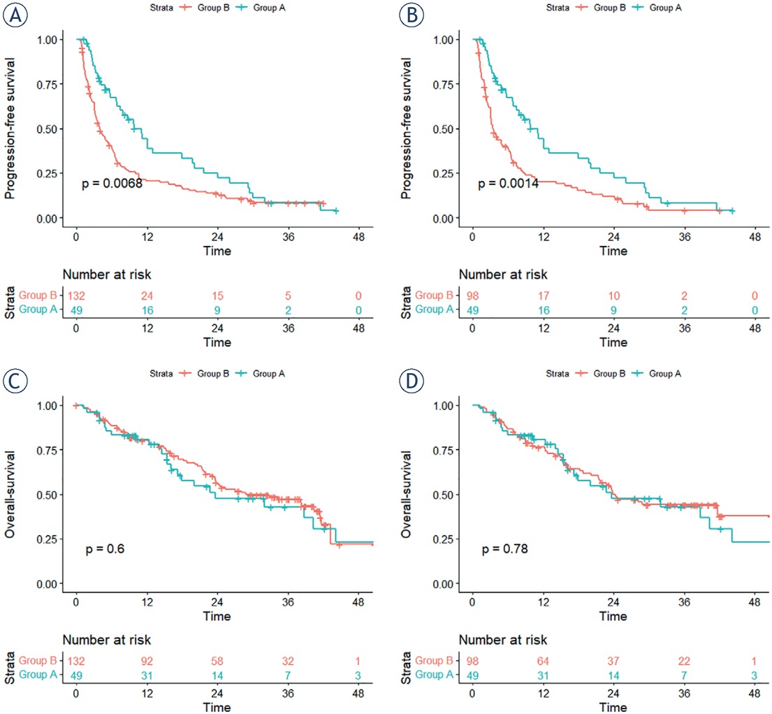
Figure 2
Kaplan-Meier curves comparing progression-free survival (PFS) and overall survival overall survival (OS) from different groups. (A) Cumulative PFS between the unmatched A and B groups. (B) Cumulative PFS between the matched A and B groups. (C) Cumulative OS between the unmatched A and B groups. (D) Cumulative OS between the matched A and B groups.
Table 3
Multivariate analyses of predictors of progression-free survival after treatment
| Risk Factor | Univariate | Multivariate | ||||
|---|---|---|---|---|---|---|
| HR | 95%CI | P value | HR | 95%CI | P value | |
| Sex | ||||||
| Female | 1 | |||||
| Male | 0.755 | 0.393–1.451 | 0.399 | |||
| Age (y) | ||||||
| < 60 | 1 | |||||
| ≥ 60 | 1.093 | 0.761–1.569 | 0.63 | |||
| Maximum diameter (tumor mm) | 1.006 | 1.000–1.013 | 0.071* | 1.003 | 0.996-1.010 | 0.458 |
| Tumor number | ||||||
| 1-2 | 1 | |||||
| ≥ 3 | 1.104 | 0.915–1.332 | 0.302 | |||
| BCLC stage | ||||||
| B | 1 | |||||
| C | 1.395 | 0.929–2.095 | 0.109 | |||
| High-risk location | ||||||
| 1 | 1 | |||||
| 2 | 0.665 | 0.456–0.970 | 0.034** | 0.846 | 0.550-1.302 | 0.447 |
| PVTT | ||||||
| None | ||||||
| Yes | 1.615 | 1.124–2.319 | 0.010** | 1.625 | 1.015-2.546 | 0.040** |
| Distant metastasis | ||||||
| None | ||||||
| Yes | 1.099 | 0.772–1.565 | 0.602 | |||
| TACE sessions | 1.062 | 0.964–1.170 | 0.223 | |||
| MWA sessions | 1.006 | 0.914–1.106 | 0.905 | |||
| Treatment | ||||||
| TACE+MWA | 1 | |||||
| TACE+MWA+ 125I | 0.527 | 0.357–0.778 | 0.002** | 0.479 | 0.328-0.733 | <0.001** |
| Cause of liver disease | ||||||
| Other | 1 | |||||
| HBV/HCV | 1.36 | 0.662–2.791 | 0.402 | |||
| ECOG score | ||||||
| 0 | 1 | |||||
| 1 | 1.269 | 0.876–1.837 | 0.208 | |||
| AFP | ||||||
| ≤ 400 ng/mL | 1 | |||||
| Continued | ||||||
| > 400 ng/mL | 1.72 | 1.164–2.542 | 0.006** | 1.403 | 0.948-2.129 | 0.090 |
| Prothrombin time (s) | 0.942 | 0.827–1.073 | 0.367 | |||
| Albumin (g/L) | 0.987 | 0.951–1.024 | 0.477 | |||
| Continued | ||||||
| Total Bilirubin (μmol/L) | 1.001 | 0.995–1.008 | 0.649 | |||
| Child-pugh class | ||||||
| A | 1 | |||||
| B | 1.216 | 0.654–2.263 | 0.536 | |||
[i] AFP = alpha fetoprotein; ALB = albumin; BCLC = Barcelona Clinic Liver Cancer; ECOG = Eastern Cooperative Oncology Group; HR = hazard ratio; MWA = microwave ablation; PVTT = portal vein tumor thrombosis; PT = prothrombin time; TACE = transarterial chemoembolization; TBIL = total bilirubin
*P value ≤ 0.1 in uni,variate were included in multivariate analysis, **P value ≤ 0.05 was considered to indicate statistical significance in multivariate analysis
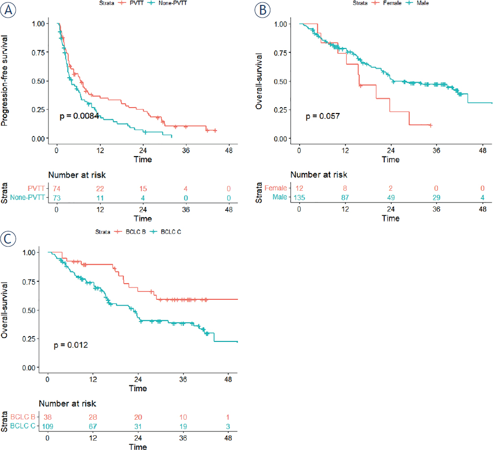
Figure 3
Kaplan-Meier curves comparing progression-free survival (PFS) and overall survival (OS) according to the statistically significant prognostic factors in the multivariate analysis after propensity score-match. (A) Cumulative PFS between portal vein tumor thrombosis (PVTT) and None-PVTT patients. (B). Cumulative OS between male and female. (C) Cumulative OS between Barcelona clinic liver cancer (BCLC) B and C stages patients.
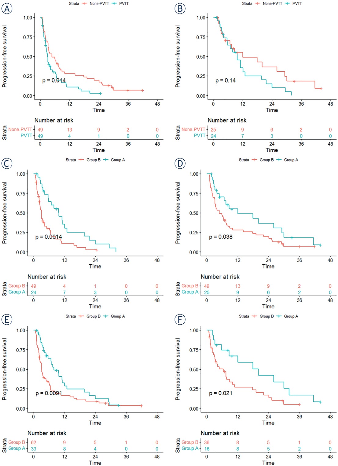
Figure 4
Subgroup analyses revealed by Kaplan-Meier curves comparing progression-free survival (PFS). (A) Comparison of the PFS between portal vein tumor thrombosis (PVTT) and None-PVTT patients in group B. (B) Comparison of the PFS between PVTT and None-PVTT patients in group A. (C) Comparison of the PFS between group A and B in patients with PVTT. (D) Comparison of the PFS between group A and B in none-PVTT patients. (E) Comparison of the PFS between group A and B in high-risk location 1. (F) Comparison of the PFS between group A and B in high-risk location 2.