Have a personal or library account? Click to login
Ultrasonography of peripheral nerve tumours: a case series Cover

Ultrasonography of peripheral nerve tumours: a case series

By: Simon Podnar  
Open Access
|Jan 2023

Figures & Tables

Figure

(A) Transverse ultrasonographic (US) view of the median (M) and ulnar (U) nerve in the axillary region showing numerous globular hypoechoic peripheral nerve tumours (PNTs) causing largely increased nerve cross sectional areas (CSAs, 148 mm2 and 101 mm2, respectively). (B) Longitudinal view of a single partially encapsulated, slightly lobulated and rather homogenous oval PNT (length 24 mm, thickness 9 mm) with central, but poorly defined nerve-tumour transition.5 Most probably these numerous PNTs are neurofibromas, although diagnosis in this 52-year-old woman presenting with peripheral neuropathy, primary lymphedema, and history of mitral and aortic valve surgery (patient #13, Tables 2–3), is not known yet.

Figure

Comparison of transverse image of the sciatic peripheral nerve tumour (PNT) on (A) magnetic resonance (MR) T1 (arrow) and (B) ultrasonography (US). (C) Longitudinal US view of PNT in the middle thigh (A – small picture on the left), affecting the tibial portion of sciatic nerve. In this 22-year-old girl MR revealed at the exit of sciatic nerve from the pelvis another PNT affecting its fibular portion, and causing right foot drop of 14 years duration (patient #10, Tables 2–3). Based on clinical and imaging features in this patient diagnosis of probable perineurioma was made.

Figure

(A) Transverse and (B) longitudinal ultrasonographic (US) view of a large peripheral nerve tumour (PNT) on the left ulnar nerve just above the elbow. Three years before this 47-year-old man noted a palpable mass that in the last 6 months on touching started to elicit electrisation spreading into the last two fingers (patient #8, Tables 2–3). Well encapsulated, slightly lobulated, predominantly cystic and highly heterogenous hypoechoic oval lesion with central and poorly defined nerve-tumour transition can be seen. Histological examination confirmed a diagnosis of schwannoma.

Figure

(A) Hands of a 26-year-old woman with 4-year history of muscle atrophy, weakness and numbness in the distal ulnar nerve territory (patient #5, Tables 2–3). Note intrinsic right palm muscle atrophy and clawing of the last two fingers. (B) On transverse ultrasonographic (US) view ulnar nerve cross sectional area (CSA) increased from 7 mm2 both proximally and distally to 20 mm2 in the middle of the lesion. (C) On longitudinal view a partially encapsulated, lobulated, fusiform hypoechoic right ulnar peripheral nerve tumour (PNT) of the forearm can be seen. Based on clinical and US features, we made a diagnosis of probable perineurioma.

Morphological features useful for differentiation between neurofibromas and schwannomas5

Peripheral nerve tumor (PNT) featureComment
Maximum to minimum diameterRatio > 3 → neurofibroma
Shape: round, oval, fusiformFusiform → neurofibroma
Contour: smooth, lobulatedLobulated → neurofibroma
Encapsulation: absent, partial, completeComplete → schwannoma
Echogenicity: hypo-, iso-, hyper-Hypoechoic → PNT
Echo texture: homogenous, heterogenousHeterogenous → schwannoma
Cystic changes: absent, focal, partial,Cystic changes →
largeschwannoma
Calcifications: absent, presentPresent → schwannoma
Target sign: absent, present
Nerve entrance: not identified, identified
Nerve-tumor position: central, eccentricCentral → neurofibroma
Nerve-tumor transition: clear, poorly defined, infiltrativeInfiltrative → neurofibroma
Vascularity: increased, normal, decreasedHypovascular → neurofibroma

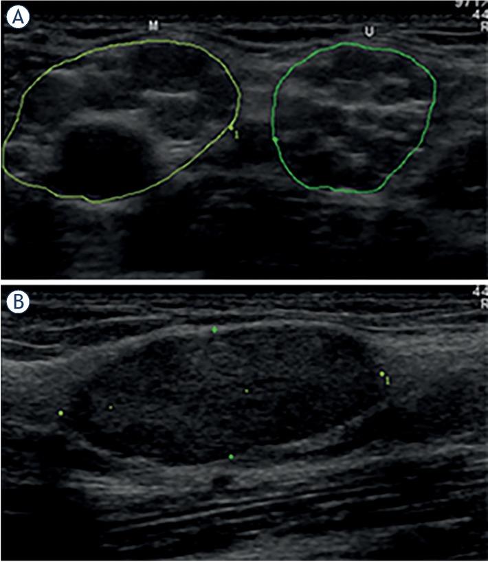
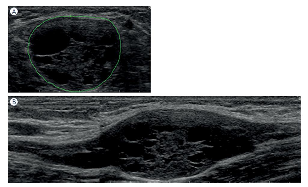
Morphological features of peripheral nerve tumors (PNTs) found on ultrasonographic (US) examination5 of individual patients

#RatioShapeContourEncapsulationEcho textureNerve positionNerve transitionNumberTumor diagnosis
15FusiformLobulatedPartialHeterogeneousCentralInfiltrativeSingleNeurofibroma
2? LobulatedPartialHeterogeneous??SeveralSchwannoma*
36FusiformSmoothWholeHeterogeneousCentralPoorly definedSingleSchwannoma
46FusiformFusiformNoneHeterogeneousCentralInfiltrativeSeveralNeurofibroma
58FusiformLobulatedPartialHeterogeneousCentralInfiltrativeSinglePerineurioma
6> 10FusiformLobulatedPartialHeterogeneous??SinglePerineurioma
76FusiformSmoothNoneHomogenousCentralInfiltrativeSinglePerineurioma
83OvalSmoothWholeHeterogeneousCentralPoorly definedSingleSchwannoma*
9> 10FusiformLobulatedPartialHeterogeneousEccentricInfiltrativeSingleNeurofibroma
105FusiformSmoothWholeHeterogeneousCentralInfiltrativeSeveralPerineurioma
111,5OvalSmoothWholeHomogenousEccentric?SingleSchwannoma*
122,5OvalSmoothWholeHeterogeneousCentralPoorly definedSingleSchwannoma*
132,5OvalSmoothWholeHeterogeneousCentralPoorly definedSeveralNeurofibroma
144FusiformLobulatedPartialHeterogeneousCentralInfiltrativeSeveralNeurofibroma*
151,7OvalSmoothWholeHeterogeneousEccentricPoorly definedSingleSchwannoma

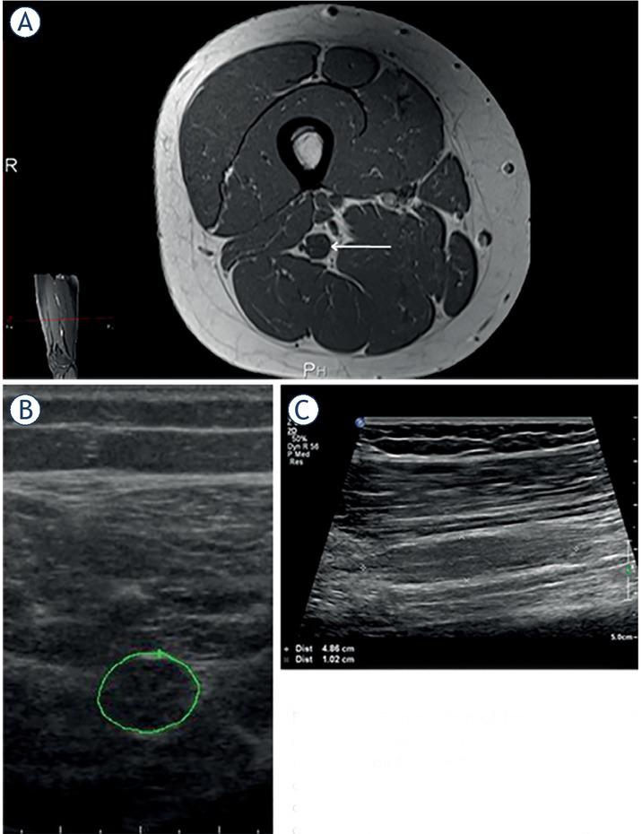
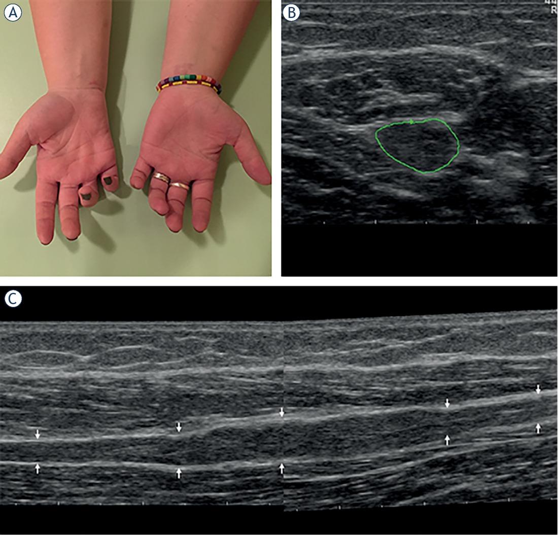
Demographic, anatomical, clinical, electrodiagnostic (EDx) and ultrasonographic (US) features of patients with peripheral nerve tumors (PNTs)

#GenderAgeSideNerveLocationSymptoms & SignsCMAP amp. (mV) SNAP amp. (μ V)Tumor CSA (mm2)Tumor diagnosisOther
1Male69RUlnarElbowAWS 43Neurofibroma
2Male24L#RadialUpper armW0.2424Schwannoma*NF2
3Male66RMedianForearmÆ6.9549Schwannoma
4Male16L#MedianUpper armWS 61NeurofibromaNF1
5Female26RUlnarForearmAWS0.2030Perineurioma
6Female18LSciaticThighAWS0.40109Perineurioma
7Female18RFibularKneeAWS0047Perineurioma
8Male47LUlnarElbowM7.63348Schwannoma*
9Female58RMedianForearmP7.61645Neurofibroma
10Female22RSciaticThighAWS0097Perineurioma
11Female34RTibialAnklePAWS10.671250Schwannoma*
12Male63LUlnarElbowL8.25368Schwannoma*
13Female52R#UlnarForearm 6.212212NeurofibromaNF?
14Male24R#MedianUpper armP6.33326Neurofibroma*NF1
15Female33LTibialAnkleL 92Schwannoma
DOI: https://doi.org/10.2478/raon-2023-0005 | Journal eISSN: 1581-3207 | Journal ISSN: 1318-2099
Language: English
Page range: 35 - 41
Submitted on: Aug 5, 2022
Accepted on: Dec 8, 2022
Published on: Jan 20, 2023
Published by: Association of Radiology and Oncology
In partnership with: Paradigm Publishing Services
Publication frequency: 4 issues per year

© 2023 Simon Podnar, published by Association of Radiology and Oncology
This work is licensed under the Creative Commons Attribution-NonCommercial-NoDerivatives 4.0 License.