
Table 1
Primers sequences and amplification conditions for genes
| Genes | Direction | Primer sequence | Amplification condition |
|---|---|---|---|
| BTG2 | Forward | 5/-CCTGGGCAGAGAGTGAAAAG-3/ | 95°C for 5 min, followed by 30 |
| Reverse | 5/-CCTTCCATCCTAACCCCAAT-3/ | cycles of 95°C for 30 s, 58°C for | |
| Forward | 5/-CCATGGAGAAGGCTGGGG-3/ | 45 s, 72°C for 45 s and 72°C for | |
| GAPDH | Reverse | 5/-CAAAGTTGTCATGGATGACC-3/ | 10 min |
| Forward | F1-5/-ATAGGGAGTCATGGAGGGTTTG-3/ | 95°C for 5 min, followed by 35 | |
| AKT1 | Reverse | R1-5/-CTTTACCAAATCCTGGTCACTGAA-3/ | cycles of 95°C for 30 s, 60°C for |
| Forward | F2-5/-AAAAAATTGATTGATGGGAGGAAG-3/ | 45 s, 72°C for 45 s and 72°C for | |
| Reverse | R2-5/-TAATCCCTGGCCTGCTCAG-3/ | 10 min |
Table 2
Demographic and clinical information of control subjects and colorectal cancer patients
| Characteristics | Cases n = 200(%) | Control n = 197(%) |
|---|---|---|
| Age* | ||
| 40 ≤ | 131 (65.5) | 120 (60.91) |
| 40 > | 69 (34.5) | 77 (39.09) |
| Sex* | ||
| Male | 119 (59.50) | 112 (57.50) |
| Female | 81 (40.50) | 85 (42.50) |
| Food consumption* | ||
| Mainly vegetables | 106 (53.00) | 97 (49.24) |
| Mixed Food | 94 (47) | 100 (50.76) |
| Smoking* | ||
| Ever | 36 (18.00%) | 29 (14.72%) |
| Never | 164 (82.00%) | 168 (85.28%) |
| Cancer family history* | ||
| Yes | 27 (13.50) | 17 (8.63) |
| No | 173 (86.50) | 180 (91.37) |
| Cancer Stages | ||
| I | 1 (0.50) | |
| II | 33 (16.50) | |
| III | 112 (56.00) | |
| IV | 54 (27.00) |
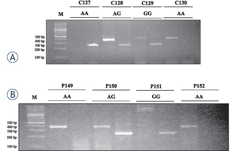
Figure 1
Akt rs1130233 single nucleotide polymorphism in (A) Control and (B) colorectal cancer patients. Representative images have been shown. AA genotype (379 bp band); AG genotype (245 and 379 bp bands); GG genotype (245 bp band). The number above the lanes indicate subjects identity.
C = Control, P = CRC Patient, M = DNA Marker
Table 3
Gene and allele frequencies of AKT1 rs1130233 polymorphism and its association with colorectal cancer
| Type of polymorphism | Genotype | n = Cases 200 (%) | n Control = 197 (%) | P value | OR (95% CI) |
|---|---|---|---|---|---|
| GG | 60 (30.00) | 90 (45.69) | Reference | ||
| Genotype Frequency | AG | 120 (60.00) | 101 (51.27) | 0.01 | 1.80 (1.18–2.74) |
| AA | 20 (10.00) | 6 (3.04) | 0.001 | 5.00 (1.90–13.18) | |
| Dominant Model | GG | 60 (30.00) | 90 (45.69) | ||
| AG+AA | 140 (70.00) | 107 (54.31) | 0.001 | 1.96 (1.30–2.96) | |
| Recessive Model | GG+AG | 180 (90.00) | 191 (96.96) | ||
| AA | 20 (10.00) | 6 (3.04) | 0.01 | 0.28 (0.11–0.72) | |
| Allele Frequency | G A | 0.6000 0.4000 | 0.7132 0.2868 |
Table 4
Frequencies of AKT1 rs1130233 polymorphism and alleles in colon cancer cases and control
| Type of polymorphism | Genotype | n = Colon 102 (%) | n Control = 197 (%) | P value | OR (95% CI) |
|---|---|---|---|---|---|
| GG | 30 (29.41) | 90 (45.69) | Reference | ||
| Genotype frequency | AG | 61 (59.80) | 101 (51.27) | 0.02 | 1.81 (1.08–3.05) |
| AA | 11 (10.79) | 6 (3.04) | 0.001 | 5.50 (1.87–16.15) | |
| Dominant model | GG | 30 (29.41) | 90 (45.69) | 0.006 | 2.02 (1.21–3.36) |
| AG+AA | 72 (70.59) | 107 (54.31) | |||
| Recessive model | GG+AG | 91 (89.21) | 191 (96.96) | 0.01 | 3.85 (1.38–10.73) |
| AA | 11 (10.79) | 6 (3.04) |
Table 5
Frequencies of AKT1rs1130233 polymorphism and alleles in rectum cancer cases and control
| Type of polymorphism | Genotype | n Rectum = 98 (%) | n Control = 197 (%) | P value | OR (95% CI) |
|---|---|---|---|---|---|
| GG | 30 (30.61) | 90 (45.69) | Reference | ||
| Genotype frequency | AG | 59 (60.20) | 101 (51.27) | 0.04 | 1.75 (1.04–2.96) |
| AA | 9 (9.19) | 6 (3.04) | 0.008 | 4.50 (1.48–13.69) | |
| Dominant model | GG | 30 (30.61) | 90 (45.69) | 0.01 | 1.91 (1.14–3.18) |
| AG+AA | 68 (69.39) | 107 (54.31) | |||
| Recessive model | GG+AG | 89 (90.81) | 191 (96.96) | 0.03 | 3.22 (1.11–9.32) |
| AA | 9 (9.19) | 6 (3.04) |
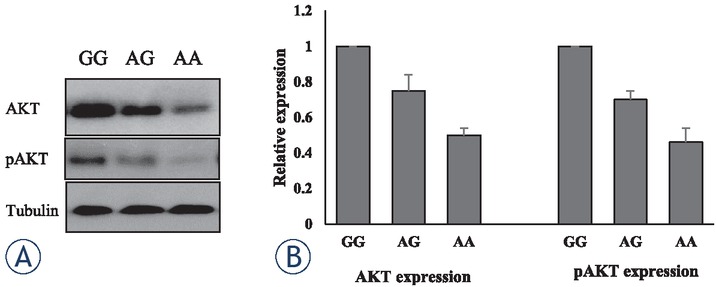
Figure 2
Association of AKT rs1130233 single nucleotide polymorphism with AKT expression and phosphorylation (A) Representative images of immunoblotting showing expression of AKT and pAKT in lymphocytes of GG, AG and AA carriers. (B) Mean densitometry profile of AKT and pAkt expression of different subjects of various genotypes. P < 0.05 GG vs AG, and P < 0.01 GG vs AA.
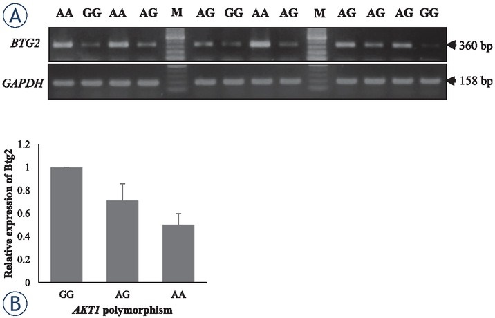
Figure 3
Association of AKT rs1130233 single nucleotide polymorphism BTG2 expression. Representative images have been shown. (A) BTG2 (360 bp) expression was determined in leukocytes of control individuals carrying GG, AG or AA genotypes of AKT1. GAPDH (158 bp) was used as a loading control. M = DNA Marker. (B) Mean densitometry profile of Btg2 mRNA expression of different subjects of various genotypes. P < 0.05 GG vs AG, and P < 0.01 GG vs AA.
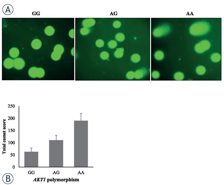
Figure 4
Association of AKT1 rs1130233 single nucleotide polymorphism with DNA damage. (A) Representative images of comet assay have been shown. (B) Comet tail was quantified in leukocytes of control individuals carrying GG, AG or AA genotypes of AKT1. P < 0.05 GG vs AG, and P < 0.01 GG vs AA.