
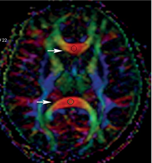
Figure 1
Fractional anisotropy value measurement at the genu and splenium of corpus callosum (circles).
Table 1
Fractional anisotropy (FA), axial diffusivity (AD), mean diffusivity (MD) and radial diffusivity (RD) values of the defined regions of interest in the patients with non-small cell lung cancer (NSCLC) and the control group
| Regions of interest | NSCLC patients (n = 25) | Control (n = 27) | ||||||
|---|---|---|---|---|---|---|---|---|
| FA | AD | MD | RD | FA | AD | MD | RD | |
| R ILF | 591.86±74.62 | 1500.75±122.17 | 863.76±65.15 | 545.27±96.29 | 575.66±79.55 | 1458.45±131.20 | 843.76±76.46 | 536.41±93.74 |
| L ILF | 600.06±59.94 | 1529.15±173.76 | 868.6±97.02 | 538.32±103.53 | 613.26±80.83 | 1543.02±136.96 | 856.6±105.47 | 513.38±125.10 |
| R SLF | 558.53±114.37 | 1299.28±201.73 | 754.18±67.60 | 481.63±88.42 | 595.02±87.69 | 1411.18±141.51 | 792.77±46.74 | 483.57±74.45 |
| L SLF | 532.01±118.55 | 1299.01±173.29 | 778.50±71.55 | 518.24±110.51 | 591.15±96.81 | 1416±149.16 | 795.97±59.44 | 485.95±82.96 |
| R FM | 554.53±100.65 | 1338.19±156.48 | 769.57±105.53 | 512.13±99.98 | 629.28±151.29 | 1508.51±288.33 | 813.40±80.35 | 465.84±139.28 |
| L FM | 507.28±109.36 | 1307.52±188.16 | 807.18±63.09 | 557.02±109.36 | 653.10±173.43 | 1605.51±322.02 | 836.1±86.54 | 451.52±168.22 |
| R ATR | 340.74±88.42 | 1103.33±81.11 | 808.82±52.68 | 661.56±75.32 | 356.29±93.06 | 1079.83±101.61 | 785.80±60.54 | 638.79±74.98 |
| L ATR | 346.5±80.36 | 1106.73±129.93 | 787.19±107.16 | 654.76±99.75 | 370.34±99.10 | 1106.84±127.16 | 795.80±78.49 | 640.28±91.63 |
| R ACR | 462.64±165.21 | 1257.80±260.67 | 796.58±212.71 | 593.33±239.64 | 474.52±112.45 | 1189.22±123.22 | 766.57±64.26 | 555.24±93.58 |
| L ACR | 426.54±118.96 | 1132.28±137.42 | 771.2±75.36 | 590.67±106.29 | 493.42±113.02 | 1204.54±150.69 | 762.29±68.51 | 541.17±86 |
| R EC | 421.5±103.19 | 1186.76±131.98 | 801.44±49.57 | 608.78±75.25 | 467.90±76.67 | 1206.38±142.25 | 785.90±67.27 | 575.67±66.99 |
| L EC | 473.82±92.65 | 1207.59±112.50 | 775.09±67.91 | 558.84±93.05 | 496.03±82.19 | 1220.38±150.32 | 766.20±69.01 | 539.11±75.26 |
| R IFOF | 492.68±71.61 | 1265.71±107.18 | 790.68±68.32 | 553.16±80.63 | 518.93±69.64 | 1319.51±149.14 | 803.93±64.48 | 546.14±61.75 |
| L IFOF | 475.18±85.00 | 1235.06±108.44 | 786.80±56.69 | 562.67±77.186 | 492.90±88.55 | 1305.21±143.83 | 817.78±68.26 | 574.07±80.16 |
| G CC | 805.78±79.27 | 1773.6±231.49 | 799.20±135.38 | 312±138.14 | 792.98±56.11 | 1810.47±167.43 | 822.28±72.58 | 328.19±75.54 |
| S CC | 806.87±83.00 | 1862.29±206.96 | 832.26±115.01 | 317.24±143.53 | 845.36±49.03 | 1823.75±243.74 | 775.42±113.42 | 251.25±81.05 |
| R FWM | 383.34±89.30 | 1171.85±148.03 | 822.22±66.41 | 647.41±81.58 | 388.92±109.23 | 1137.18±106.56 | 798.74±61.26 | 629.51±98.77 |
| L FWM | 377.06±89.53 | 1143.71±126.45 | 810.22±55.30 | 643.47±69.93 | 399.48±116.43 | 1147.24±114.15 | 798.29±78.91 | 623.81±114.37 |
| R POWM | 353.36±76.49 | 1155.84±130.45 | 837.16±70.62 | 677.82±70.49 | 394.78±91.19 | 1182.94±193.23 | 818.38±108.45 | 636.1±103.87 |
| L POWM | 344.49±75.64 | 1142.25±122.83 | 834.81±74.07 | 681.09±75.55 | 388.00±89.02 | 1254.38±148.62 | 871.37±93.75 | 679.86±102.15 |
[i] ACR = anterior corona radiata; ATR = anterior thalamic radiation; CC = splenium of corpus callosum; EC = external capsule; FM = forceps minor; G CC = genu of corpus callosum; IFOF = inferior fronto occipital fasciculus; ILF = inferior longitudinal fasciculus; L = left; POWM = parieto-occipital white matter; R = right; S CC = superior corpus callosum; S FWM = superior frontal white matter; SLF = superior longitudinal fasciculus
Table 2
Comparison of diffusion tensor imaging (DTI) values of the defined anatomic locations before and after chemotherapy (the significant results are in italics)
| Group 1 (n:7) | Group 2 (n:18) | ||||||
|---|---|---|---|---|---|---|---|
| Region of interest | Pre-chemotherapy values (Median (min-max)) | Post-chemotherapy values (Median (min-max)) | P value | Regions of interest | Pre-chemotherapy values (Median (min-max) | Post-chemotherapy values (Median (min-max) | P value |
| R ILF FA | 623 (514–703.3) | 522 (418.7–679.8) | 0.028 | R ILF FA | 600 (395.3–694.5) | 593 (336–707) | 0.89 |
| R FM FA | 539 (448–591) | 471 (379.7–615.8) | 0.31 | R FM FA | 525 (454.5–864) | 487 (322.3–712) | 0.022 |
| R FM RD | 487.4 (465.5–605.65) | 619.4 (439.5–667.85) | 0.062 | R FM RD | 533.275 (201–689) | 580.35 (367.8–819.15) | 0.011 |
| R FWM AD | 1123.5 (912.8–1244) | 1135.5 (988.3–1265.5) | 0.39 | R FWM AD | 1157.65 (1011.3–1556.5) | 1104.15 (854.8–1256.3) | 0.008 |
| R FWM MD | 786.1 (699.1–871.26) | 823.26 (658.93–855.1) | 0.31 | R FWM MD | 793.01 (658.2–955.36) | 812.33(704.53–1006.9) | 0.029 |
Table 3
Comparison of the diffusion tensor imaging (DTI) results of the two chemotherapy regimens on the defined anatomic locations (only the significant results are shown)
| Region of interest | Cisplatin | Carboplatin | P |
|---|---|---|---|
| Left POWM FA | 389 (273.3–531) | 319 (167.3–423) | 0.034 |
| Left POWM MD | 767.6 (727.53–925.27) | 837.13 (737.2–1071.77) | 0.034 |
| Left POWM RD | 606.1 (507.9–726.65) | 705.92 (592.80–905.15) | 0.029 |