
Figure 1
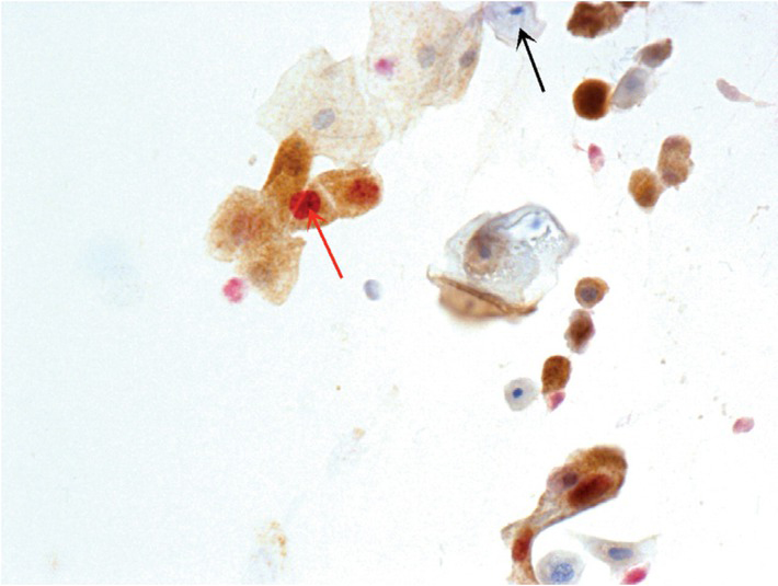
Positive reaction was defined as p16 brown signal and Ki-67 red signal (red arrow) present in the same cell with red stained nucleus and brown stained cytoplasm. Note: negative p16/Ki-67 dual immunostaining (p16/Ki-67 DS) reaction (black arrow) (p16/Ki-76 DS, magnification 400x).
Table 1
p16/Ki-67 dual immunostaining (p16/Ki-67 DS) positivity and number of positive cells among different smear results
| 0 (Negative) | 1 | 2 | 3 | 4 | ≥ 5 | 1+ (Total Positive) | ||
|---|---|---|---|---|---|---|---|---|
| Normal | 70 (69.3) | 13 (12.9) | 9 (8.9) | 1 (1.0) | 1 (1.0) | 7 (6.9) | 31 (30.7) | 101 |
| ASC-US | 14 (58.3) | 0 (0.0) | 1 (4.2) | 2 (8.3) | 1 (4.2) | 6 (25.0) | 10 (41.7) | 24 |
| LSIL | 7 (50.0) | 3 (21.4) | 0 (0.0) | 0 (0.0) | 1 (7.1) | 3 (21.4) | 7 (50.0) | 14 |
| AGC-N | 0 (0.0) | 0 (0.0) | 0 (0.0) | 0 (0.0) | 0 (0.0) | 1 (100.0) | 1 (100.0) | 1 |
| HSIL | 0 (0.0) | 1 (3.4) | 0 (0.0) | 0 (0.0) | 0 (0.0) | 28 (96.6) | 29 (100.0) | 29 |
| ASC-H | 0 (0.0) | 0 (0.0) | 0 (0.0) | 1(25.0) | 0 (0.0) | 3 (75.0) | 4 (100.0) | 4 |
| Inv. cancer | 0 (0.0) | 0 (0.0) | 0 (0.0) | 0 (0.0) | 0 (0.0) | 1(100.0) | 1 (100.0) | 1 |
| Total | 91 (52.3) | 17 (9.8) | 10 (5.7) | 4 (2.3) | 3 (1.7) | 49 (28.2) | 83 (47.7) | 174 |
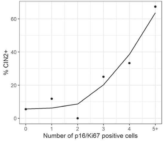
Figure 2
The association between the number of p16/Ki-67 dual immunostaining (p16/Ki-67 DS) positive cells and the risk for cervical intraepithelial neoplasia (CIN2+). Observed values are marked as points. Smoothed line (Method spline) is added for better trend representation.
Table 2
Cervical intraepithelial neoplasia (CIN2+) according to the number of p16/Ki-67 dual immunostaining (p16/Ki-67 DS) positive cells
| p16/Ki-67 | Histology | ||
|---|---|---|---|
| Positive cells | n | < CIN2 n (%) | CIN2+ n (%) |
| 0 | 91 | 86 (94.5) | 5 (5.5) |
| 1 | 17 | 15 (88.2) | 2 (11.8) |
| 2 | 10 | 10 (100.0) | 0 (0.0) |
| 3 | 4 | 3 (75.0) | 1 (25.0) |
| 4 | 3 | 2 (66.7) | 1 (33.3) |
| ≥ 5 | 49 | 16 (32.7) | 33 (67.3) |
| Total | 174 | 132 (75.9) | 42 (24.1) |
Table 3
Diagnostic performance of p16/Ki-67 dual immunostaining (p16/Ki-67 DS) according to cytology results and according to different cut-offs (number of positive cells) in detecting cervical intraepithelial neoplasia (CIN2+)
| Sensitivity (%, 95% CI) | Specificity (%, 95% CI) | PPV† (%, 95% CI) | NPV‡ (%, 95% CI) | ||
|---|---|---|---|---|---|
| Cytology result | |||||
| Negative | 66.7 | 70.4 | 6.5 | 98.6 | |
| (n = 101) | 3 | (9.4–99.2) | (60.3–79.2) | (0.8–21.4) | (92.3–100.0) |
| ASC-US | 6 | 50.0 | 61.1 | 30.0 | 78.6 |
| (n = 24) | (11.8–88.2) | (35.7–82.7) | (6.7–65.2) | (49.2–95.3) | |
| LSIL | 2 | 50.0 | 50.0 | 14.3 | 85.7 |
| (n = 14) | (1.3–98.7) | (21.1–78.9) | (0.4–57.9) | (42.1–99.6) | |
| HSIL | 100.0 | 0.0 | 89.7 | ||
| (n = 29) | 26 | (86.8–100.0) | (0.0–70.8) | (72.6–97.8) | / |
| Number of positive p16/Ki-67 cells cut-off | |||||
| 1+ | 88.1 | 65.2 | 44.6 | 94.5 | |
| (n = 174) | 42 | (74.4–96.0) | (56.4–73.2) | (33.7–55.9) | (87.6–98.2) |
| 2+ | 42 | 83.3 | 76.5 | 53.0 | 93.5 |
| (n = 174) | (68.6–93.0) | (68.4–83.5) | (40.3–65.4) | (87.1–97.4) | |
| 3+ | 42 | 83.3 | 84.1 | 62.5 | 94.1 |
| (n = 174) | (68.6–93.0) | (76.7–89.9) | (48.5–75.1) | (88.2–97.6) | |
| 4+ | 42 | 81.0 | 86.4 | 65.4 | 93.4 |
| (n = 174) | (65.9–91.4) | (79.3–91.7) | (50.9–78.0) | (87.5–97.1) | |
| 5+ | 78.6 | 87.9 | 67.3 | 92.8 | |
| (n = 174) | 42 | (63.2–89.7) | (81.1–92.9) | (52.5–80.1) | (86.8–96.7) |
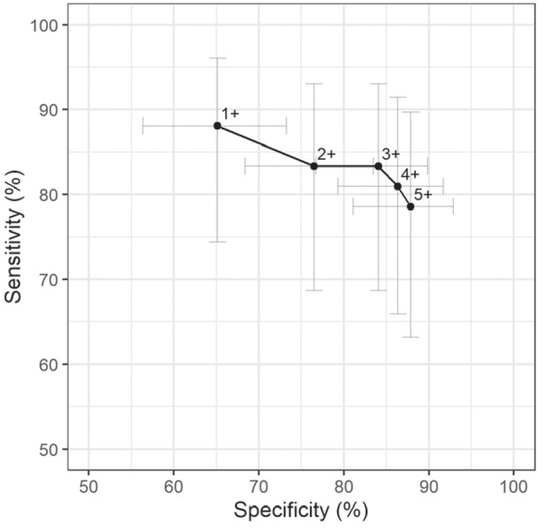
Figure 3
Diagnostic performance of p16/Ki-67 dual immunostaining (p16/Ki-67 DS) at different cut-offs (number of positive cells).