
Figure 1
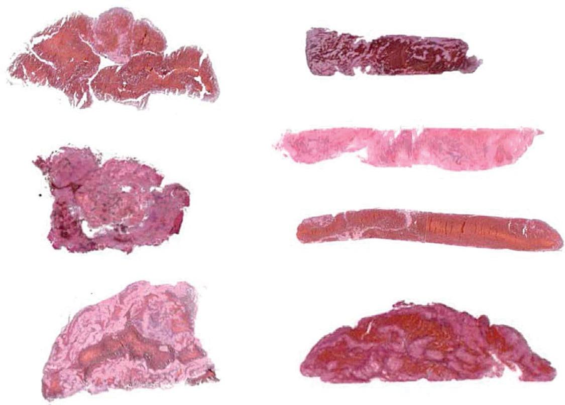
Figure 2
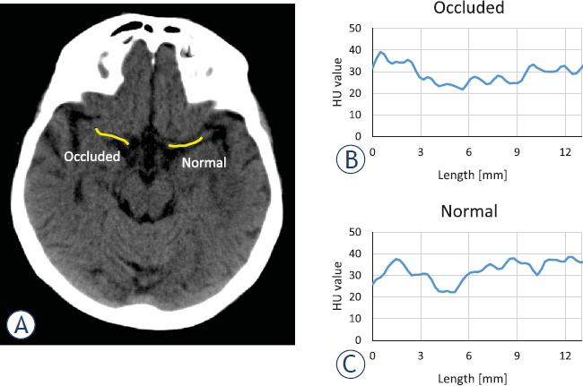
Figure 3
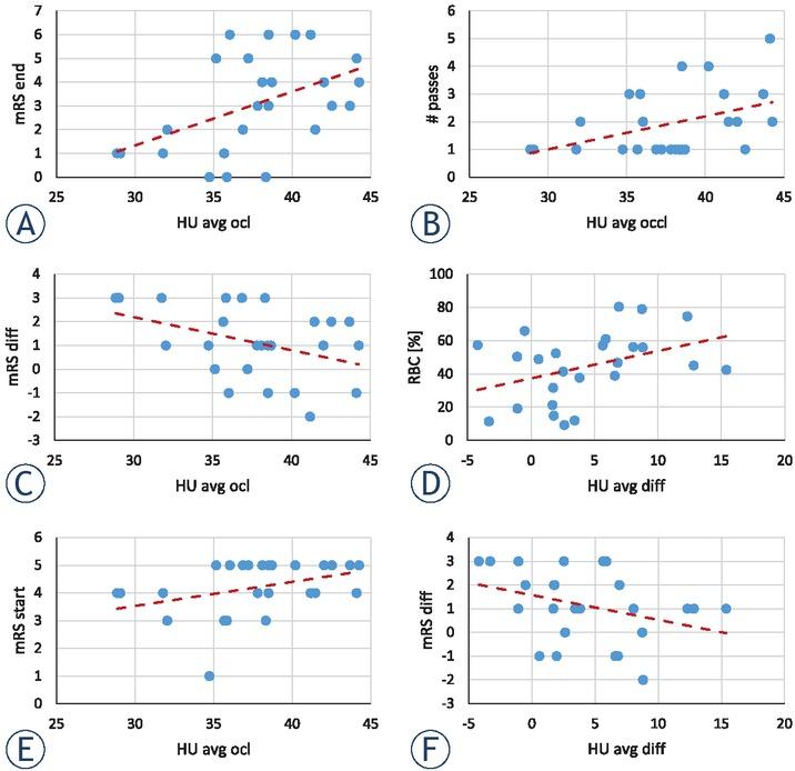
Linear regression and Pearson correlation coefficient analysis of data group pairs with statistically most significant correlations and linear regression parameters
| Data group pair | Linear regression | Pearson coefficient | |||
|---|---|---|---|---|---|
| x-values | y-values | k | p-value | R2 | ρ |
| HU avg occl | mRS end | 0.227 ± 0.086 | 0.015 | 0.233 | 0.483 |
| HU avg occl | # passes | 0.119 ± 0.052 | 0.031 | 0.186 | 0.432 |
| HU avg occl | mRS diff | -0.140 ± 0.067 | 0.049 | 0.158 | -0.398 |
| HU avg diff | RBC [%] | 1.646 ± 0.809 | 0.053 | 0.153 | 0.391 |
| HU avg occl | mRS start | 0.087 ± 0.045 | 0.065 | 0.140 | 0.374 |
| HU avg diff | mRS diff | -0.104 ± 0.059 | 0.093 | 0.118 | -0.343 |
Experimental data of patients qualified for the study, which include CT, histological, clinical and procedure parameters
| CT parameters | Histology | Clinical parameters | Procedure parameters | |||||||||||||||||
|---|---|---|---|---|---|---|---|---|---|---|---|---|---|---|---|---|---|---|---|---|
| # | HU avg | HU var | Stroke etiology | Therapy before stoke | NIHSS | mRS | ||||||||||||||
| occl | nor | diff | diff | occl | nor | diff | diff | L | RBC | strt | end | diff | strt | end | diff | Rd | # pass | |||
| 1. | 29.1 | 32.4 | -3.3 | -11.4 | 4.20 | 4.42 | -0.22 | -5.0 | 20.8 | 11.5 | AT | / | 21 | 1 | 20 | 4 | 1 | 3 | 42 | 1 |
| 2. | 42.0 | 29.2 | 12.8 | 30.5 | 4.58 | 8.98 | -4.40 | -96.3 | 16.3 | 45.1 | CE | / | 17 | 8 | 9 | 5 | 4 | 1 | 62 | 2 |
| 3. | 38.5 | 35.1 | 3.4 | 8.9 | 3.56 | 2.75 | 0.80 | 22.6 | 17.0 | 12.0 | CE | / | 23 | 7 | 16 | 4 | 3 | 1 | 65 | 1 |
| 4. | 36.9 | 34.3 | 2.5 | 6.8 | 11.66 | 4.58 | 7.08 | 60.7 | 17.5 | 41.4 | CE | AA | 18 | 3 | 15 | 5 | 2 | 3 | 77 | 1 |
| 5. | 43.7 | 44.2 | -0.5 | -1.2 | 3.55 | 3.86 | -0.31 | -8.7 | 17.9 | 65.9 | AT | / | 26 | 6 | 20 | 5 | 3 | 2 | 97 | 3 |
| 6. | 35.8 | 30.0 | 5.9 | 16.4 | 4.17 | 2.23 | 1.95 | 46.7 | 16.3 | 61.0 | CE | / | 7 | 0 | 7 | 3 | 0 | 3 | 38 | 3 |
| 7. | 31.8 | 36.0 | -4.2 | -13.2 | 2.77 | 3.26 | -0.49 | -17.8 | 22.2 | 57.3 | AT | / | 13 | 3 | 10 | 4 | 1 | 3 | 90 | 1 |
| 8. | 37.8 | 38.9 | -1.1 | -2.9 | 2.82 | 4.45 | -1.63 | -58.0 | 26.9 | 50.3 | CE | / | 14 | 3 | 11 | 4 | 3 | 1 | 77 | 1 |
| 9. | 32.0 | 24.0 | 8.1 | 25.1 | 9.50 | 4.90 | 4.60 | 48.4 | 22.7 | 56.3 | CE | ACAA | 5 | 2 | 3 | 3 | 2 | 1 | 115 | 2 |
| 10. | 38.5 | 36.5 | 2.0 | 5.1 | 3.20 | 2.80 | 0.40 | 12.5 | 19.7 | 52.4 | CE | / | 26 | 42 | -16 | 5 | 6 | -1 | 82 | 4 |
| 11. | 38.3 | 39.4 | -1.1 | -2.8 | 5.49 | 3.66 | 1.83 | 33.4 | 17.1 | 19.1 | AT | / | 12 | 0 | 12 | 3 | 0 | 3 | 60 | 1 |
| 12. | 35.7 | 33.9 | 1.7 | 4.9 | 3.56 | 5.39 | -1.83 | -51.2 | 13.9 | 31.8 | CE | AA | 6 | 3 | 3 | 3 | 1 | 2 | 69 | 1 |
| 13. | 44.3 | 31.9 | 12.3 | 27.8 | 4.66 | 5.11 | -0.46 | -9.8 | 22.5 | 74.7 | AT | / | 19 | 12 | 7 | 5 | 4 | 1 | 108 | 2 |
| 14. | 44.1 | 37.5 | 6.6 | 15.0 | 2.60 | 3.92 | -1.32 | -50.9 | 19.3 | 38.9 | CE | / | 18 | 20 | -2 | 4 | 5 | -1 | 63 | 5 |
| 15. | 36.0 | 35.4 | 0.6 | 1.6 | 3.48 | 2.67 | 0.81 | 23.4 | 17.9 | 48.8 | CE | / | 42 | 42 | 0 | 5 | 6 | -1 | 75 | 2 |
| 16. | 38.1 | 36.4 | 1.7 | 4.5 | 4.16 | 5.45 | -1.30 | -31.2 | 18.6 | 21.3 | AT | / | 15 | 4 | 11 | 5 | 4 | 1 | 77 | 1 |
| 17. | 42.5 | 40.7 | 1.8 | 4.2 | 3.11 | 3.74 | -0.63 | -20.4 | 13.5 | 14.8 | CE | AA | 14 | 2 | 12 | 5 | 3 | 2 | 53 | 1 |
| 18. | 38.7 | 23.3 | 15.4 | 39.8 | 5.66 | 4.79 | 0.87 | 15.3 | 19.1 | 42.4 | CE | AC | 22 | 3 | 19 | 5 | 4 | 1 | 43 | 1 |
| 19. | 34.7 | 30.9 | 3.8 | 11.0 | 5.61 | 4.43 | 1.18 | 21.1 | 20.9 | 37.7 | AT | / | 3 | 1 | 2 | 1 | 0 | 1 | 60 | 1 |
| 20. | 41.2 | 32.4 | 8.8 | 21.4 | 5.14 | 4.58 | 0.56 | 10.9 | 19.3 | 56.0 | AT | AC | 11 | 40 | -29 | 4 | 6 | -2 | 76 | 3 |
| 21. | 37.2 | 28.5 | 8.7 | 23.5 | 3.29 | 5.27 | -1.99 | -60.4 | 29.2 | 79.1 | AT | / | 19 | 10 | 9 | 5 | 5 | 0 | 61 | 1 |
| 22. | 35.2 | 32.5 | 2.6 | 7.5 | 3.02 | 2.79 | 0.24 | 7.9 | 13.6 | 9.3 | CE | / | 16 | 9 | 7 | 5 | 5 | 0 | 95 | 3 |
| 23. | 41.5 | 34.5 | 6.9 | 16.7 | 5.14 | 4.47 | 0.67 | 13.0 | 21.5 | 80.4 | AT | / | 16 | 3 | 13 | 4 | 2 | 2 | 65 | 2 |
| 24. | 28.9 | 23.2 | 5.6 | 19.5 | 3.52 | 4.72 | -1.20 | -34.1 | 12.1 | 57.1 | CE | / | 13 | 3 | 10 | 4 | 1 | 3 | 55 | 1 |
| 25. | 40.2 | 33.4 | 6.8 | 17.0 | 2.91 | 4.30 | -1.39 | -47.8 | 18.7 | 46.8 | AT | AA | 42 | 42 | 0 | 5 | 6 | -1 | 90 | 4 |