
Figure 1
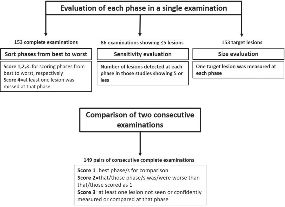
Flowchart of the reading protocol. There were four steps: sort of phases from best to worst, sensitivity study based only on the reading of examinations having ≤5 metastases, measurement of one target lesion in each examination and evaluation of phases for comparing metastases in 2 consecutive follow-up scans.
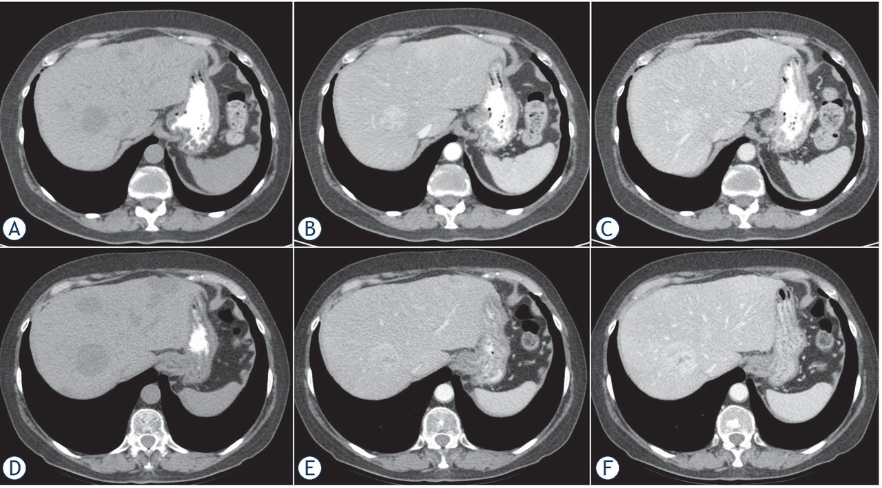
Figure 2
48-year-old woman with hepatic metastases from breast cancer. Upper row corresponds to previous study and lower row is follow-up. Previous unenhanced phase (A) show more lesions than either the arterial (B) and portal (C) phases. At follow-up 6 weeks later, worsening seen in unenhanced phase (D) cannot be confidently confirmed only by arterial (E) and portal (F) phases.
Table 1
Distribution of scores of each phase by both readers in 153 examinations
| Reader 1 | |||||||
|---|---|---|---|---|---|---|---|
| UNENHANCED | |||||||
| 1 | 2 | 3 | 4 | ||||
| 1 | 32 | 2 | 2 | 0 | |||
| 1 | ARTERIAL | 2 | 16 | 20 | 10 | 1 | |
| 3 | 9 | 10 | 0 | 0 | |||
| 4 | 4 | 0 | 0 | 0 | |||
| 1 | 1 | 0 | 2 | 0 | |||
| PORTAL | 2 | ARTERIAL | 2 | 6 | 0 | 0 | 0 |
| 3 | 15 | 0 | 0 | 1 | |||
| 0 | 0 | 0 | |||||
| 1 | 0 | 1 | 0 | 0 | |||
| 3 | ARTERIAL | 2 | 4 | 0 | 0 | 0 | |
| 3 | 4 | 0 | 0 | 0 | |||
| 4 | 0 | 0 | 0 | 0 | |||
| 2 | 1 | 0 | 0 | 0 | |||
| 4 | ARTERIAL | 4 | 9 | 0 | 0 | 0 |
| Reader 2 | |||||||
|---|---|---|---|---|---|---|---|
| UNENHANCED | |||||||
| 1 | 2 | 3 | 4 | ||||
| 1 | 30 | 5 | 1 | 0 | |||
| 2 | 18 | 20 | 7 | 1 | |||
| 1 | ARTERIAL | 3 | 7 | 12 | 0 | 0 | |
| 4 | 6 | 0 | 0 | 0 | |||
| 1 | 2 | 0 | 2 | 1 | |||
| 2 | ARTERIAL | 2 | 6 | 0 | 0 | 1 | |
| PORTAL | 3 | 14 | 0 | 0 | 0 | ||
| 4 | 3 | 0 | 0 | 0 | |||
| 1 | 1 | 0 | 0 | 0 | |||
| 3 | ARTERIAL | 2 | 3 | 0 | 0 | 0 | |
| 3 | 3 | 0 | 0 | 0 | |||
| 4 | ARTERIAL | 2 | 1 | 0 | 0 | 0 | |
| 4 | 9 | 0 | 0 | 0 |
Table 2
Distribution of scores of each phase for comparing metastatic lesions in two consecutive scans by both readers in 149 examinations
| Reader 1 | ||||||
|---|---|---|---|---|---|---|
| UNENHANCED | ||||||
| 1 | 2 | 3 | ||||
| 1 | 35 | 6 | 1 | |||
| 1 | ARTERIAL | 2 | 42 | 20 | 0 | |
| 3 | 2 | 0 | 0 | |||
| PORTAL | 1 | 3 | 0 | 1 | ||
| 2 | ARTERIAL | 2 | 23 | 0 | 0 | |
| 3 | 2 | 0 | 0 | |||
| 3 | ARTERIAL | 2 | 1 | 0 | 0 | |
| 3 | 13 | 0 | 0 |
| Reader 2 | ||||||
|---|---|---|---|---|---|---|
| UNENHANCED | ||||||
| 1 | 2 | 3 | ||||
| 1 | 39 | 4 | 1 | |||
| 1 | ARTERIAL | 2 | 34 | 24 | 0 | |
| 3 | 2 | 0 | 0 | |||
| PORTAL | 1 | 1 | 0 | 2 | ||
| 2 | ARTERIAL | 2 | 26 | 0 | 0 | |
| 3 | 2 | 0 | 0 | |||
| 2 | 1 | 0 | 0 | |||
| 3 | ARTERIAL | 3 | 13 | 0 | 0 |
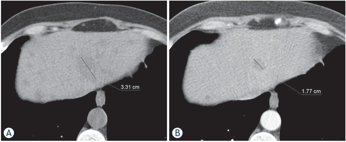
Figure 3
56-year-old woman with hepatic metastases from breast cancer. Measurement of target lesion in unenhanced phase (A) is 33 mm and in the portal phase (B) is 18 mm, that represent a 45% difference. A part of the metastatic lesion posteriorly is scarcely seen as a subtle increase attenuation in the portal phase, but both readers failed to consider that area as part of the lesion, and measured only the hypoattenuating component. Note how unenhanced phase also shows other small lesions not seen in the portal phase.