
Table 1
Overview of the included patients
| Localization | n (%) |
|---|---|
| Tonsil | 7 (24.1) |
| Tongue | 7 (24.1) |
| Hypopharynx | 6 (20.7) |
| Oropharynx | 5 (17.2) |
| Larynx | 4 (13.9) |
| Grading | |
| Moderate (G2) | 12 (37.5) |
| Poor (G3) | 17 (62.5) |
| Stage | |
| T2 | 6 (20.7) |
| T3 | 10 (34.5) |
| T4 | 13 (44.8) |
| Nodal status | |
| Nodal positive | 25 (86.2) |
| Nodal negative | 4 (13.8) |
Table 2
Overview of the investigated apparent diffusion coefficient (ADC) histogram parameters
| Parameter | Mean ± standard deviation, x10-3 mm2/s | Range, x10-3 mm2/s |
|---|---|---|
| Mean | 1.14 ± 0.22 | 0.78-1.68 |
| Min | 0.70 ± 0.25 | 0.17-1.24 |
| Max | 1.78 ± 0.32 | 1.35-2.39 |
| P10 | 0.90 ± 0.21 | 0.54-1.41 |
| P25 | 1.0 ± 0.21 | 0.64-1.50 |
| P75 | 1.27 ± 0.24 | 0.87-1.82 |
| P90 | 1.41 ± 0.26 | 0.94-2.03 |
| Median | 1.12 ± 0.22 | 0.76-1.64 |
| Mode | 0.97 ± 0.28 | 0.78-1.55 |
| Kurtosis | 3.74 ± 1.40 | 2.23-7.93 |
| Skewness | 0.49 ± 0.49 | -0.54-1.49 |
| Entropy | 2.44 ± 0.49 | 1.67-3.75 |
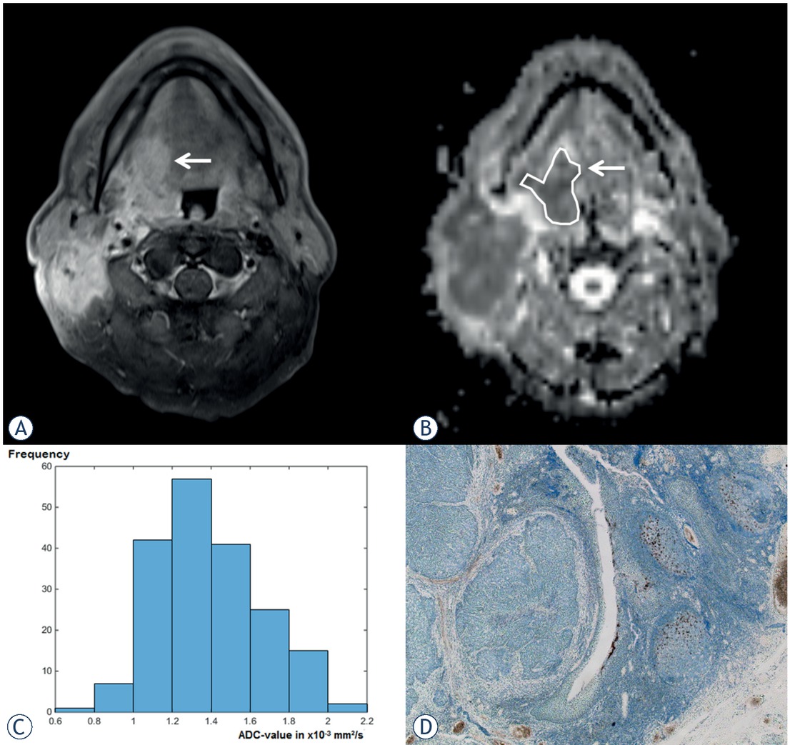
Figure 1
Representative case with a negative programmed cell death ligand 1 (PD-L1) score. (A) Postcontrast T1-weighted image shows a large enhancing tumor of the right tongue base (arrow). A large lymph node metastasis on the right side can also be appreciated. (B) Apparent diffusion coefficient (ADC) map of the same slide. Both lesions show a marked hypointensity consistent with a diffusion restriction. The tumor (arrow) is surrounded with a region of interest, which is drawn on every tumor showing slide within the margins of the tumor in accordance to the corresponding T1-weighted images. (C) The resulting ADC histogram of the tumor. The histogram analysis parameters (× 10-3 mm2/s) are as follows: ADCmin = 0.79, ADCmean = 1.38, ADCmax = 2.05, P10 = 1.05, P25 = 1.19, P75 = 1.53, P90 = 1.78, median = 1.35,mode = 1.27, kurtosis = 2.61, skewness = 0.41 and entropy = 2.44. (D) The corresponding PD-L1 stained specimen. No staining can be appreciated resulting in a value of 0 in all calculated scores.
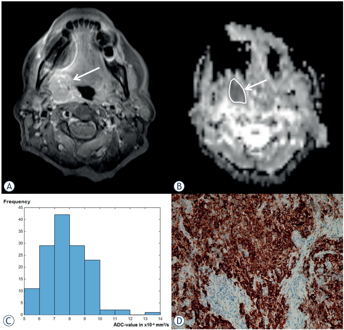
Figure 2
Representative case with a positive programmed cell death ligand 1 (PD-L1) score. (A) On the postcontrast T1-weighted image a large enhancing tumor of the side within the right palatine tonsil (arrow). Susceptibility artifacts due to dental material can also be appreciated. (B) Apparent diffusion coefficient (ADC) map on the corresponding slide. A marked hypointensity can be seen consistent with a diffusion restriction. (C) The resulting ADC histogram of the tumor. The histogram analysis parameters (× 10-3 mm2/s) are as follows: ADCmin = 0.51, ADCmean = 0.78, ADCmax = 1.36, P10 = 0.63, P25 = 0.69, P75 = 0.87, P90 = 0.94, median = 0.76, mode = 0.69, kurtosis = 4.57, skewness = 0.72, and entropy = 2.41. (D) The corresponding PD-L1 stained specimen. A weak staining of PD-L1 for tumor cells and associating immune cells can be appreciated. The resulting score values are 5 for the combined positive score and 3 for the immune cell score.
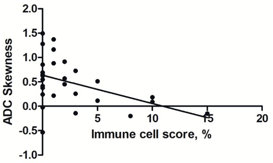
Figure 3
Correlation analysis between skewness derived from apparent diffusion coefficient (ADC) and the immune cell score (ICS). The resulting Spearman’s correlation coefficient is r = -0.38, p = 0.04.