
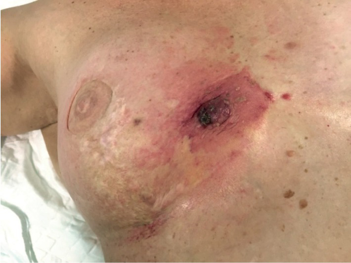
Figure 1
Clinical presentation of patient with angiosarcoma of right reconstructed breast.
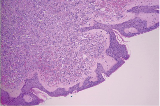
Figure 2
Pathologic hematoxylin and eosin (HE) specimen showing radiotherapy-associated angiosarcoma (RAA) involving dermis.
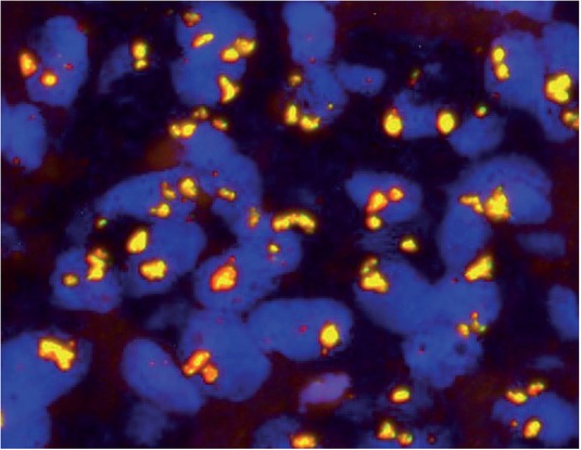
Figure 3
Amplification of the MYC oncogene was confirmed using FISH method on tumor specimen.
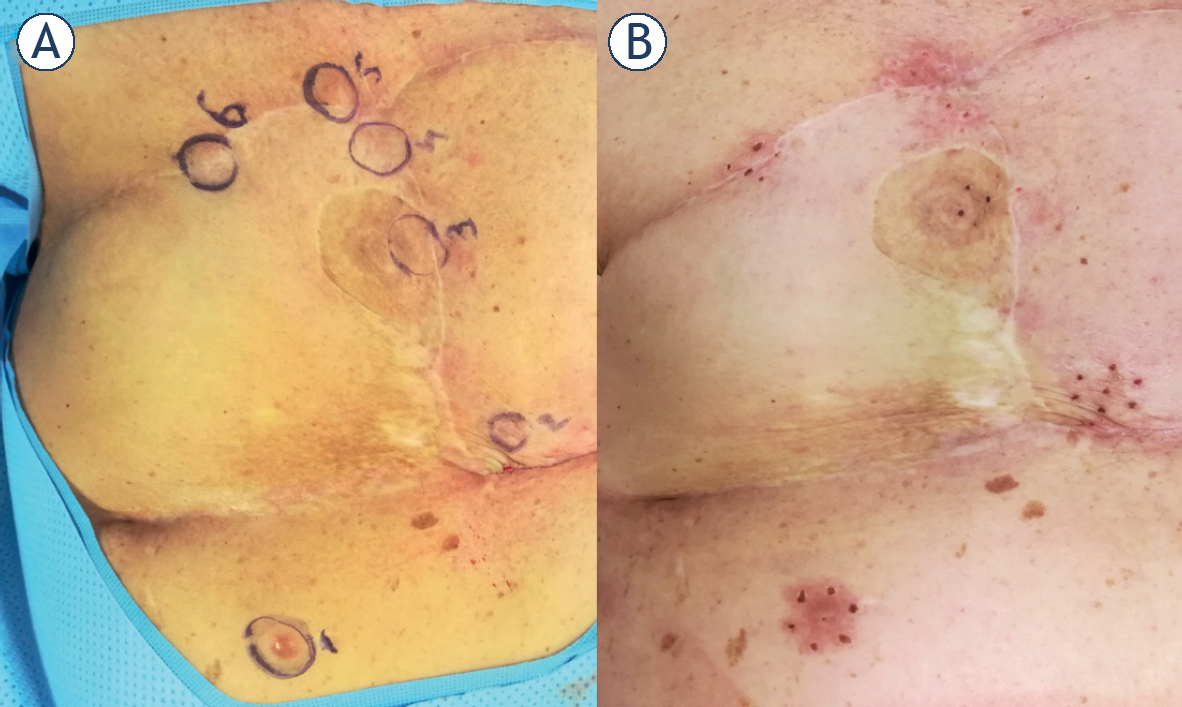
Figure 4
Treatment with electrochemotherapy. (A) Just before the treatment nodules were marked for evaluation; (B) 1 month after the treatment all nodules were in complete response.
Table 1
Case reports with radiotherapy-associated angiosarcoma (RAA) after autologous breast reconstruction
| First author | Year published | Case # | Years after RT | Type of cancer surgery | Type of breast reconstruction |
|---|---|---|---|---|---|
| Hanasono et al.4 | 2005 | 1 | 6 | mastectomy | DIEP flap |
| Aljarrah et al.5 | 2014 | 2 | 6 | mastectomy | TRAM flap |
| Yip et al.6 | 2019 | 3 | 5 | mastectomy and axillary lymph node dissection | LD flap |
| Yip et al.6 | 2019 | 4 | 10 | mastectomy and axillary lymph node dissection | DIEP flap |