
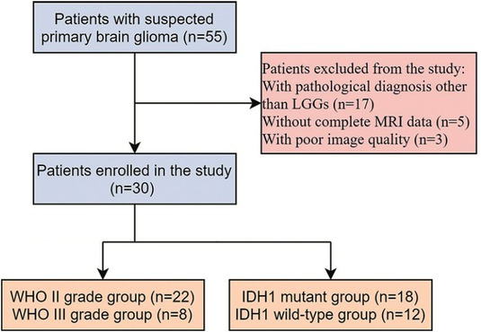
Figure 1
Flowchart of study design.
Table 1
Patient characteristics
| Characteristic | IDH1 mutants (n = 18) | IDH1 wild-type (n = 12) |
|---|---|---|
| Mean age (y)a | 42.8 (22–67) | 47.9 (19–78) |
| Sex distribution (M/F)b | 10/8 | 7/5 |
| WHO grade | ||
| II | 15 | 7 |
| III | 3 | 5 |
| Histologic type | ||
| Astrocytoma | 12 | 5 |
| Oligodendroglioma | 3 | 0 |
| Oligoastrocytoma | 0 | 1 |
| Anaplastic astrocytoma | 1 | 3 |
| Anaplastic oligodendroglioma | 1 | 2 |
| Anaplastic oligoastrocytoma | 1 | 1 |
[i] * Mean (range) or count is reported; a = significant difference in age was noted between isocitrate dehydrogenase 1 (IDH1) mutant and wild-type groups (P = 0.020); b = no significant difference in sex distribution was noted between IDH1 mutant and wild-type groups (P = 0.769)
F = female; M = male
Table 2
Parameters derived from dynamic contrast-enhanced (DCE) MRI and diffusion-weighted imaging (DWI) between WHO grade II and III gliomas
| Parameter | Grade II | Grade III | P-value |
|---|---|---|---|
| Ktrans (min−1) | 0.067 ± 0.048 | 0.116 ± 0.064 | 0.013 |
| ve | 0.071 ± 0.057 | 0.401 ± 0.344 | 0.018 |
| vp | 0.036 ± 0.020 | 0.051 ± 0.018 | 0.035 |
| ADC0,1000 (×10−3 mm2/s) | 1.093 ± 0.203 | 0.904 ± 0.184 | 0.028 |
| SPF (%) | 10.78 ± 4.378 | 16.391 ± 5.471 | 0.012 |
| D (×10−3 mm2/s) | 1.194 ± 0.261 | 0.949 ± 0.169 | 0.021 |
| D* (×10−3 mm2/s) | 6.692 ± 1.564 | 8.618 ± 2.215 | 0.037 |
| f (%) | 3.315 ± 1.536 | 6.380 ± 3.419 | 0.020 |
[i] * P-values are considered significant at P < 0.05.
ADC = apparent diffusion coefficient; D = diffusion coefficient; D* = pseudo-diffusion coefficient; f = perfusion fraction; Ktrans = volume transfer constant; ve = extravascular extracellular volume fraction; vp = vascular plasma volume fraction; SPF = simplified perfusion fraction
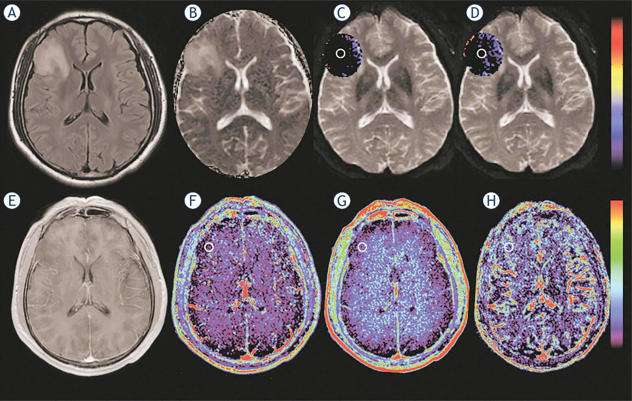
Figure 2
Images obtained in a 44-year-old man with astrocytoma (isocitrate dehydrogenase 1 [IDH1] mutant glioma). (A) Fluid-attenuated inversion recovery (FLAIR) image shows a heterogeneous hyperintense lesion in the right frontal lobe. (B) Apparent diffusion coefficient (ADC)0,1000 map shows increased ADC value in the lesion. (C, D) Intravoxel incoherent motion (IVIM) perfusion fraction (f) and simplified perfusion fraction (SPF) maps show no increased values in the corresponding area of the hyperintense lesion as shown in (A). (E) On contrast-enhanced T1-weighted image, the lesion is non-enhancing. (F–H) Dynamic contrast-enhanced (DCE) MRI parametric maps of volume transfer constant (Ktrans), extravascular extracellular volume fraction (ve) and vascular plasma volume fraction (vp) show no increased values in the lesion. Regions of interest are marked on parametric maps.
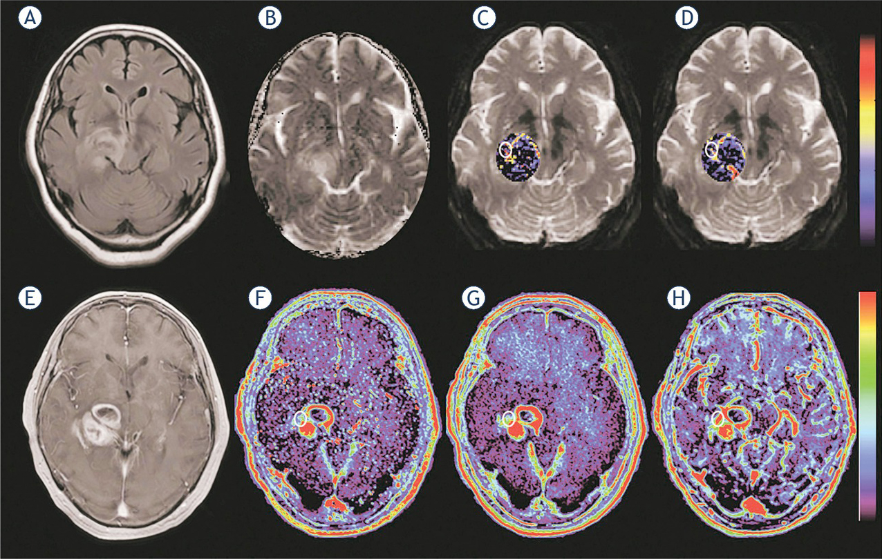
Figure 3
Images obtained in a 72-year-old woman with astrocytoma (isocitrate dehydrogenase 1 [IDH1] wildtype glioma). (A) FLAIR shows a heterogeneous hyperintense lesion in the right hemisphere. (B) Apparent diffusion coefficient (ADC)0,1000 map shows a mixed pattern of high and intermediate ADC values in the lesion. (C, D) Intravoxel incoherent motion (IVIM) perfusion fraction (f) and simplified perfusion fraction (SPF) maps show markedly increased f and SPF values in the corresponding area of the contrast-enhanced lesion as shown in (E). (E) On contrast-enhanced T1-weighted image, the lesion is vividly enhanced. (F–H) Dynamic contrast-enhanced (DCE) MRI parametric maps of volume transfer constant (Ktrans), extravascular extracellular volume fraction (ve) and vascular plasma volume fraction (vp) show obviously increased values in the corresponding area of the contrast-enhanced lesion. Regions of interest are marked on parametric maps.
Table 3
Parameters derived from dynamic contrast-enhanced (DCE) MRI and diffusion-weighted imaging (DWI) between isocitrate dehydrogenase 1 (IDH1) mutant and wild-type gliomas
| Parameter | IDH1 mutant | IDH1 wild-type | P-value* |
|---|---|---|---|
| Ktrans (min−1) | 0.054 ± 0.024 | 0.123 ± 0.073 | 0.007 |
| ve | 0.052 ± 0.035 | 0.121 ± 0.080 | 0.007 |
| vp | 0.032 ± 0.015 | 0.051 ± 0.022 | 0.015 |
| ADC0,1000 (×10−3 mm2/s) | 1.123 ± 0.185 | 0.923 ± 0.199 | 0.009 |
| SPF (%) | 9.572 ± 3.437 | 16.332 ± 4.925 | < 0.001 |
| D (×10−3 mm2/s) | 1.108 ± 0.245 | 0.959 ± 0.146 | 0.047 |
| D* (×10−3 mm2/s) | 6.546 ± 1.757 | 8.196 ± 1.794 | 0.020 |
| f (%) | 3.080 ± 1.581 | 5.712 ± 2.924 | 0.005 |
[i] * P-values are considered significant at P < 0.05
ADC = apparent diffusion coefficient; D = diffusion coefficient; D* = pseudo-diffusion coefficient; Ktrans = volume transfer constant; f = perfusion fraction; SPF = simplified perfusion fraction; v= e extravascular extracellular volume fraction; vp = vascular plasma volume fraction
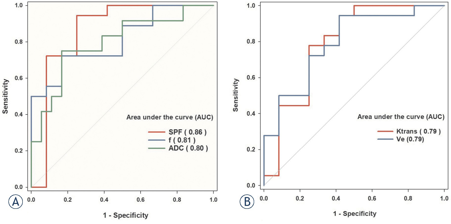
Figure 4
Receiver operating characteristic (ROC) curves and corresponding area under the curve values for (A) diffusion-weighted imaging (DWI) parameters (simplified perfusion fraction [SPF], perfusion fraction [f], apparent diffusion coefficient [ADC]0,1000) and (B) dynamic contrast-enhanced (DCE) MRI parameters (transfer constant [Ktrans], extravascular extracellular volume fraction [ve] and vascular plasma volume fraction [vp]) in the differentiation of isocitrate dehydrogenase 1 (IDH1) mutant and wildtype gliomas. SPF showed the highest diagnostic performance with the area under the curve value of 0.86.
Table 4
Diagnostic performance of parameters for differentiation between isocitrate dehydrogenase 1 (IDH1) mutant and wild-type gliomas
| Parameter | AUC (95% CI) | Sensitivity (%) | Specificity (%) | Cutoff value |
|---|---|---|---|---|
| Ktrans (min−1) | 0.773 (0.563–0.983) | 77.8 | 75.0 | > 0.062 |
| ve | 0.760 (0.569–0.951) | 94.4 | 58.3 | > 0.119 |
| vp | 0.680 (0.451–0.909) | 55.6 | 91.7 | > 0.029 |
| ADC0,1000 (×10−3 mm2/s) | 0.718 (0.531–0.904) | 83.3 | 75.0 | ≤ 1.002 |
| SPF (%) | 0.861 (0.686–0.959) | 94.4 | 75.0 | > 14.500 |
| D (×10−3 mm2/s) | 0.727 (0.541–0.913) | 72.2 | 83.3 | > 1.065 |
| D* (×10−3 mm2/s) | 0.690 (0.493–0.886) | 44.4 | 91.4 | ≤ 5.959 |
| f (%) | 0.810 (0.658–0.963) | 72.2 | 83.3 | > 3.617 |