
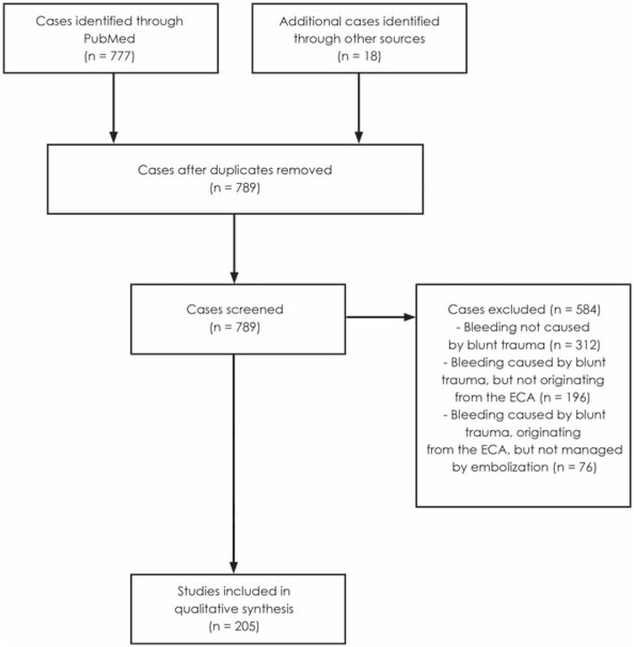
Figure 1
A flow diagram based on simplified Preferred Reporting Items for Systematic Reviews and MetaAnalyses (PRISMA) guidelines depicting the number of cases identified, included and excluded. The reasons for the exclusions are also noted.13
Table 1
Data from studies, case series and case reports pertaining to TAE of the ECA or its branches in the treatment of haemorrhage caused by blunt maxillofacial trauma
| First author | Year Published | Case number | Vessel injured | Vessel embolized | Embolization agents used | efficacy (complete cessation of bleeding following TAE of the ECA or its branches) | complications |
|---|---|---|---|---|---|---|---|
| 1 | none identified | RL ECA | partial necrosis tongue | ||||
| 2 | R IMA | R IMA | none | ||||
| 3 | R IMA | RL IMA | none | ||||
| 4 | none identified | RL ECA above LA | none | ||||
| 5 | none identified | RL ECA above LA | C | groin | |||
| Bynoe2 | 2003 | GF | 100% | hematoma | |||
| 6 | none identified | RL ECA above LA | PVA | none | |||
| 7 | L IMA | L IMA | none | ||||
| 8 | L IMA | L IMA | groin hematoma | ||||
| 9 | L IMA | R ECA above LA, L IMA | none | ||||
| 10 | none identified | R ECA above LA | none | ||||
| 11 | R IMA, R STA | not specified | C GF | could assessed not be | |||
| 12 | R IMA | not specified | CGF | none | |||
| 13 | L IMA | not specified | GF | could assessed not be | |||
| Chen12 | 2009 | 14 | R IMA | not specified | GF | 100% | none |
| 15 | L IMA | not specified | GF | none | |||
| 16 | RL IMA | not specified | GF | could assessed not be | |||
| 17 | L ECA | not specified | CGF | none | |||
| 18 | L IMA | not specified | GF | none | |||
| Cogbill48 | 2008 | 19–39 | not discernible due to the merging of blunt and penetrating trauma patients' data | not discernible due to the merging of blunt and penetrating trauma patients' data | C GF | 85% | none |
| Kim49 | 2011 | 40 | not specified | L IMA | PVA | success | none |
| 41 | not specified | R SPA | none | ||||
| 42 | not specified | RL SPA, IOA, FA | none | ||||
| 43 | not specified | L SPA | none | ||||
| RL SPA, LPA, ADTA6, | |||||||
| 44 | not specified | IAA7, FA, LA | C | none | |||
| Komiyama50 | 1998 | 45 | not specified | RL SPA, FA | GF | 100% | none |
| 46 | not specified | BL SPA, AAA8 | PVA | could assessed not be | |||
| 47 | not specified | R STA, FA | none | ||||
| 48 | not specified | RL GPA9, EA | none | ||||
| 49 | not specified | RL SPA, SPAA10 | none | ||||
| 12 x IMA | |||||||
| 6 x FA | |||||||
| 6 x LA | |||||||
| Kuan47 | 2015 | 50–76 | 5 x MMA11 3 x ECA 1 x APA12 | not specified | C GF PVA NBCA | 92.3%; data includes one penetrating injury | no serious systemic neurologic or complications |
| 5x other vessels | |||||||
| Note: this statistics also includes one penetrating trauma. | |||||||
| Langel | 2020 | 77 | LIMA + R SPA | L ECA + R SPA | CPHIL 25 | success | none |
| 25 x IMA | |||||||
| 5 x MMA | |||||||
| 4 x ECA | |||||||
| 4 x SPA | |||||||
| 4 x STA | |||||||
| Liao40 | 2007 | 78–112 | 2 x APA | not specified | not specified | 79.4% | none reported |
| 1 x FA | |||||||
| 1 x SALA | |||||||
| 1 x IALA | |||||||
| 1 x DPA | |||||||
| 5 x other | |||||||
| (observed contrast pooling) | |||||||
| Liu3 | 2008 | 113 | L STA + L IMA | not specified | GF | success | none reported |
| Maiorello51 | 2011 | 114 | L FA | L FA | Onyx 18 | success | none |
| Mauldin52 | 1989 | 115 | R ECA | R ECA | C | success | none reported |
| Mehringer52 | 1982 | 116–194 | not discernible due to the merging of blunt and penetrating trauma patients' data | not discernible due to the merging of blunt and penetrating trauma patients' data | GF PVA silastic spheres | 100% | 1x cerebral infarction 2x transient occulomotor nerve palsy |
| Mehrotra6 | 1984 | 195–196 | R IMA | R IMA | GF PVA | success | none reported |
| L IMA | L IMA | GF | success | none reported | |||
| Noy38 | 2007 | 197 | LIMA, RL FA | LIMA, RL FA | MS NBCA | success | none |
| Remonda54 | 2000 | 198 | RL IMA | not specified | C PVA NBCA | success | Transient trismus |
| Thiex33 | 2011 | 199 | L FA | not specified | Onyx | success | none |
| 200 | L STA | not specified | C | none | |||
| 201 | L FA | not specified | C | none | |||
| Wang55 | 2015 | 202 | R STA | not specified | C | 100% | none |
| 203 | R STA | not specified | C | none | |||
| 204 | L STA | not specified | C | none | |||
| Wong56 | 2013 | 205 | R IMA | R IMA | NBCA | success | none |
[i] Anatomical abbreviations: AAA = anterior auricular artery; ADTA = anterior deep temporal artery; APA = ascending pharyngeal artery; DPA = descending palatine artery; ECA = external carotid artery; FA = facial artery; GPA = greater palatine artery; IAA = inferior alveolar artery; IALA = inferior alveolar artery; IMA = internal maxillary artery; IOA = infraorbital artery; L = left; LPA = lesser palatine artery; MMA = middle meningeal artery; R = right; SALA = superior alveolar artery; SPA = sphenopalatine artery; SPAA = superior posterior alveolar artery; STA = superficial temporal artery
Embolization agent abbreviations: C = coils; GF = Gelfoam; MS = microspheres; PHIL = precipitating hydrophobic injectable liquid; PVA = polyvinyl alcohol particles
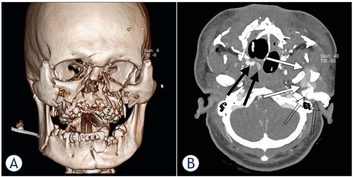
Figure 2
(A) A 3D CT reconstruction showing multiple maxillo-facial fractures. (B) An aortocervical CTA showing two small hematomas in the region of the right pterygopalatine fossa and nasal cavity (black arrows) and a cm 3 × 4 cm hematoma in the region of the left masticatory space and deep parotid space (white arrows). Also visible is a hyperdense material used in left ear tamponade (empty arrows).
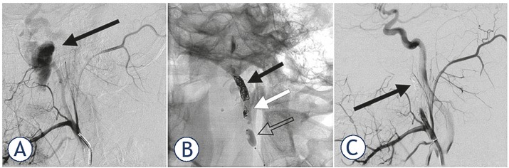
Figure 3
(A) A lateral left ECA angiogram showing ECA laceration with 3 × 4 cm pseudoaneurysm continuing into the proximal part of the left IMA. Contrast extravasation can be observed in the vicinity of the pseudoaneurysm. (B) A fluoroscopic view showing left ECA embolization using coils (black arrow) and PHIL 25 liquid embolization agent (white arrow) under balloon flow control (empty arrow). Also of note area small number of stray coils anchored in the vessel in the region of PHIL application (white arrow). (C) A post-embolization lateral left ECA angiogram showing complete exclusion of the ECA distally to the facial artery (black arrow).
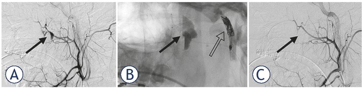
Figure 4
(A) A lateral right ECA angiogram showing two pseudoaneurysms in the region of the right pterygopalatine fossa and nasal cavity (black arrows). (B) Fluoroscopy showing microcatether proximally to the hematoma in the right pterygopalatine fossa (black arrow) prior to PHIL 25 application. Also visible is the embolized contralateral ECA (empty arrow). (C) A post-embolization right ECA angiogram showing complete sphenopalatine artery occlusion (black arrow). Also visible are the patent vessels proximally to the embolization.