
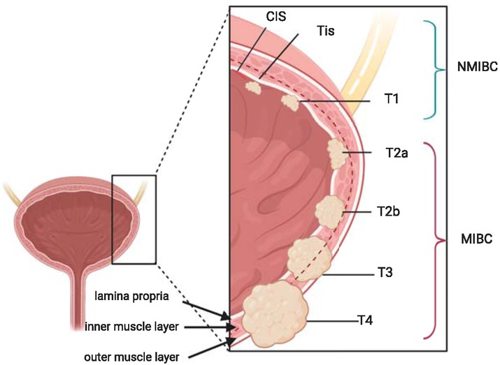
Figure 1
Classification of bladder cancer.
MIBC = Muscle invasive bladder cancer; NMIBC = Non muscle invasive bladder cancer
Table 1
2017 TNM classification of urinary bladder cancer 5
| T - primary tumour |
|---|
| TX Primary tumour cannot be assessed |
| T0 No evidence of primary tumour |
| Ta Non-invasive papillary carcinoma |
| Tis Carcinoma in situ: ‘flat tumour’ |
| T1 Tumour invades subepithelial connective tissue |
| T2 Tumour invades muscle |
| T2a Tumour invades superficial muscle (inner half) |
| T2b Tumour invades deep muscle (outer half) |
| T3 Tumour invades perivesical tissue |
| T3a Microscopically |
| T3b Macroscopically (extravesical mass) |
| T4 Tumour invades any of the following: prostate stroma, seminal vesicles, uterus, vagina, pelvic wall, abdominal wall |
| T4a Tumour invades prostate stroma, seminal vesicles, uterus or vagina |
| T4b Tumour invades pelvic wall or abdominal wall |
| N - regional lymph nodes |
| NX Regional lymph nodes cannot be assessed |
| N0 No regional lymph node metastasis |
| N1 Metastasis in a single lymph node in the true pelvis (hypogastric, obturator, external iliac, or presacral) |
| N2 Metastasis in multiple regional lymph nodes in the true pelvis (hypogastric, obturator, external iliac, or presacral) |
| N3 Metastasis in common iliac lymph node(s) |
| M - distant metastasis |
| M0 No distant metastasis |
| M1a Non-regional lymph nodes |
| M1b Other distant metastases |
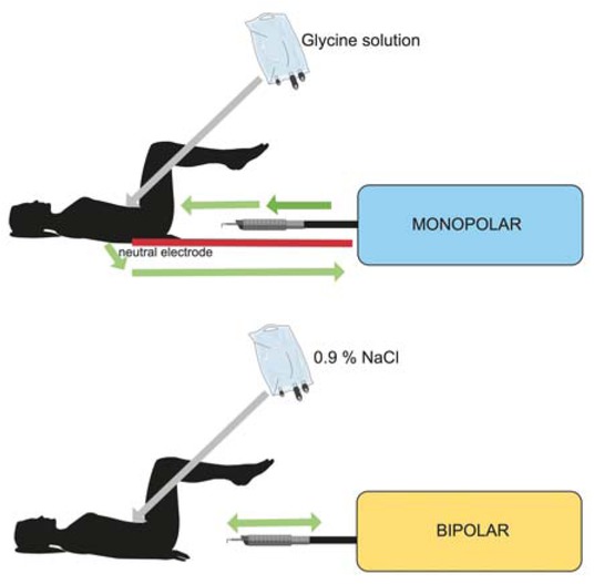
Figure 2
Monopolar vs. bipolar transurethral resection of bladder tumor.
Table 2
Comparison of monopolar and bipolar current for TURB1.9-14
| Variable | Monopolar | Bipolar |
|---|---|---|
| Dispersive electrode pad | yes | no |
| Energy | high | low |
| Voltage | high | low |
| Working medium | glycine | saline |
| Temperature at thermal effect (°C) | 400 | 40–70 |
| Time of resection | limited | (not extended strictly limited) |
| TUR syndrome | common | rare |
| Obturator jerk | common | rare |
| Quality of haemostasis and coagulum | poor | good |
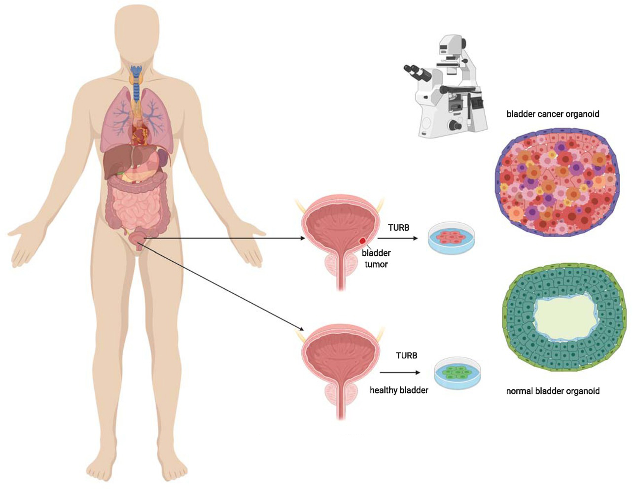
Figure 3
Shematic presentation of bladder organoid preparation.
Sample of bladder wall (healty and /tumorous) is taken with transurethral resection of bladder tumour. The cells are cultivated under special conditions in laboratory to form organoids. Organoids are used for studying the characteristics of normal and tumorous bladder wall, pathogenesis, response to different treatment approaches. This is step toward personalized treatment of bladder cancer.