
Table 1
Comparing 6 reported cases of mechanical thrombectomy in acute bilateral ICA and/or MCA occlusions
| Author, (Year), | Sex/age | Site of occlusion | Mechanical | Clinical | ||
|---|---|---|---|---|---|---|
| reference | Clinical presentation | (years) | ICA | MCA | thrombectomy (technique) | outcome |
| Dietrich et al. (2014)5 | left hemiparesis, progressing to coma | M/72 | - | + (M1) | aspiration+stent- retriever | minor deficit |
| Pop et al. (2014)6 | impaired consciousness | F/78 | + | + (M2) | stent-retriever | no deficit |
| Pop et al.(2014)6 | right sided weakness | F/66 | + | + (M1) | stent-retriever | severe deficit |
| Braksick et al. (2018)7 | coma | F/76 | - | + (M1) | - (no data) | coma |
| Larrew (2019) et al. 8 | coma | - (no data) / middle age | + | + - | aspiration | fatal |
| Storey et al. 2019)9 | hemiparesis / hemiplegia | F/64 | + | + (M1,M2) | aspiration+stent-retriever | minor deficit |
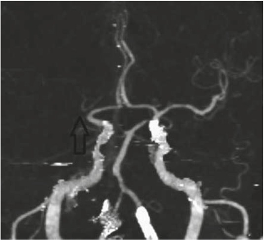
Figure 1
Initial imaging workup upon arrival at the general hospital. CT angiography (CTA) shows right M1 occlusion (arrow).
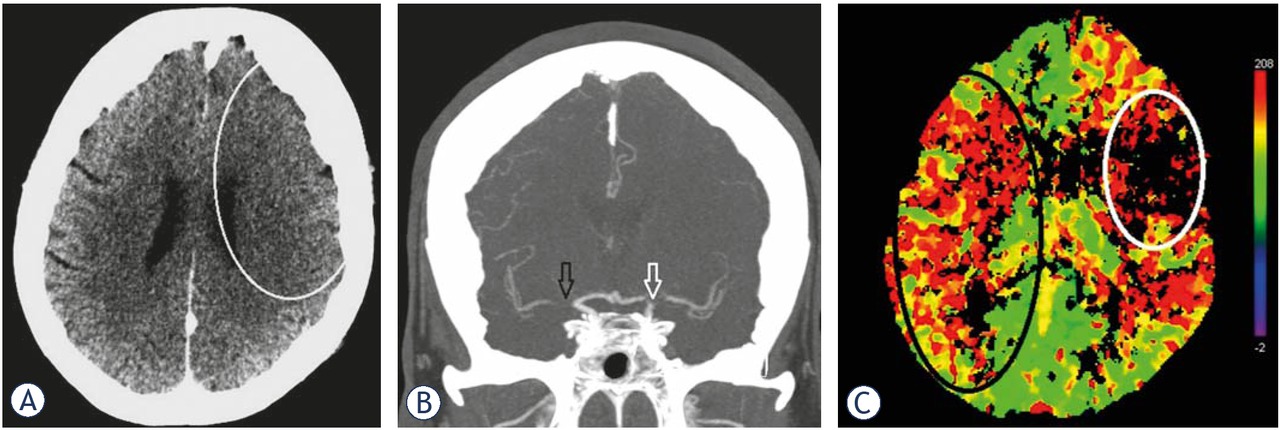
Figure 2
(A) Control images taken in the general hospital after clinical deterioration during intravenous thrombolysis and before the transfer to the tertiary institution. There were still no signs of ischaemic brain damage in the right cerebral hemisphere but there were subtle signs of stroke in the left middle cerebral artery (MCA) territory (white line delineates loss of cortical grey matter – white matter differentiation in the frontoparietal lobe with sulcal effacement). (B) Control images taken in the general hospital after clinical deterioration during intravenous thrombolysis and before the transfer. CT angiography (CTA) showed persistent right M1 occlusion (black arrow) but also left carotid “T” occlusion (white arrow). (C) Control images taken in the general hospital after clinical deterioration during intravenous thrombolysis and before the transfer. CT perfusion imaging (CTP) showed a penumbra in the right MCA territory (black circle) and irreversible brain damage in the left MCA territory (white circle).
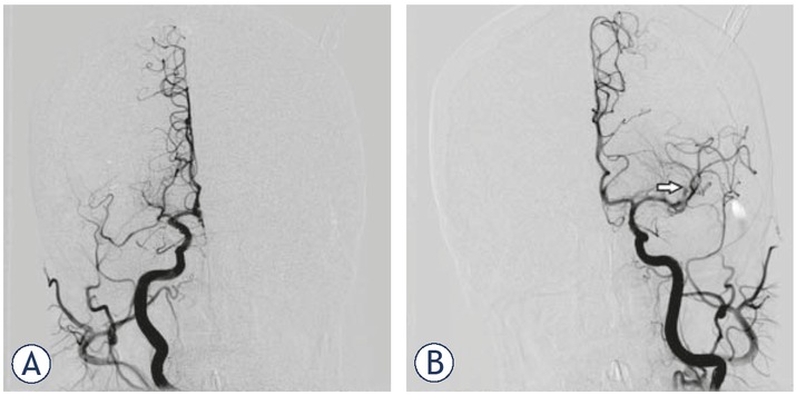
Figure 3
(A) Digital subtraction angiography (DSA) at the beginning of mechanical recanalization. Right internal carotid contrast injection confirming right M1 occlusion. (B) DSA at the beginning of mechanical recanalization. Left side contrast injection showing complete spontaneous recanalization of the carotid “T” occlusion with thrombembolar distal migration (occlusion of the proximal M2 segment of the major MCA branch) (arrow).
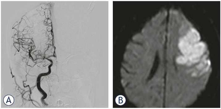
Figure 4
(A) Digital subtraction angiography (DSA) after mechanical recanalization. Right M1 mechanical recanalization (aspiration device) led to complete flow restoration. (B) MR diffusion weighted imaging (DWI) scan taken 6 days after mechanical recanalization: complete salvage of the affected right middle cerebral artery (MCA) brain parenchyma (recanalization at 7 hours after stroke onset). In contrast, subsequent persistent left M2 occlusion without collateral flow resulted in significant stroke within 3 hours after stroke onset.