
Table 1
WHO criteria for staging canine oral melanoma
| Stage | Tumor diameter | Lymph node involvement | Metastasis |
|---|---|---|---|
| I | < 2 cm | No | No |
| II | 2 - 4 cm | No | No |
| III | > or = 4 cm | No | No |
| Any | Yes | No | |
| IV | Any | Yes or No | Yes |
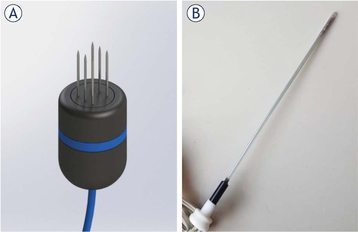
Figure 1
Electrodes used to treat patients. In (A) a render of the six-needle electrode used. It consists of two rows of needles separated 4 mm from each other. In (B) a picture of the Single Needle Electrode for nasal duct treatment.
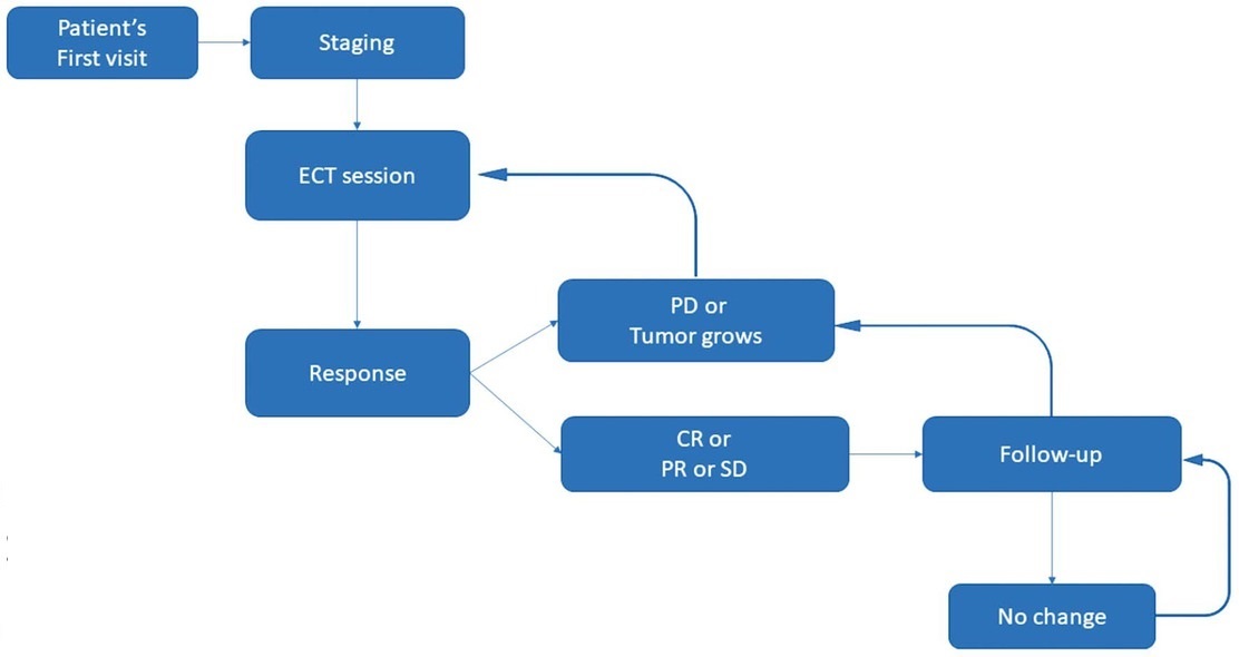
Figure 2
Treatment plan algorithm.
CR = complete response; ECT = electrochemotherapy; PD = progressive disease; PR = partial response; SD = stable disease
Table 2
Patients’ demographics
| Characteristics of 67 patients | Mean (range) (%) |
|---|---|
| Age (years) | 11.7 (6–16) |
| Sex ratio (M:F) | 37:30 |
| Body weight (kg) | 22.6 (3.5–58) |
| Breeds | |
| Crossbreed | 24 (35.8%) |
| Labrador retriever | 9 (13.4%) |
| Cocker spaniel | 7 (10.4%) |
| Golden retriever | 6 (8.9%) |
| Beagle | 4 (5.9%) |
| Poodle | 4 (5.9%) |
| Rottweiler | 3 (4.4%) |
| Doberman | 2 (2.9%) |
| Shar-Pei | 2 (2.9%) |
| Dogo | 1 (1.4%) |
| English Mastiff | 1 (1.4%) |
| Chow-Chow | 1 (1.4%) |
| Basset Hound | 1 (1.4%) |
| Pekingese | 1 (1.4%) |
| Dalmatian | 1 (1.4%) |
| Location (Rostral : Caudal) | 36 : 31 |
| Stage | |
| I | 11 (16.5%) |
| II | 19 (28.3%) |
| III | 26 (38.8%) |
| IV | 11 (16.4%) |
| Histologic subtype | |
| Epithelioid | 17 (25.4%) |
| Mixed | 17 (25.4%) |
| Spindle cell | 17 (25.4%) |
| Anaplastic | 9 (13.4%) |
| Others | 7 (10.4%) |
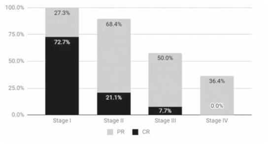
Figure 3
The bars show the sum of partial and complete response percentages obtained on each stage. For stage I: complete responses (CR) 72.7% (8) partial responses (PR) 27.3% (3) stable diseases (SD) 0% progressive diseases (PD) 0%, stage II: CR 21.1%(4) PR 68.4% (13) SD 0% PD 10.5% (2), stage III: CR 7.7% (2) PR 50% (13) SD 26.9% (7) PD 15.4% (4) and stage IV: CR 0% PR 36.4% (4) SD 36.4% (4) PD 27.3% (3).
Table 3
Univariate (Fisher exact test) and multivariate (Logistic regression analysis) test for analyzing the association between OR and different factors
| Characteristics | Number of patients with OR | Univariate Fisher exact test | Multivariate OR 95% CI Logistic Regression Analysis | p |
|---|---|---|---|---|
| Sex (M/F) | 37/30 | p = 0.602 | – | – |
| Location (Rostral/Caudal) | 36/31 | p = 0.062 | – | – |
| Bone Involvement (Yes/No) | 43/24 | p = 0.026 | automatically excluded | – |
| Metastasis present, excluding lymph nodes (Yes/No) | 10/57 | p = 0.150 | – | – |
| Early-stage (stages I+II/stages III+IV) | 30/37 | p = 0.00016 | 0.08(0.02-0.36) | 0.0013 |
Table 4
Univariable (Kaplan-Meier) and multivariable (Cox proportional-hazards) analysis of patient-related factors for time to progression and overall survival time. Hazard Ratio and 95% Confidence interval is reported
| N. of patients | Overall Survival [Univariable] | P | [Multivariable] | P | TTP [Univariable] | P | [Multivariable] | P | |
| Sex | |||||||||
| Male | 35 | 1.58 (0.95–2.63) | 0.0634 | – | – | 1.67 (0.99–2.75) | 0.0433 | – | – |
| Female | 30 | – | – | – | – | ||||
| Location | |||||||||
| Rostral | 34 | 0.71 (0.43–1.16) | 0.1596 | – | – | 0.77 (0.47–1.26) | 0.2921 | – | – |
| Caudal | 31 | – | – | – | – | ||||
| Metastasis | |||||||||
| No (or lynph node only) | 55 | 0.29 (0.09–0.88) | 0.0001 | – | – | 0.34 (0.12–0.96) | 0.0009 | – | – |
| Yes | 10 | – | – | – | – | ||||
| Bone Involvement | |||||||||
| Yes | 42 | 2.45 (1.50–4.00) | 0.0004 | ||||||
| No | 23 | (0.24–0.88) | 0.0184 | 0.42 (0.26–0.68) | 0.0005 | (0.28–0.98) | 0.043 | ||
| Histology | |||||||||
| Epithelioid vs. Fusocellular | 16 / 16 | 0.59 (0.31–1.14) | 0.1768 | – | – | 0.60 (0.32–1.14) | 0.0534 | – | – |
| Epithelioid vs Mixed. | 16 / 17 | 0.54 (0.28–1.05) | – | – | 0.49 (0.25–0.96) | – | – | ||
| Epithelioid vs. Anaplastic | 16 / 9 | 0.46 (0.19–1.11) | – | – | 0.35 (0.14–0.93) | – | – | ||
| Fusocellular vs. Mixed | 16 / 17 | 0.92 (0.44–1.93) | – | – | 0.82 (0.39–1.72) | – | – | ||
| Fusocellular vs. Anaplastic | 16 / 9 | 0.77 (0.30–1.99) | – | – | 0.59 (0.22–1.64) | – | – | ||
| Mixed vs. Anaplastic | 17 / 9 | 0.84 (0.33–2.18) | – | – | 0.73 (0.26–2.04) | – | – | ||
| Stage | |||||||||
| I vs. II | 10 / 18 | 1.86 (1.03–3.34) | <0.0001 | Stage I: (0.13–0.74) | 0.0083 | 0.53 (0.30–0.96) | 0.0001 | Stage I: (0.07 –0.47) | 0.0005 |
| I vs. III | 10 / 26 | 3.28 (1.76–6.09) | 0.28 (0.15–0.53) | Stage II: (0.24–0.82) | 0.0094 | ||||
| I vs. IV | 10 / 11 | 9.05 (2.58–31.75) | 0.19 (0.07–0.51) | ||||||
| II vs. III | 18 / 26 | 0.57 (0.30–1.06) | 0.52 (0.27–0.99) | ||||||
| II vs. IV | 18 / 11 | 0.21 (0.06–0.72) | Stage IV:(2.04–9.29) | 0.0001 | 0.35 (0.13–0.96) | ||||
| III vs IV | 26 / 11 | 0.36 (0.10–1.29) | 0.67 (0.24–1.90) |
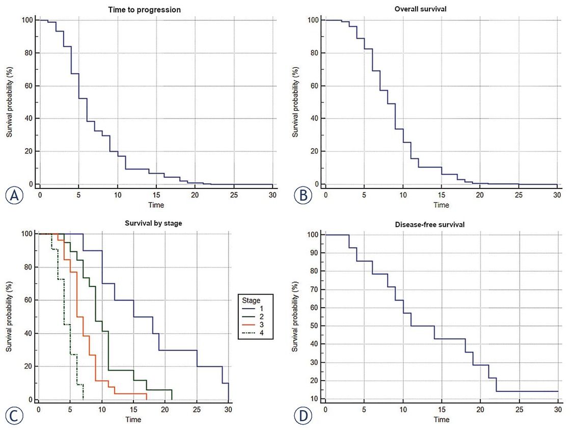
Figure 4
(A) Time to progression curve (Cox proportional-hazards regression, n = 67, p < 0.0001). (B) Overall survival time (Cox proportional-hazards regression, n = 67, p < 0.0001). (C) Survival time by stage (Kaplan-Meier curves of survival, n = 67, p < 0.0001). (D) Disease-free survival time (Kaplan-Meier, n = 14).
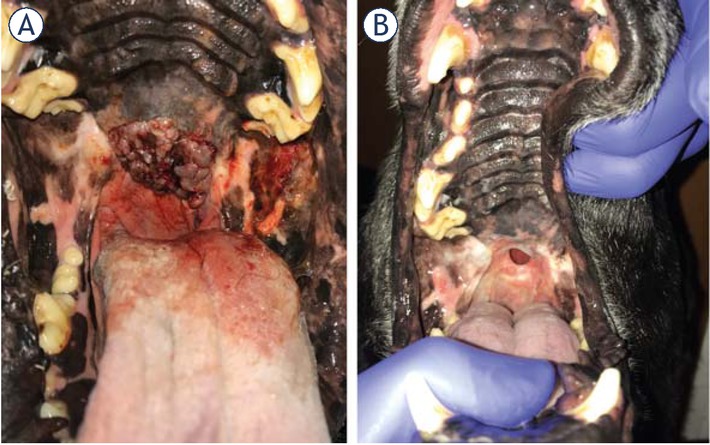
Figure 5
(A) Stage II patient with melanoma in the soft palate, before the treatment. (B) Final response obtained 30 days after third round of ECT. Completely absent tumoral tissue and a fistula can be seen. Quality of life of the patient improved in spite of the fistula, which was asymptomatic.