
Table 1
Biological actions of parathyroid hormone (PTH) in the body. PTH increases the serum calcium concentration and lowers the serum phosphate concentration
| Organ system | Function of PTH |
|---|---|
| Kidneys (leading role) | It increases calcium and decreases phosphate reabsorption, stimulates calcitriol production by increasing the synthesis of the enzyme 1-α hydroxylase in proximal tubules. |
| Skeletal | It raises calcium levels in blood by increasing bone destruction (via osteoblast-mediated activation of osteoclasts) and decreasing the formation of new bone. |
| Gastrointestinal system | It increases calcium absorption by stimulating the production of 1,25-dihydroxycholecalciferol. |
| Other (minor role, experimental) | Metabolic effects (reduced glucose tolerance, changes in fat metabolism), effects on the liver, adipose tissue, cardiovascular system, neuromuscular function. |
Table 2
Clinical presentation of developed primary hyperparathyroidism (PHPT). Symptoms and clinical signs are associated with an elevated serum calcium concentration and/or increased secretion of parathyroid hormone (PTH)
| Organ system | Symptoms and clinical signs |
|---|---|
| General | anorexia, polyuria, polydipsia, weight gain, anaemia |
| Skeletal | osteitis fibrosa cystica (bone pain, decreased bone density or generalized osteoporosis, pathological fractures) |
| Kidney | kidney stones, renal parenchymal calcifications, nephrocalcinosis, chronic renal impairment |
| Neuromuscular | proximal muscle weakness, depression, decline in cognitive ability, psychosis |
| Cardiovascular | arterial hypertension, arrhythmias, left ventricular hypertrophy, vascular wall and myocardial calcification |
| Gastrointestinal | nausea, vomiting, constipation, ulcer disease, pancreatitis |
| Rheumatological | gout, pseudogout |
Table 3
The 2014 Fourth International Guidelines for the Management of Asymptomatic PHPT. Patients need to meet at least one of the following criteria to be advised to have surgery
| Measurement | Criteria |
|---|---|
| Age of patient | < 50 years |
| Serum calcium concentration (above the upper reference value) | > 0.25 mmol/L (1.0 mg/dL) |
| Skeletal injury | bone mineral density (DXA): T-score < -2.5 SD* |
| spinal fracture (proven by XR, CT, MRI or VFA) | |
| Renal impairment | creatinine clearance < 60 mL/min |
| kidney stones or nephrocalcinosis (proven by XR, US or CT) | |
| 24-hour calcium in urine > 10 mmol/L (400 mg/day) or increased risk for kidney stones based on biochemical analysis |
[i] * measured on the lumbar spine, hip, femoral neck or distal third of the radius
CT = computed tomography; DXA = dual-energy x-ray absorptiometry; MRI = magnetic resonance imaging; SD = standard deviation; US = ultrasound; VFA = vertebral fracture assessment; XR = x-ray imaging
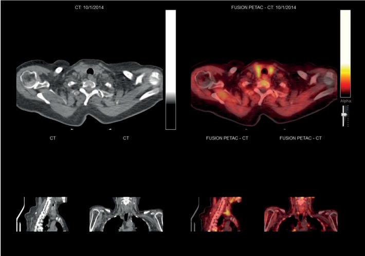
Figure 1
18F-fluorocholine (FCH) PET-CT fusion images of patient with pathologic uptake in the right lower parathyroid gland (solitary adenoma).
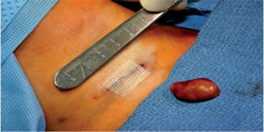
Figure 2
Directed parathyroidectomy. The image shows the incision site and the removed parathyroid tissue.
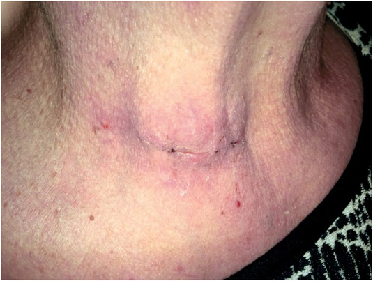
Figure 3
Appearance of the incision and surrounding skin 10 days after performing directed parathyroidectomy.
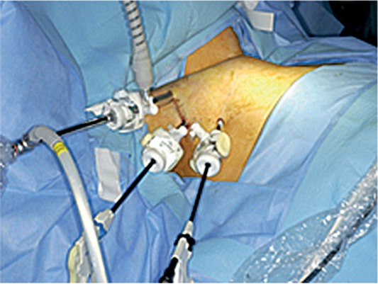
Figure 4
Total endoscopic parathyroidectomy.