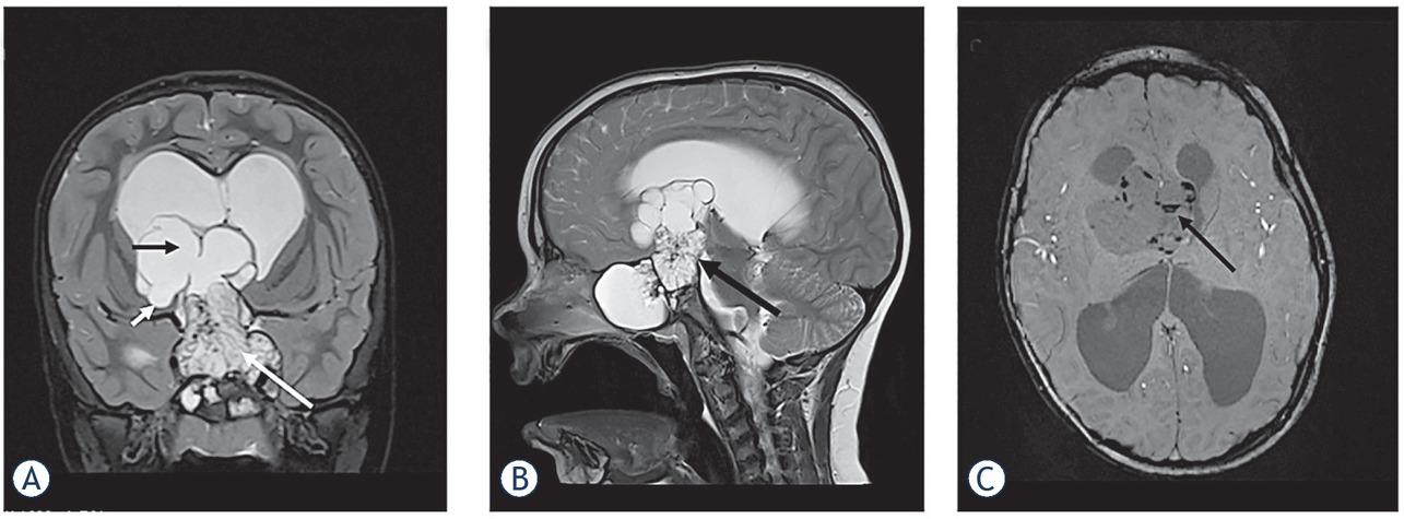
Have a personal or library account? Click to login
Advances in the management of craniopharyngioma in children and adults Cover

Advances in the management of craniopharyngioma in children and adults

Open Access
|Oct 2019

Figures & Tables

Figure 1

Large, partially solid, partially multicystic adamantinomatous craniopharyngioma in a 5-year old boy. (A) Coronal T2 sequence through sellar region; solid part of the tumor (white arrow) involves enlarged sella turcica, parasellar regions, occupies the suprasellar cistern and the third ventricle. The cystic portion of the tumor (black arrow) extends into the lateral ventricles and on the right side it infiltrates the adjacent brain parenchym (small arrow). Lateral ventricles are enlarged with a band of periventricular transependimal edema as a sign of acute hydrocephalus. (B) Sagital T2 sequence through the sellar region; Hypothalamus and mammillary bodies are not visible (arrow). (C) SWI sequence through the multicystic portion of the tumor shows multiple calcifications in the cyst walls (arrow).

Comparison of pediatric and adult-onset craniopharyngioma characteristics

Pediatric-onsetAdult-onset
30–50 % of all CPs
Age at presentationPeak at 5–14 years1Peak at 40–44 years2
Gender distribution (m/f)Equal8,21Equal8,27
Most frequent presentationHeadache (68–85%)Visual impairment (40–84%)
Visual impairment (36– 55%)Menstrual irregularities (57%)
Growth failure (7–36%)9,35,66Headache (42–56%)13,26,27
Pathohistological typeAdamantinomatous 99%33
Papillary extremely rare*Papillary 14–50%
Initial hypothalamic involvement42–66%8,9,3542%18
Endocrine deficits at diagnosis
Any40–87%8,13,21,35,65,6641–73%8
GH41–75%8,13,21,35,65,6618–86%8,13
FSH/LH20–56%8,13,21,35,65,6629–74%8,13
ACTH8–68%8,13,21,35,65,6635–58%8,13
TSH15–32%8,13,21,35,65,6635–56%8,13
ADH7–17%8,13,21,35,65,666–17%8,13
Pituitary hormone deficiencies after treatment
Any64–100%8,6448–97%8,64
GH93–96%8,18,2152–68%8,18
FSH/LH59–95%8,18,2170–94%8,18
ACTH78–100%8,18,2174–88%8,18
TSH86–100%8,18,2181–92%8,18
ADH65–96%8,18,2143–70%8,18
Panhypopituitarism***43–100%8,18,6459–74%8,18,64
Obesity**44–64%8,9,19,6441–47%8,19,64
DOI: https://doi.org/10.2478/raon-2019-0036 | Journal eISSN: 1581-3207 | Journal ISSN: 1318-2099
Language: English
Page range: 388 - 396
Submitted on: Jun 21, 2019
|
Accepted on: Jul 11, 2019
|
Published on: Oct 25, 2019
In partnership with: Paradigm Publishing Services
Publication frequency: 4 issues per year

© 2019 Mojca Jensterle, Soncka Jazbinsek, Roman Bosnjak, Mara Popovic, Lorna Zadravec Zaletel, Tina Vipotnik Vesnaver, Barbara Faganel Kotnik, Primoz Kotnik, published by Association of Radiology and Oncology
This work is licensed under the Creative Commons Attribution-NonCommercial-NoDerivatives 3.0 License.