
Table 1
Baseline characteristics of the study population
| Age: median; range (years) | 33; 16–49 | |||||
| Age of start of prophylaxis: mean (years) | 17.4 | |||||
| age group: 0–9 (patient count) | 7 | |||||
| age group: 10–19 (patient count) | 14 | |||||
| age group: 20+ (patient count) | 9 | |||||
| Duration of prophylaxis: mean (years) | 15.4 | |||||
| Ankles | Knees | Elbows | ||||
| Right | Left | Right | Left | Right | Left | |
| No. of joints | 30 | 29 | 25 | 28 | 28 | 28 |
| No. of lifetime joint bleeds: | 5 | 5 | 12 | 15 | 14 | 13 |
| 0–5 (joint count) | 12 | 11 | 7 | 10 | 4 | 2 |
| 6–20 (joint count) | ||||||
| > 20 (joint count) | 13 | 12 | 5 | 3 | 9 | 12 |
| Unknown (joint count) | 0 | 1 | 1 | 0 | 1 | 1 |
| HJHS 2.1 score: mean; max* | 3.3; 12 | 2.6; 11 | 1.4; 7 | 1.2; 8 | 1.9; 9 | 1.9; 8 |
[i] * Minimum was 0 for all the scores
Table 2
Correlation between the HEAD-US and IPSG MRI scores
| Elbows | Knees | Ankles | All joints | |
|---|---|---|---|---|
| Overall score (r) | 0.949 | 0.941 | 0.838 | 0.921 |
| Detailed scores: | ||||
| Synovial hypertrophy (r) | 0.840 | 0.710 | 0.561 | |
| Cartilage degradation (r) | 0.734 | 0.812 | 0.537 | |
| Bone changes (r) | 0.883 | 0.741 | 0.725 |
[i] Notes: all the reported correlations are statistically significant (p<0.001); the values for the elbows, knees and ankles are the averages over the right and left side values (the differences between them were negligible); the correlations are averaged using Fisher-z transformation.
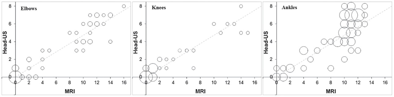
Figure 1
Concordance bubble-plot for depicting agreement between HEAD-US and MRI score for all three joints. The circles are centered at the observed combinations of the HEAD-US and MRI scores; their size is proportional to the number of the patients with a given combination. Dashed line represents a perfect agreement.
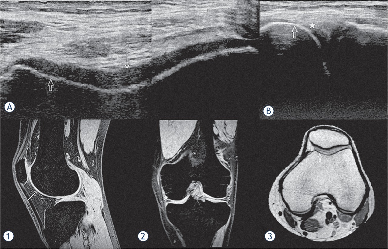
Figure 2
An example of a good concordance between HEAD-US and MRI. US images of the femoral trochlea in the transverse plane (A) and the medial femorotibial space in the coronal plane (B) are shown. T2* weighted MR images in the sagittal (1) and coronal (2) planes and a PD weighted MR image in the transverse plane (3) of the same knee are shown for comparison of the corresponding structures. The smooth surface, normal thickness and homogenous structure of the trochlear joint cartilage are shown in the US image (A) (white arrow); the corresponding intact cartilage is shown on MR image (1). The intact cortical bone of the medial femoral condyle (hollow arrow) is shown by US in the images (A) and (B); the corresponding cortical bone is depicted by MRI in the image (3). No signs of synovium hypertrophy are shown by US in the medial femorotibial recess (white star) in the image B; the corresponding recess confirming no synovium hypertrophy is shown by MRI in the image (2). On MRI, there were also no arthropathic changes in the parts of the joint not visualized by US. The images show a perfect concordance between US and MRI findings in this knee with no signs of haemophilic arthropathy.
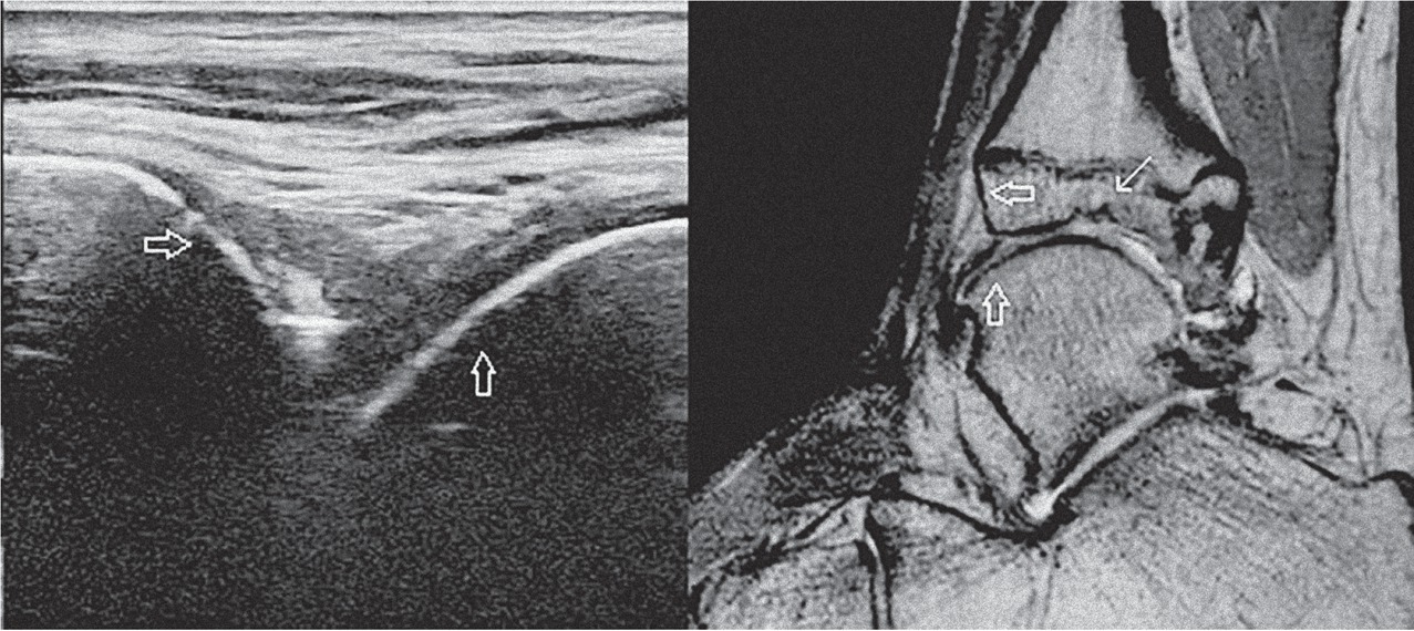
Figure 3
An example of a discordance between US and MRI. An US image of the tibiotalar joint in the sagittal plane is shown on the left, a PD weighted MR image of the same ankle in the sagittal plane is shown on the right for comparison of the corresponding structures. In both images, the smooth surface of the tibial cortical bone is marked by the horizontal hollow arrow and the smooth surface of the talar cortical bone is marked by the vertical hollow arrow. MRI demonstrates an osteochondral defect at the tibial side of the talocrural joint (white arrow), which is outside of the visualization area of US.