
TABLE 1
List of the analysed miRNAs with known functions in glioma pathogenesis
| MicroRNAs | Invasion | Proliferation | Tu growth | Migration | Cell cycle | cell Survival/death | Other functions |
|---|---|---|---|---|---|---|---|
| miR-7 | + | + | + | nd | + | + | Differentiation, Vasculogenic mimicry |
| miR-9 | nd | + | nd | + | nd | nd | Self-renewal, Vasculogenic mimicry |
| miR-15b | + | + | + | nd | + | + | Angiogenesis |
| miR-21 | + | + | + | + | + | + | / |
| miR-26b | + | + | + | + | nd | + | Vasculogenic mimicry |
| miR-124 | + | + | + | + | + | + | Differentiation, Angiogenesis |
| miR-199a | nd | + | nd | nd | nd | nd | / |
| let-7a | nd | + | + | + | nd | nd | / |
| let-7b | + | nd | nd | + | nd | nd | / |
| let-7d | nd | nd | nd | nd | nd | nd | / |
| let-7f | + | + | nd | + | nd | nd | / |
TABLE 2
Demographic and clinicopathological characteristics of patients included in the study
| Histopathological diagnosis | GBM, grade WHO IV | |
|---|---|---|
| Number of cases | 83 | |
| Mean age (Year) | 50 (min ± 12.8 8, max 71) | |
| Male | 50 (60%) | |
| Gender | ||
| Female | 33 (40%) | |
| Karnofsky performance status ≥ 80 | 83 | |
| radiotherapy after first surgery | 83/83 (100%) | |
| chemotherapy after first surgery | 54/83 (65%) | |
| Adjuvant treatment | ||
| radiotherapy after second surgery | 15/83 (18%) | |
| chemotherapy after second surgery | 47/83 (56%) | |
| MGMT (tested methylated in a subset of patients treated with TMZ) | Primary GBM | 38/47 (81%) |
| Recurrent GBM | 46/47 (98%) | |
| Time to recurrence (months) | 0.84–67.30 | |
| Survival after second surgery (months) | 0.03–25.75 | |
| Overall survival (months) | 0.84–70.48 | |
[i] MGMT = methyl guanine methyl transferase; TMZ = temozolamide
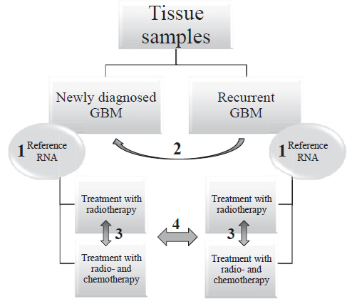
FIGURE 1
Schematic representation of the workflow and statistical comparisons.
GBM = glioblastoma; Reference RNA = Human Brain Reference RNA; 1 = Willcoxon Signed Rank Test between primary glioblastoma and corresponding recurrent glioblastoma using ΔCt; 2 = normalization to Human Brain reference RNA resulting in ΔΔCt; 3 and 4 = Mann-Whitney test between independent groups of samples using ΔΔCt (normalization of glioblastoma samples to Human Brain Reference RNA)
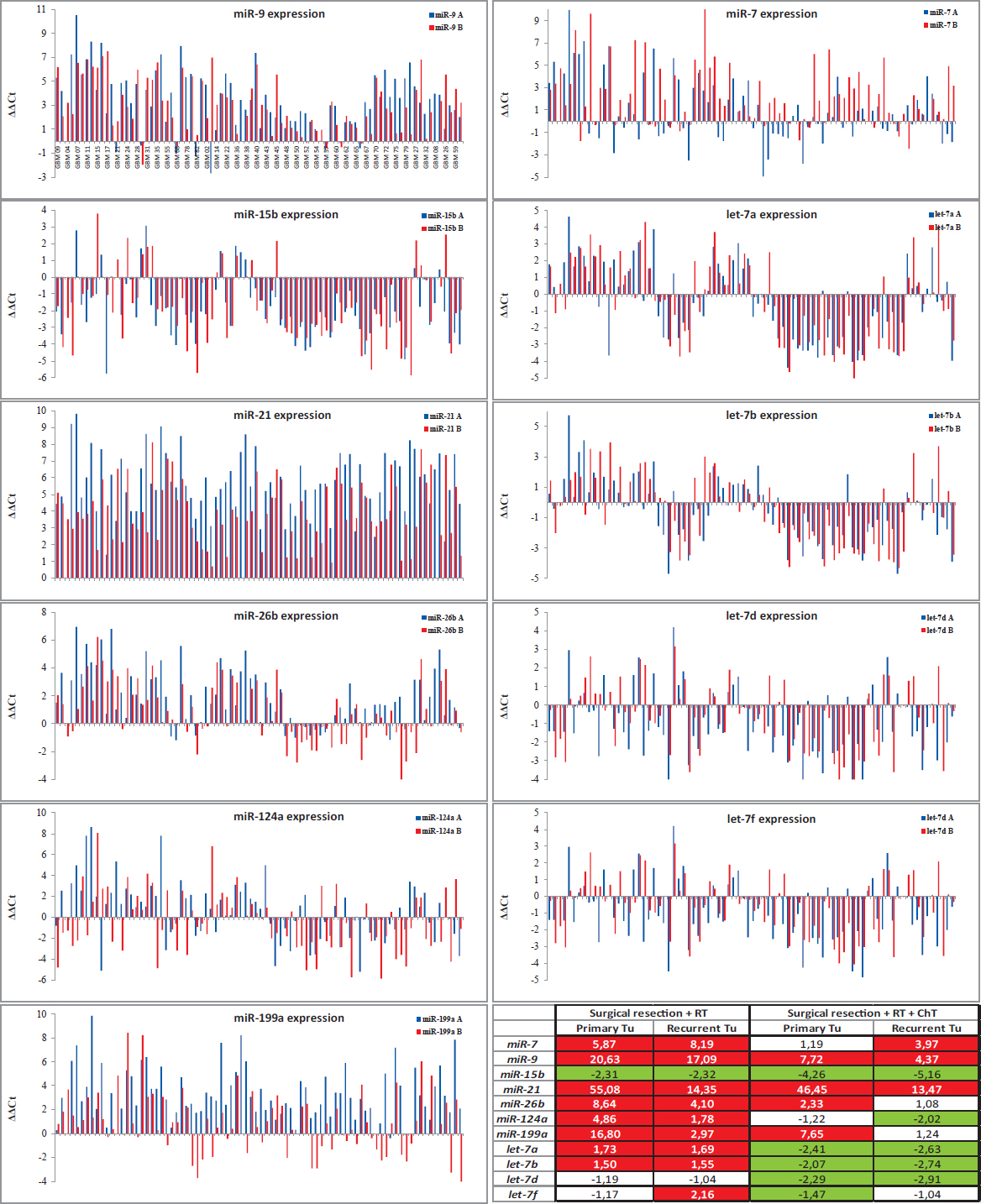
FIGURE 2
Expression of miRNAs in primary and recurrent GBM (compared to the Human Brain Reference RNA, ΔΔCt) with Table presenting Average Fold Changes.
ChT = chemotherapy; RT = radiotherapy; A = primary GBM; B = recurrent GBM
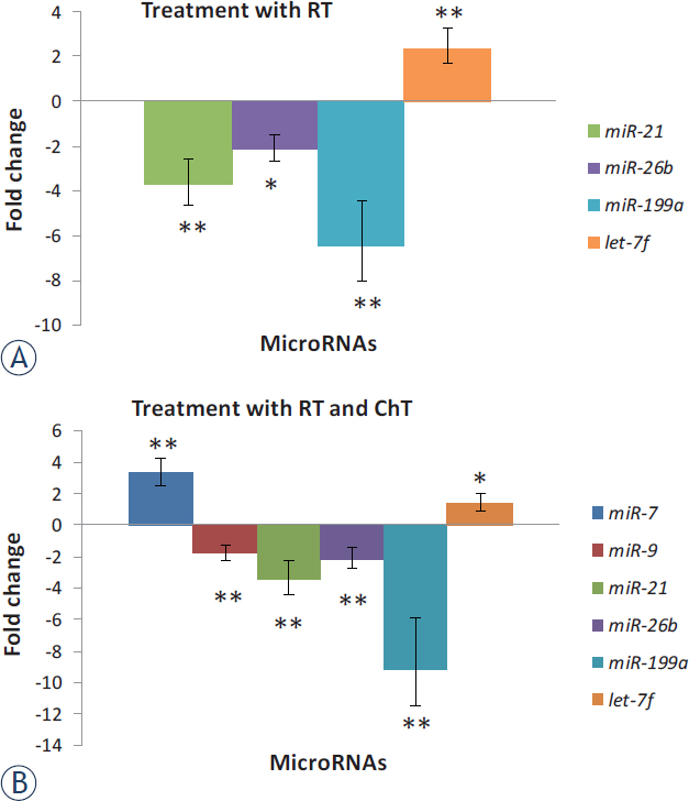
FIGURE 3
Expression of miRNAs in recurrent glioblastoma (GBM) after treatment compared to expression in primary GBM. Results are represented separately for patients treated with RT and those treated with RT and ChT.
ChT = chemotherapy; RT = radiotherapy; * = significant differences in expression of miRNAs, p < 0.05; ** = p < 0.01
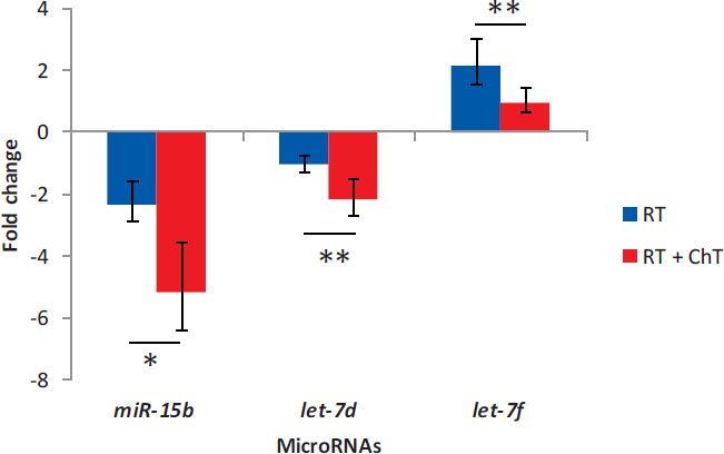
FIGURE 4
Distinctive expression of miRNAs after different treatment options in recurrent glioblastoma
ChT = chemotherapy; RT = radiotherapy; * = p < 0.05; ** = p < 0.01
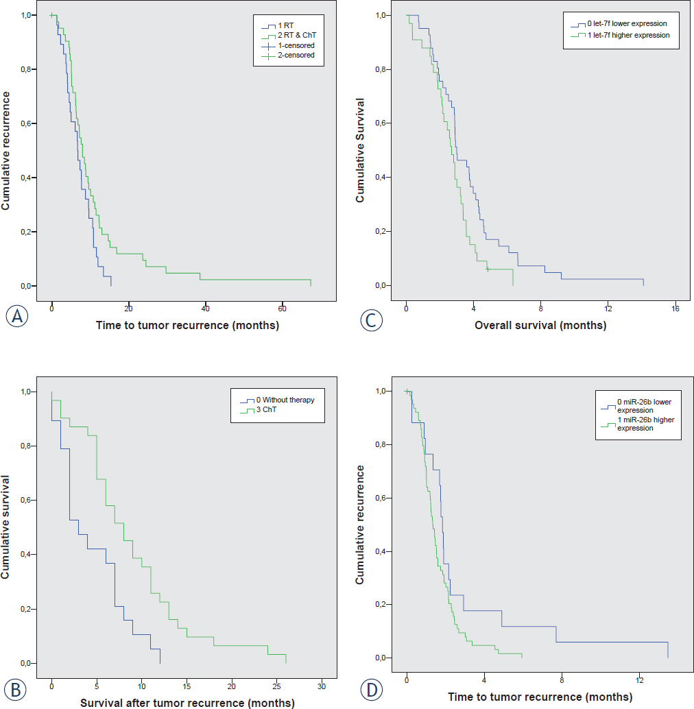
FIGURE 5
Survival curve analyses; (A) Progression free survival between patients that received RT alone or both, RT and ChT; (B) Survival time after second surgery for patients who received no therapy versus patients who received ChT; (C) Progression free survival between patients with up- and downregulated miR-26b; (D) Progression free survival between patients with up- and downregulated let-7f.
ChT = chemotherapy; RT = radiotherapy
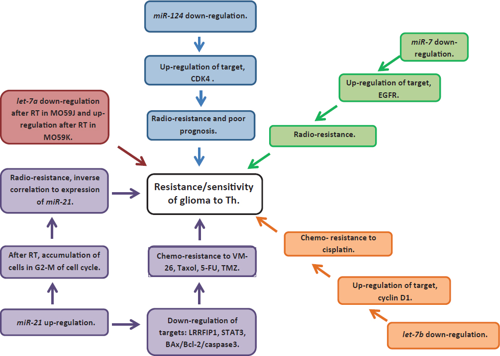
FIGURE 6
Summary of known functions in radio- and chemoresistance and sensitivity of miRNAs analyzed in our study.