
Table 1
Dosimetry of PTV
| IGBT | VMAT | ||
|---|---|---|---|
| Parameters | n=38 | n=38 | p-value |
| mean S.D | mean S.D | ||
| V95 (%) | 92.1 3.0 | 98.4 0.9 | <0.05 |
| V98 (%) | 90.2 3.2 | 90.4 3.7 | >0.05 |
| V100 (%) | 89.0 3.4 | 76.7 8.9 | <0.05 |
| D90 (%) | 98.6 4.7 | 98.2 0.8 | <0.05 |
| D100 (%) | 58.6 9.0 | 87.0 3.2 | <0.05 |
[i] Abbreviations: S.D., standard deviation; PTV, planning target volume; IGBT, image guided high-dose-rate brachytherapy; VMAT, volumetric modulated arc therapy; n, number of patients for analysis; VX, relative volume of the PTV receiving at least X% of the PD; DX, minimum relative dose of the PD delivered to X% of the PTV.
Table 2
Dosimetry of mandible and spinal cord
| IGBT | VMAT | ||||
|---|---|---|---|---|---|
| Parameters | p-value | ||||
| mean | S. D | mean | S.D | ||
| Mandible, n=38 | |||||
| D0.1cm3 (%) | 77.0 | 17.2 | 85.4 | 7.9 | <0.05 |
| D1cm3 (%) | 56.9 | 13.4 | 74.5 | 9.6 | <0.05 |
| D2cm3 (%) | 48.4 | 12.2 | 68.4 | 9.5 | <0.05 |
| Spinal cord, n=37* | |||||
| D0.1cm3 (%) | 9.7 | 2.6 | 12.3 | 4.1 | <0.05 |
| D1cm3 (%) | 6.8 | 2.0 | 10.8 | 3.7 | <0.05 |
| D2cm3 (%) | 5.9 | 1.9 | 10.0 | 3.6 | <0.05 |
Abbreviations: S.D., standard deviation; IGBT, image guided high-dose-rate brachytherapy; VMAT, volumetric modulated arc therapy; n, number of patients for analysis; DXcm3, minimum relative dose of the PD delivered to most exposed X cm3 of the organs.
Table 3
Dosimetry of parotid gland
| IGBT | VMAT | ||||
|---|---|---|---|---|---|
| Parameters | p-value | ||||
| mean | S.D | mean | S. D | ||
| ipsilateral side (n=34*) | |||||
| Dmean (%) | 4.6 | 1.4 | 4.6 | 2.6 | >0.05 |
| D0.1cm3 (%) | 11.2 | 2.8 | 18.0 | 7.1 | <0.05 |
| D1cm3 (%) | 8.1 | 2.1 | 12.9 | 6.3 | <0.05 |
| D2cm3 (%) | 7.0 | 2.0 | 10.5 | 5.7 | <0.05 |
| D10 (%) | 7.3 | 2.1 | 11.2 | 5.6 | <0.05 |
| D30 (%) | 5.5 | 1.6 | 5.5 | 4.0 | >0.05 |
| D50 (%) | 4.5 | 1.3 | 3.0 | 3.1 | <0.05 |
| V10 (%) | 3.1 | 4.0 | 18.0 | 16.9 | <0.05 |
| V30 (%) | 0.0 | 0.0 | 0.7 | 3.9 | N.A |
| V50 (%) | 0.0 | 0.0 | 0.1 | 0.5 | N.A |
| contralateral side (n=35**) | |||||
| Dmean (%) | 3.0 | 0.9 | 3.9 | 2.1 | >0.05 |
| D0.1cm3 (%) | 8.9 | 2.1 | 15.3 | 5.9 | <0.05 |
| D1cm3 (%) | 5.9 | 1.6 | 10.8 | 5.9 | <0.05 |
| D2cm3 (%) | 4.9 | 1.5 | 9.1 | 5.4 | <0.05 |
| D10 (%) | 5.4 | 1.3 | 9.9 | 4.4 | <0.05 |
| D30 (%) | 4.0 | 1.0 | 4.9 | 3.7 | >0.05 |
| D50 (%) | 3.2 | 0.8 | 2.2 | 2.3 | <0.05 |
| V10 (%) | 0.5 | 0.3 | 14.2 | 13.2 | <0.05 |
| V30 (%) | 0.0 | 0.0 | 0.8 | 4.7 | N.A |
| V50 (%) | 0.0 | 0.0 | 0.2 | 1.3 | N.A |
Abbreviations: S.D., standard deviation; IGBT, image guided high-dose-rate brachytherapy; VMAT, volumetric modulated arc therapy; n, number of patients for analysis; Dmean, mean relative dose of the PD delivered to the organs; DXcm3, minimum relative dose of the PD delivered to most exposed X cm3 of the organs; DX, minimum relative dose of the PD delivered to X% of the organs; VX, relative volume of the organs receiving at least X% of the PD; N.A., not available.
Table 4
Dosimetry of submandibular gland
| IGBT | VMAT | ||||
|---|---|---|---|---|---|
| Parameters | p-value | ||||
| mean | S.D | mean | S. D | ||
| ipsilateral side | |||||
| Dmean (%), n=20* | 16.4 | 10.7 | 21.9 | 19.9 | >0.05 |
| D0.1cm3 (%), n=20* | 27.4 | 15.6 | 39.9 | 23.3 | >0.05 |
| D1cm3 (%), n=18** | 19.4 | 11.2 | 27.2 | 17.6 | >0.05 |
| D2cm3 (%), n=17** | 16.1 | 9.4 | 20.8 | 14.7 | >0.05 |
| D10 (%), n=20* | 22.6 | 13.9 | 33.2 | 23.0 | >0.05 |
| D30 (%), n=20* | 18.3 | 11.7 | 25.8 | 21.9 | >0.05 |
| D50 (%), n=20* | 15.7 | 10.4 | 21.0 | 20.9 | >0.05 |
| V10 (%), n=20* | 61.3 | 38.6 | 63.9 | 33.6 | >0.05 |
| V30 (%), n=20* | 14.5 | 28.0 | 30.1 | 38.7 | >0.05 |
| V50 (%), n=20* | 1.4 | 3.7 | 14.1 | 27.4 | >0.05 |
| contralateral side | |||||
| Dmean (%), n=34*** | 8.2 | 4.8 | 16.9 | 10.8 | <0.05 |
| D0.1cm3 (%), n=34*** | 13.4 | 5.3 | 29.7 | 12.0 | <0.05 |
| D1cm3 (%), n=32**** | 9.4 | 3.2 | 21.7 | 8.6 | <0.05 |
| D2cm3 (%), n=32*** | 8.1 | 2.9 | 18.3 | 8.3 | <0.05 |
| D10 (%), n=34*** | 11.1 | 5.6 | 25.3 | 12.2 | <0.05 |
| D30 (%), n=34*** | 9.2 | 5.1 | 20.4 | 11.4 | <0.05 |
| D50 (%), n=34*** | 8.0 | 4.7 | 16.7 | 11.4 | <0.05 |
| V10 (%), n=34*** | 26.0 | 30.2 | 69.9 | 27.5 | <0.05 |
| V30 (%), n=34*** | 1.2 | 7.0 | 14.1 | 25.1 | <0.05 |
| V50 (%), n=34*** | 0.0 | 0.0 | 2.7 | 12.1 | N.A |
Abbreviations: S.D., standard deviation; IGBT, image guided high-dose-rate brachytherapy; VMAT, volumetric modulated arc therapy; n, number of the patients for analysis; Dmean, mean relative dose of the PD delivered to the organs; DXcm3, minimum relative dose of the PD delivered to most exposed X cm3 of the organs; DX, minimum relative dose of the PD delivered to X% of the organs; VX, relative volume of the organs receiving at least X% of the PD, N.A., not available.
* in fifteen patients the submandibular glands were resected by operation and three patients were not evaluated because of central implantation.
* in fifteen patients the submandibular glands were resected by operation and three patients were not evaluated because of central implantation.
** two and three patients, respectively, were additionally excluded from the analysis of D1cm3 and D2cm3 because the volume was too small.
** two and three patients, respectively, were additionally excluded from the analysis of D1cm3 and D2cm3 because the volume was too small.
* in fifteen patients the submandibular glands were resected by operation and three patients were not evaluated because of central implantation.
* in fifteen patients the submandibular glands were resected by operation and three patients were not evaluated because of central implantation.
* in fifteen patients the submandibular glands were resected by operation and three patients were not evaluated because of central implantation.
* in fifteen patients the submandibular glands were resected by operation and three patients were not evaluated because of central implantation.
* in fifteen patients the submandibular glands were resected by operation and three patients were not evaluated because of central implantation.
* in fifteen patients the submandibular glands were resected by operation and three patients were not evaluated because of central implantation.
*** in one patient the submandibular gland was resected by operation and three patients were not evaluated because of central implantation.
*** in one patient the submandibular gland was resected by operation and three patients were not evaluated because of central implantation.
**** two and two patients, respectively, were additionally excluded from the analysis of D1cm3 and D2cm3 because the volume was too small.
*** in one patient the submandibular gland was resected by operation and three patients were not evaluated because of central implantation.
*** in one patient the submandibular gland was resected by operation and three patients were not evaluated because of central implantation.
*** in one patient the submandibular gland was resected by operation and three patients were not evaluated because of central implantation.
*** in one patient the submandibular gland was resected by operation and three patients were not evaluated because of central implantation.
*** in one patient the submandibular gland was resected by operation and three patients were not evaluated because of central implantation.
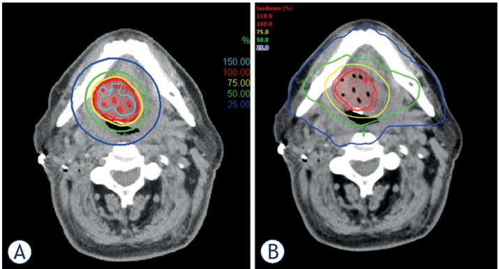
Figure 1
Representative dose distribution of (A) image guided high-dose-rate brachytherapy (IGBT) and (B) volumetric modulated arc therapy (VMAT).