
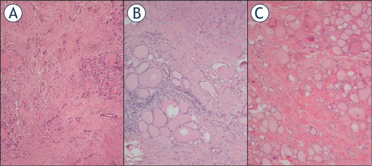
Figure 1
Haematoxylin and eosin stained sections of Case 4 (A), Case 5 (B) and Case 6 (C). Follicular atrophy and fibrosis, fibrosis accompanied by chronic inflammatory cells and fibrosis are seen, respectively.
Table 1
Demographic characteristics and laboratory findings at disease onset of patients with subacute thyroiditis and papillary thyroid cancer
| Cases | Age | Sex | FT3 (pmol/L) (3.1–6.8) | FT4 (pmol/L) (12–22) | TSH (miU/L) (0.27–4.2) | CRP (mg/L) (0–5) | ESR (mm/h) (0–20) | ∗Tc 99m /RAI Uptake (%) (0.3–3 vs. 20–50) | Ultrasonography |
|---|---|---|---|---|---|---|---|---|---|
| 1 | 42 | F | NA | 19.9 | 0.2 | NA | 55 | Low/0.2 | 3.2 cm hypoechoic nodule |
| 2 | 56 | F | 6.73 | 26.3 | 0.009 | NA | 34 | 0.06/1.35 | Diffuse HEAs, 2.2 cm hypo-isoechoic nodule |
| 3 | 56 | F | 4.5 | 21.1 | 0.68 | 125.0 | 100 | 0.59/NA focal hypoactivity | 1.8 cm focal HEA, 0.7 cm hypoechoic nodule with microcalcification |
| 4 | 51 | M | NA | 44.2 | 0.01 | NA | 91 | Low/NA | Focal HEAs, 1.6 cm isoechoic nodule |
| 5 | 52 | F | 7.01 | 24.7 | 0.02 | 15.6 | 60 | Low/NA | 2.4 cm heterogenous nodule with calcification and 1.1 cm isoechoic nodule |
| 6 | 45 | F | 13 | 45 | 0.005 | 138.8 | 132 | Low/NA | 2.2 cm hypoechoic, 1.9 cm isoechoic nodules |
Table 2
Presurgical ultrasonographic findings and histopathologic features of subacute thyroiditis patients with papillary thyroid cancer
| Cases | Op.Time(mo) | Nodule size in USG (cm)∗ | Sonographic features of nodules | FNAB | Tumor subtype/Histology | Tumor size (cm) | Stage (8th TNM) | Treatment |
|---|---|---|---|---|---|---|---|---|
| 1 | 107 | 0.55 and 0.50 | Hypoechoic, indefinite margins | Suspicious for malignancy | Papillary-tall cell and classical Focal fibrosis | 0.5 and 0.05 | I | TT+RAI |
| 2 | 13 | 2.4 and 1.0 | Hypo-isoechoic, calcification | Dyskaryotic thyrocytes | Papillary-classical Chronic lymphocytic thyroiditis | 1.0 | I | TT+RAI |
| 3 | 29 | 0.7 | Hypoechoic, microcalcification | FLUS Suspicious for malignancy | Papillary-follicular variant Fibrosis, chronic lymphocytic thyroiditis | 0.6 | I | TT |
| 4 | 16 | 1.9 | Isoechoic | Suspicious for malignancy | Papillary-follicular variant Fibrosis, focal follicular atrophy | 0.4 | I | Lobectomy |
| 5 | 13 | 1.1 and 0.73 | Isoechoic, microcalcification | Suspicious for malignancy | Papillary-classical and follicular | 1.1, 0.7, 0.3, 0.2 | I | TT+ RAI |
| Fibrosis, chronic lymphocytic thyroiditis | ||||||||
| 6 | 37 | 1.7 and 0.9 | Hypoechoic and isoechoic | AUS Papillary carcinoma | Papillary-classical and follicular Fibrosis | 1.2, 0.3, 0.2 | I | TT+RAI |