
Table 1A
Patient characteristics.
| Characteristic | abs. (rel.) 87 (100) |
|---|---|
| Age (mean/median) years | 58.4/59.0 |
| Male | 67 (77.0) |
| Female | 20 (23.0) |
| Primary tumour site | |
| Oropharynx | 33 (37.9) |
| Oral cavity | 30 (34.5) |
| Larynx | 9 (10.3) |
| Hypopharynx | 15 (17.2) |
| Localization | |
| right | 42 (48.3) |
| left | 40 (46.0) |
| midline | 5 (5.7) |
Table 1B
Pre- and postoperative staging results.
| Stage | Pathological staging | Clinical staging | p-value | |
|---|---|---|---|---|
| T | ||||
| 1 | 19 (21.8) | 19 (21.8) | 0.000a | |
| 2 | 42 (48.3) | 34 (39.1) | ||
| 3 | 14 (16.1) | 19 (21.8) | ||
| 4 | 12 (13.8) | 15 (17.2) | ||
| N | ||||
| 0 | 21 (24.1) | 17 (19.5) | 0.000a | |
| 1 | 15 (17.2) | 15 (17.2) | ||
| 2a | 2 (2.3) | 6 (6.9) | ||
| 2b | 34 (39.1) | 24 (27.6) | ||
| 2c | 13 (14.9) | 25 (28.7) | ||
| 3 | 2 (2.3) | 0 (0) | ||
| UICC | ||||
| I | 4 (4.6) | 2 (2.3) | 0.000a | |
| II | 12 (13.8) | 6 (6.9) | ||
| III | 11 (12.6) | 17 (19.5) | ||
| IV | 60 (69) | 62 (71.3) |
Table 1C
Clinical and pathological lymph node staging results in relation to the type of neck dissection performed.
| Stage | Bilateral neck dissection | Unilateral neck dissection | Total |
|---|---|---|---|
| pN0 | 15 | 6 | 21 |
| pN1 | 13 | 2 | 15 |
| pN2a | 2 | 0 | 2 |
| pN2b | 32 | 2 | 34 |
| pN2c | 13 | 0 | 13 |
| pN3 | 2 | 0 | 2 |
| Total | 77 | 10 | 87 |
| cN0 | 14 | 3 | 17 |
| cN1 | 11 | 4 | 15 |
| cN2a | 6 | 0 | 6 |
| cN2b | 21 | 3 | 24 |
| cN2c | 25 | 0 | 25 |
| Total | 77 | 10 | 87 |
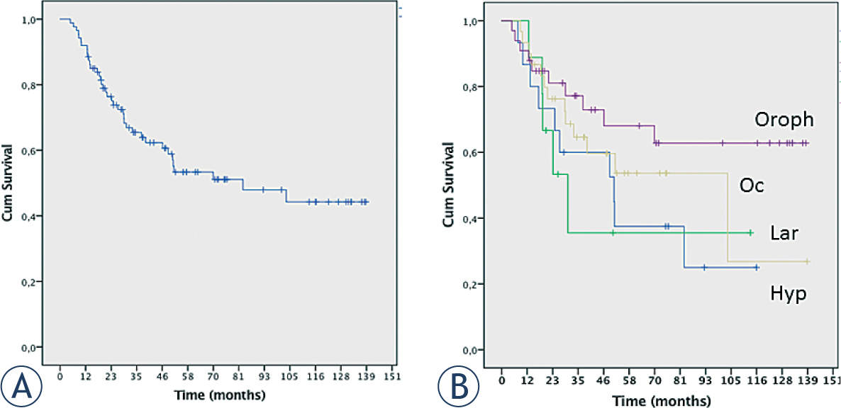
Figure 1
Kaplan Meier curves for the complete patient group (A) and according to primary tumour sites (B).
Hyp = hypopharyngeal carcinoma; Lar = laryngeal carcinoma; Oc = oral cavity carcinoma; Oroph = oropharyngeal carcinoma
Table 2A
Discrepancies of pathological and clinical findings according to imaging type
| Number of discrepant results per patient | CT (n = 55) | MRI (n = 21) | CT/MRI (n = 11) | Total (n = 87) |
|---|---|---|---|---|
| 0 | 43.6% | 38.1% | 36.4% | 36 (41.4%) |
| 1 | 25.5% | 23.8% | 45.5% | 24 (27.6%) |
| 2 | 18.2% | 23.8% | 9.1% | 16 (18.4%) |
| 3 | 5.5% | 9.5% | 0.0% | 5 (5.7%) |
| 4 | 7.3% | 4.8% | 0.0% | 5 (5.7%) |
| 5 | 0.0% | 0.0% | 9.1% | 1 (1.1%) |
Table 2B
Concordance of pathological and clinical findings according to N stage
| Clinical stage | Pathological stage | |||||
|---|---|---|---|---|---|---|
| N = 87 | pN0 | pN1 | pN2a | pN2b | pN2c | pN3 |
| cN0 (N = 17) | 11 | 3 | 0 | 1 | 2 | 0 |
| cN1 (N = 15) | 4 | 6 | 0 | 3 | 2 | 0 |
| cN2a (N = 6) | 1 | 2 | 1 | 2 | 0 | 0 |
| cN2b (N = 24) | 3 | 2 | 0 | 18 | 1 | 0 |
| cN2c (N = 25) | 2 | 2 | 1 | 10 | 8 | 2 |
| cN3 (N = 0) | 0 | 0 | 0 | 0 | 0 | 0 |
Table 3A
Prevalence of regional lymph node involvement according to tumour site
| Primary tumour localization | |||||
|---|---|---|---|---|---|
| Lymph Node Level | Hypopharynx (n = 15) | Larynx (n = 9) | Oral cavity (n = 30) | Oropharynx (n = 33) | % of total collective |
| I | 2 (13.3%) | 1 (11.1%) | 5 (16.7%) | 6 (18.2%) | 14 (16.1%) |
| II | 8 (53.3%) | 4 (44.4%) | 22 (73.3%) | 18 (54.4%) | 52 (59.8%) |
| III | 3 (20%) | 4 (44.4%) | 13 (43.3%) | 15 (45.5%) | 35 (40.2%) |
| IV | 4 (26.7%) | 2 (22.2%) | 7 (23.3%) | 5 (15.2%) | 18 (20.7%) |
| V | 2 (13.3%) | 1 (11.1%) | 4 (13.3%) | 1 (3%) | 8 (9.2%) |
Table 3B
Statistical analysis of the correlation of involvement of adjacent lymphnode levels
Table 4
List of all cases associated with up- or down-staging according to the preand postoperative regional lymph-node status
| No. | cT | cN | Overall clinical stage | pT | pN | Overall pathological stage |
|---|---|---|---|---|---|---|
| Down-staging according to pN status n = 10 (11.5%) | ||||||
| 1 | 2 | 1 | III | 1 | 0 | I |
| 2 | 1 | 2a | IV | 1 | 0 | I |
| 3 | 2 | 2b | IV | 1 | 0 | I |
| 4 | 2 | 1 | III | 2 | 0 | II |
| 5 | 2 | 1 | III | 2 | 0 | II |
| 6 | 2 | 2b | IV | 2 | 0 | II |
| 7 | 1 | 2b | IV | 2 | 0 | II |
| 8 | 3 | 2c | IV | 2 | 0 | II |
| 9 | 3 | 2b | IV | 2 | 1 | III |
| 10 | 2 | 2c | IV | 2 | 1 | III |
| Up-staging according to pN status N = 9 (10.3%) | ||||||
| 1 | 2 | 0 | II | 2 | 1 | III |
| 2 | 3 | 0 | III | 4 | 1 | IV |
| 3 | 2 | 1 | III | 2 | 2b | IV |
| 4 | 2 | 1 | III | 2 | 2b | IV |
| 5 | 3 | 0 | III | 3 | 2b | IV |
| 6 | 2 | 1 | III | 2 | 2b | IV |
| 7 | 2 | 1 | III | 3 | 2c | IV |
| 8 | 1 | 0 | I | 1 | 2c | IV |
| 9 | 1 | 2b | III | 1 | 2c | IV |