
Introduction
Chronic kidney disease (CKD) is a major global health burden, progressively leading to end-stage renal disease (ESRD) and necessitating renal replacement therapies (1). Cardiovascular disease remains the leading cause of morbidity and mortality in this population (2). Among the spectrum of cardiovascular complications, pulmonary hypertension (PH) has emerged as a highly prevalent and severe complication in patients with CKD (3, 4).
The pulmonary circulation and the kidneys exhibit a constant, complex and bidirectional interaction. While some patients develop PH secondary to established left heart or respiratory comorbidities, a significant proportion develop PH driven directly by the uraemic milieu, severe endothelial dysfunction, volume overload and the haemodynamic burden of arteriovenous fistulas (AVFs) used for haemodialysis (3, 4). Reflecting this complex aetiology, current PH guidelines classify CKD-PH within group 5—PH with unclear and/or multifactorial mechanisms (5–7).
The development of PH in patients with CKD is independently associated with poorer outcomes, including a higher risk of death. Similarly, renal dysfunction in patients with PH and specifically with pulmonary arterial hypertension (PAH) is an independent predictor of mortality (3). Given the growing recognition of this cardiopulmonary–renal axis, this narrative review aims to summarise the current literature regarding the definition, epidemiology and intricate pathophysiological mechanisms linking PH and CKD. Furthermore, we outline the diagnostic, clinical and therapeutic implications of this dual burden, highlighting the critical need for multidisciplinary collaboration between pulmonologists, nephrologists and cardiologists to optimise patient outcomes.
Definition and classification of PH
PH is defined as a mean pulmonary artery pressure (mPAP) >20 mmHg, measured using right heart catheterisation (RHC) (5–7). PH can be subdivided into three subtypes depending on other haemodynamic variables: pre-capillary PH, isolated post-capillary PH (IpcPH) and combined pre- and postcapillary PH (CpcPH). Pre-capillary PH is present when pulmonary artery wedge pressure (PAWP) is ≤15 mmHg and pulmonary vascular resistance (PVR) is >2 Wood units (WU). IpcPH is defined as PH with PAWP >15 mmHg and PVR ≤2 WU. In CpcPH, both PAWP and PVR are elevated (5–7). Although RHC is the gold standard for PH diagnosis, echocardiography is widely used for non-invasive assessment of right heart morphology and function, as well as estimating systolic pulmonary artery pressure (sPAP) (5, 6). Doppler echocardiography can be used to measure the peak tricuspid regurgitation velocity (TRV) and derive the tricuspid regurgitation pressure gradient (TRPG). Other echocardiographic variables such as inferior vena cava (IVC) diameter and collapsibility can be used to estimate right atrial pressure (RAP). The sum of TRPG and RAP is the estimated sPAP (5, 6). RAP approximation on echocardiography can be inaccurate when compared with directly measured RAP on invasive RHC (8), while TRPG is an indirectly derived value. For these reasons, echocardiographic estimates of sPAP correlate only moderately with direct measurements on RHC (9).
Current PH guidelines recommend that modern echocardiographic screening assigns a low, intermediate or high probability of PH based primarily on TRV rather than estimated sPAP (5, 6). Specifically, a TRV >2.8 m/s, particularly when accompanied by other indirect echocardiographic signs of right ventricular (RV) pressure overload (such as right ventricle dilation or flattened interventricular septum), indicates an intermediate or high probability of PH (5, 6). Despite these recommendations, estimated sPAP has been widely used in older clinical studies to define PH, especially in cohorts without strict indications for RHC, such as CKD or ESRD patients (10). Most of these studies used estimated sPAP >35 mmHg as a cut-off for PH, as this value was shown to correlate with elevated mPAP on RHC (11).
PH is classified into five groups, based on pathophysiological mechanisms, clinical features and haemodynamic profile: PAH (group 1), PH associated with left heart disease (group 2), PH associated with chronic lung disease or hypoxia (group 3), PH associated with pulmonary artery obstruction (group 4) and PH with unclear and/or multifactorial mechanisms (group 5) (5–7). Group 5 includes several disorders that can lead to PH through various mechanisms, including CKD with or without haemodialysis. Patients with PH can present with features of more than one group; in those cases, clinicians are encouraged to classify them based on the dominant pathophysiological mechanism (7).
Definition and classification of CKD
CKD is defined as a clinically relevant abnormality in the structure or function of the kidneys, which is persistent for at least 3 months (1). These abnormalities include changes in urine sediment, haematuria, albuminuria, electrolyte imbalances due to tubular dysfunction and structural anomalies detected by imaging or histological examination (1).
CKD is classified using the “cause”, “glomerular filtration rate” and “albuminuria” (CGA) system, which includes cause, glomerular filtration rate (GFR) category (G1-G5) and albuminuria category (A1-A3) (1).
Epidemiology of PH in CKD
PH is frequent in CKD patients and is associated with poorer outcomes, with decreased survival and a higher risk of cardiovascular events (3). Most studies on CKD cohorts have used echocardiography to define PH (10), but there are several reports that confirm the same results in RHC-confirmed PH (Table 1). The prevalence of PH is higher in more advanced stages of CKD, with the highest prevalence in patients with ESRD undergoing renal replacement therapy (RRT) (4, 10, 12, 13). Among RRT modalities, haemodialysis is consistently associated with a higher risk of PH compared with peritoneal dialysis (10, 14–16). In a large retrospective study of 2351 CKD patients, the prevalence values for PH in CKD stages G1, G2, G3a, G3b, G4 and G5 were 2.2%, 6.7%, 9.4%, 6.6%, 15.2% and 20%, respectively (17). In this same study, haemodialysis patients had a PH prevalence of 37.5% (17).
Table 1.
Studies investigating renal function and RHC-confirmed PH
| Study | Population | Key findings |
|---|---|---|
| Leuchte et al. 2007 (18) | 118 patients with PH undergoing RHC, excluding those with left-heart disease or ESRD on haemodialysis | eGFR <60 mL/min/1.73 m2 has a prevalence of 19% and is associated with increased mortality. NT-proBNP lost correlation with haemodynamics in patients with renal impairment. |
| Shah et al. 2008 (19) | 500 patients with PAH (1982–2006). Excluded patients without baseline serum creatinine or ESRD. Only 460 patients had baseline RHC | Serum creatinine ≥1 mg/dL was independently associated with increased mortality. The association between renal function and death was most prominent in patients with RAP ≤10 mmHg. |
| Pabst et al. 2012 (20) | 62 patients with CKD stage 4 or 5, with or without haemodialysis, presenting with unexplained dyspnoea. Excluded patients with left ventricular ejection fraction <50%, significant valvular disease and severe lung disease | Post-capillary PH prevalence: - 65% in dialysis - 71% in non-dialysis Pre-capillary PH prevalence: - 13% in dialysis - 6% in non-dialysis All cases of pre-capillary PH in dialysis were unmasked only after haemodialysis. |
| Navaneethan et al. 2014 (13) | 1088 patients with mPAP ≥25 mmHg. Excluded patients on chronic dialysis or with kidney transplants | 36% of the PH cohort had eGFR <60 mL/min/1.73 m2 Lower eGFR was independently associated with higher allcause mortality. |
| O’Leary et al. 2017 (21) | 4635 patients undergoing RHC, with a baseline eGFR measurement | In patients with CKD, 68% had PH. Post-capillary PH was the most frequent (76% of all PH). CKD severity and PH were associated with increased mortality. |
| Bitker et al. 2018 (22) | 179 patients with RHC-confirmed PAH | 29% had CKD (eGFR <60 mL/min/1.73 m2). CKD was independently associated with increased mortality. Lower CI and higher RAP were key haemodynamic determinants of eGFR decline during follow-up. |
| Chakinala et al. 2018 (23) | 2368 PAH patients from the REVEAL registry with baseline and follow-up eGFR | A ≥10% decline in eGFR over 1 year independently predicted higher risk of death and clinical worsening. Lower baseline eGFR correlated significantly with higher mean RAP and lower CO. |
| Nickel et al. 2019 (24) | 283 patients with PAH, with measured urinary ACR | Albuminuria (ACR >30 mg/g) was present in 20.1% of patients and associated with higher mortality. Albuminuria did not correlate with right heart haemodynamics. |
| Edmonston et al. 2020 (25) | 12,618 patients with RHC (all indications), with baseline serum creatinine | 74% of patients with CKD had PH, predominantly isolated post-capillary (39.0%) or CpcPH (38.3%). CpcPH was associated with the highest mortality risk among patients with CKD. |
| Meservey et al. 2025 (26) | 6694 patients from 18 phase III clinical trials in PAH and CTEPH | 13.5% had a baseline eGFR <60 mL/min/1.73 m2. Lower eGFR correlated with higher mean RAP and lower CI. PH therapy resulted in a minimal but statistically significant improvement in eGFR (+2.0 mL/min/1.73 m2 at 12–16 weeks). |
1 ACR, albumin to creatinine ratio; ADMA, asymmetric dimethylarginine; CI, cardiac index; CKD, chronic kidney disease; CO, cardiac output; CpcPH, combined pre- and post-capillary PH; CTEPH, chronic thromboembolic pulmonary hypertension; eGFR, estimated glomerular filtration rate; ESRD, end-stage renal disease; NT-proBNP, N-terminal pro B-type natriuretic peptide; PH, pulmonary hypertension; RAP, right atrial pressure; REVEAL, Registry to Evaluate Early and Long-term PAH Disease Management; RHC,
This observation was first reported in 2003, in a study of 58 haemodialysis patients which showed a PH prevalence of 39.7% (16). PH was associated with poorer survival (16). Notably, patients with potential causes of PH, such as cardiac or pulmonary disease, were excluded from this cohort, suggesting an effect of either CKD or dialysis on the pulmonary vasculature (16). Based on these findings, the fourth World Symposium on PH from 2008 included ‘chronic renal failure on dialysis’ among the causes of PH with unclear and/or multifactorial mechanisms (group 5 of the PH classification) (27).
Mechanisms of PH in CKD
Comorbidities
Some cases of PH in CKD may be explained by other causes (Figure 1). There is epidemiological overlap between CKD and several known aetiologies of PH (4). Connective tissue diseases which cause PAH (group 1 PH), such as systemic sclerosis and systemic lupus erythematosus, frequently involve the kidneys as well (28, 29). Cardiovascular comorbidities such as heart failure, atrial fibrillation and ischaemic heart disease, which cause group 2 PH, are frequent in CKD (2, 30). In particular, heart failure with preserved ejection fraction is exceptionally common in this cohort due to CKD-induced systemic inflammation, arterial stiffness and left ventricular hypertrophy, making it a primary driver of post-capillary PH (group 2), which is the most common type of PH in CKD (11, 29). Chronic obstructive pulmonary disease is one of the causes of group 3 PH and it is an important comorbidity in CKD (31). Obesity, which is linked to hypoventilation syndromes—another cause of group 3 PH—is also frequent in CKD (32, 33). Venous thromboembolism, which can cause group 4 PH, has a higher prevalence in patients with reduced estimated glomerular filtration rate (eGFR), especially in ESRD (34–36). However, not all associations between CKD and PH can be explained by comorbidities. Some CKD patients have ‘unexplained PH’, a term first introduced by Yigla et al. (37) in 2000. It is also plausible that in some patients who do have an identified cause of PH, renal impairment or dialysis may contribute to the pathophysiology of PH.
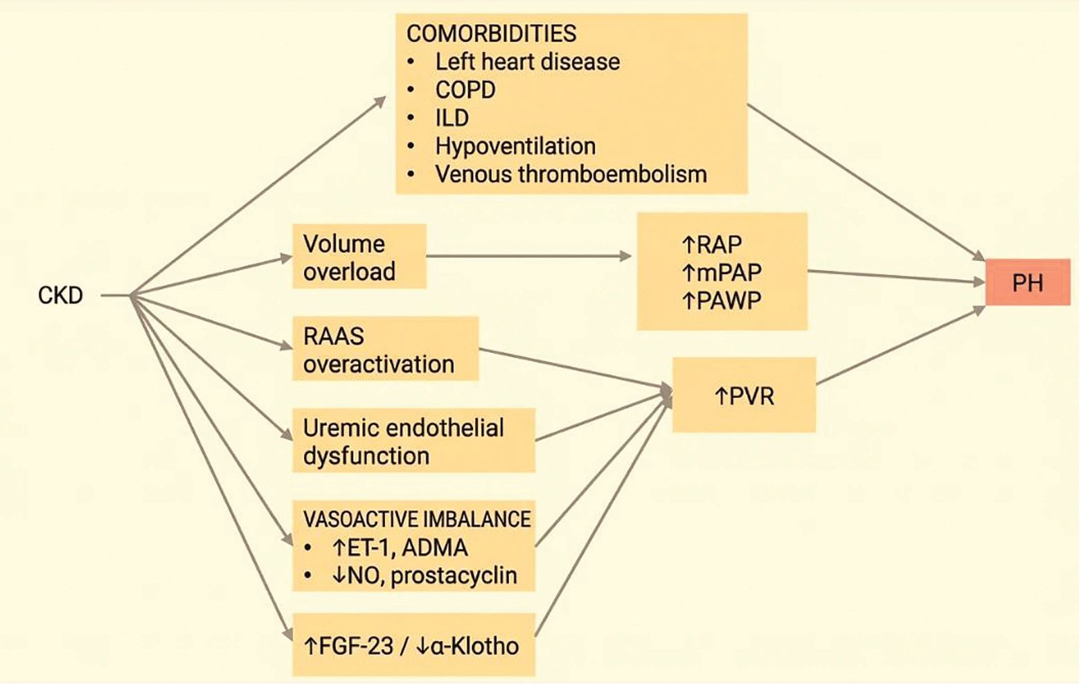
Figure 1.
Pathophysiological mechanisms of pulmonary hypertension in chronic kidney disease. ADMA, asymmetric dimethylarginine; CKD, chronic kidney disease; COPD, chronic obstructive pulmonary disease; ET-1, endothelin-1; FGF-23, fibroblast growth factor 23; ILD, interstitial lung disease; mPAP, mean pulmonary artery pressure; NO, nitric oxide; PAWP, pulmonary artery wedge pressure; PH, pulmonary hypertension; PVR, pulmonary vascular resistance; RAP, right atrial pressure; RAAS, renin-angiotensin-aldosterone system.
Volume overload
Volume overload is frequent in patients with ESRD (38). Haemodialysis patients in particular can experience significant changes in volemia in the interdialytic period (38). Even in the absence of overt left heart disease, volume overload is an important driver of PH in CKD via increased left-heart filling pressures, leading to post-capillary PH (3). In echocardiographic studies in large cohorts of CKD patients, markers of volume overload such as increased left atrial volume are significantly associated with PH (25). A higher RAP is associated with poorer outcomes in patients with CKD and PH, independent of mPAP and other variables (3, 13, 21). In patients with CKD, volume overload can cause increased renal venous congestion, leading to increased renal intraparenchymal pressure, tubular collapse and a decline in renal function (3). The direct link between volume status and PH in ESRD can be further demonstrated during dialysis, where fluid removal results in significant reduction in pulmonary artery pressure (20, 39).
Uraemia and endothelial dysfunction
Uraemia plays a central role in the pathophysiology of PH in CKD by promoting vasoconstriction, endothelial dysfunction and vascular remodelling (4). The uraemic environment disrupts normal pulmonary vascular tone by shifting the balance of vasoactive substances towards vasoconstriction. Uraemic patients exhibit impaired production of, and decreased sensitivity to, vasodilators like nitric oxide (NO) and prostacyclin, as well as elevated levels of vasoconstrictors such as endothelin-1 (ET-1), asymmetric dimethylarginine (ADMA) and thromboxane (4). This imbalance leads to persistent pulmonary vasoconstriction and decreased compliance of the pulmonary vasculature (4). In addition to vasoconstriction, high concentrations of uraemic solutes may promote pulmonary vascular remodelling and endothelial dysfunction (3). For example, high blood urea nitrogen levels correlate with increased PVR in patients with CKD (40). In vitro studies have shown that urea concentrations similar to those found in kidney failure directly induce endothelial cell dysfunction (3). Beyond its effects on the pulmonary vasculature, uraemia also has direct toxic effects on the heart muscle. This uraemia-mediated dysregulation of cardiomyocytes contributes to left ventricular dysfunction (3, 4). This cardiac impairment subsequently elevates left-sided filling pressures, leading to pulmonary venous congestion and post-capillary (group 2) PH (3).
ET-1
The endothelin (ET) signalling pathway plays a crucial role in the pathobiology of PAH (41) and ET receptor antagonists are a cornerstone of PAH treatment (5, 6, 42). Plasma levels of ET-1 are elevated in CKD, regardless of cause, and are correlated with CKD severity (43). Higher levels of ET-1 are associated with a higher risk of death, cardiovascular events and progressive renal dysfunction in CKD (44). However, there are no studies specifically addressing the association between ET-1 and PH in CKD.
ADMA
ADMA is an endogenous inhibitor of nitric oxide (NO) synthase (45). Inhibition of NO signalling is one of the main mechanisms involved in the pathogenesis of PAH (41). High levels of ADMA have been linked to PH in both mice (46) and humans (47). CKD have higher serum ADMA, due to increased synthesis, reduced degradation and impaired excretion through the kidneys (48, 49). Elevated ADMA is associated with a higher risk of death and cardiovascular events in CKD (50).
The renin–angiotensin–aldosterone system
Overactivation of the renin–angiotensin–aldosterone system (RAAS) promotes vasoconstriction, inflammation and fibrosis in both the systemic and pulmonary vessels (51). In CKD patients, RAAS activity is increased and plays an important role in the pathogenesis and progression of renal dysfunction (52). In idiopathic PAH, systemic RAAS activity is also elevated and associated with an increased risk of disease progression, death or lung transplantation (53). Increased production of angiotensin II in pulmonary endothelial cells and overexpression of angiotensin II type 1 receptor mediate pulmonary vascular remodelling in PAH (53, 54). Treatment with angiotensin converting enzyme inhibitors or angiotensin receptor blockers (ARB) are associated with improved outcomes in pre- and post-capillary PH (55). In an experimental model of PAH, treatment with ARB delayed disease progression, reduced pulmonary vascular remodelling and RV afterload and improved RV diastolic function and coupling between the RV and the pulmonary artery (53).
Fibroblast growth factor 23 and α-Klotho
Fibroblast growth factor 23 (FGF-23) is a bone-derived, endocrine hormone that regulates phosphate and vitamin D homeostasis by acting on the renal proximal tubule and parathyroid glands via FGF receptors and the co-receptor α-Klotho (56, 57). FGF-23 rises early in the course of CKD (58) and elevated levels are independently associated with CKD progression, development of ESRD and mortality (59). FGF-23 also influences cardiovascular physiology, as elevated levels are associated with heart failure and left ventricular hypertrophy (60). In dialysis patients, elevated levels of FGF-23 correlate with elevated sPAP on echocardiography (61). In patients with PAH and chronic thromboembolic PH (CTEPH), higher levels of FGF-23 are associated with higher mPAP and PVR and lower cardiac index, independent of eGFR (62), suggesting a role on the pulmonary circulation.
α-Klotho is a transmembrane protein expressed in the renal tubules, parathyroid gland and choroid plexus, with soluble form found in blood, urine and cerebrospinal fluid (63, 64). It acts as an obligatory co-receptor for FGF-23, regulating phosphate and vitamin D metabolism (63). Overexpression of this molecule extends the lifespan of mice, while its disruption causes premature ageing, hence the name derived from the Fate in Greek mythology who spins the thread of life (64, 65). CKD is a state of α-Klotho deficiency (65) and lower levels are associated with CKD progression, ESRD and mortality (66). Rats with monocrotaline-induced PAH have significantly lower levels of α-Klotho expression in lung tissue and restoration of α-Klotho expression is associated with reduced pulmonary artery pressure, reverse remodelling of pulmonary vessels and reduced RV hypertrophy (67). Infants with PH associated with bronchopulmonary dysplasia have lower levels of α-Klotho in the umbilical cord (68). Overexpression of α-Klotho in a bronchopulmonary dysplasia rodent model of PH reduces pulmonary artery pressure and pulmonary vascular remodelling (68).
Parathyroid hormone
The parathyroid hormone (PTH) is secreted by the parathyroid glands in response to imbalances in serum calcium and phosphorus. In CKD, serum PTH levels are chronically increased due to secondary parathyroidism and the increase is proportional to the severity of renal dysfunction (69, 70). PTH is an important risk factor for cardiovascular morbidity and mortality in CKD (71). In a recent study on animal models of PH, exogenous PTH administration worsened PH and RV hypertrophy, while inhibition of PTH signalling improved PH (72). In humans with PH, serum PTH levels are correlated with mPAP and PVR (72). In cultured human pulmonary vascular smooth muscle cells, PTH signalling promotes proliferation and migration (72). These findings suggest that PTH plays a role in pulmonary vascular remodelling and could represent a pathophysiological mechanism for PH in CKD.
Arterio-venous fistulas
AVFs used for vascular access in haemodialysis create a shunt between a systemic artery and a systemic vein, bypassing smaller arteries, capillaries and veins. The consequence is a reduction in systemic vascular resistance and a subsequent increase in the pre-load of the right ventricle, leading to a high cardiac output (CO) state which can increase PAP (3, 73). In addition, blood shunting through the AVF can reduce blood pressure, followed by an increase in heart rate and contractility via sympathetic activation (3). These effects are stronger in proximal upper arm AVFs compared with forearm AVFs, since proximal AVFs are characterised by higher flow rates (74, 75). High AVF flow (Qa) ≥2 L/min and Qa to CO ratios (>0.3) are strongly associated with high-output cardiac failure and PH (74, 75). In patients with Qa > 1.5 L/min, surgical reduction of Qa leads to a decrease in sPAP and improvements in RV morphology and function (76).
PH and kidney transplantation
Kidney transplantation (KT) is the definitive treatment of ESRD as it restores normal glomerular filtration, reverses uraemia-induced endothelial dysfunction, normalises volume status and mitigates bone-mineral metabolism dysregulation (77). In addition, after successful KT, AVFs can be ligated, as haemodialysis is no longer necessary. KT thus resolves many of the mechanisms of PH in CKD. Indeed, patients with CKD and PH who undergo KT survive longer than those who remain on dialysis (77). A significant proportion of patients with PH who undergo KT experience normalisation of PAP after KT (16, 78, 79). The resolution of PH post-KT is associated with improved survival, similar to that of patients without PH before KT (79).
Clinical implications of PH in patients with CKD
The presence of PH in patients with CKD poses significant diagnostic and therapeutic challenges that require multidisciplinary collaboration. From a diagnostic perspective, echocardiography remains the primary non-invasive screening tool, but its timing is critical in patients undergoing haemodialysis. Because volume fluctuations between dialysis sessions affect haemodynamics, echocardiography should ideally be performed post-dialysis, when the patient has achieved their dry weight, in order to avoid overestimation of sPAP (39). If the clinical probability of PH is high and its confirmation would alter management, RHC is needed to accurately differentiate between pre-capillary and postcapillary phenotypes (20, 39).
Pulmonary vasodilators used in the treatment of PAH are not approved in group 5 CKD-associated PH (5, 6). Given the significant contribution of volume overload and left heart disease in CKD-PH, the off-label use of pulmonary vasodilators in these patients may cause harm and should be reserved strictly for patients who have an overlapping group 1 PAH diagnosis or, occasionally, as an off-label ’bridge to transplant’ in carefully selected candidates with severe pre-capillary haemodynamics (5, 6, 39). When prescribing PAH drugs in severe CKD, rigorous monitoring is mandatory, as many of these agents or their active metabolites require dose adjustments in the setting of reduced eGFR to avoid systemic toxicity (39).
The management of CKD-PH relies primarily on addressing underlying conditions and modifiable mechanisms (3, 39). Strict volume control by optimising dialysis and ensuring adequate ultrafiltration can lead to marked reductions in pulmonary artery pressures (20, 39). Furthermore, clinicians must proactively screen for and treat comorbid left heart disease (PH group 2) and chronic lung disease (PH group 3), which are highly prevalent in CKD patients (4). For patients with high flow AVFs, surgical flow reduction should be considered to offload the right ventricle (76). If alternative therapies like peritoneal dialysis or KT are feasible, complete AVF ligation is also a viable option.
PH should be taken into consideration when establishing the indication and timing of KT. Current guidelines do not take PH into account and severe PH is sometimes considered a contraindication for KT due to increased peri-operative mortality (3, 79). However, evidence suggests that KT may improve or resolve PH in a substantial number of patients, an effect which is associated with improved survival (79).
Renal dysfunction in PH
Just as PH is frequently identified in CKD patients, renal dysfunction is also frequent in patients with PH (3). The prevalence of renal dysfunction, typically defined as eGFR <60 mL/min/1.73 m2, ranges between 13.5% and 36% depending on study population, PH aetiology and diagnostic criteria for PH. In a cohort of 1088 patients with all-cause PH, the prevalence of eGFR <60 mL/min/1.73 m2 was over 30% (13). In a cohort of 2386 PAH patients from the American Registry to Evaluate Early and Long-term PAH Disease Management (REVEAL), the prevalence was 28.3% (23). Even when eGFR is normal, 20% of PAH patients exhibit elevated urinary albumin-to-creatinine ratios (24). In a meta-analysis of 6694 participants from 18 phase 3 clinical trials for PAH and CTEPH, the prevalence was only 13.5%, but this was likely due to the fact that renal dysfunction was an exclusion criterion in half of the trials included in the meta-analysis (26). At any rate, the prevalence of renal dysfunction in PH is higher than the estimated 9.1% prevalence in the general population (3).
Across multiple large cohorts, worsening renal function is consistently associated with higher PH disease severity. Patients with lower eGFR have a worse haemodynamic profile on RHC, in particular higher mPAP, RAP and PVR and lower CO (19, 26). They have a more severe World Health Organization (WHO) dyspnoea functional class (19, 23) and walk shorter distances at the 6-min walk test (19, 23, 26). They also have higher levels of brain natriuretic peptide (BNP) and N-terminal pro-BNP (NTproBNP) (18, 23, 26). NTproBNP is particularly elevated in renal dysfunction because it is primarily cleared by the kidneys (18).
Renal dysfunction is independently associated with poorer outcomes in PH (3). Lower eGFR at baseline predicts shorter survival across all groups of PH and the risk of death increases progressively with worsening kidney function (13, 23). In PAH and CTEPH, a 10 mL/min/1.73 m2 decrease in baseline eGFR is associated with a 16% increased risk of mortality or clinical worsening (26). Beyond baseline renal function, a longitudinal decline in eGFR of ≥10% also predicts mortality and all-cause hospitalisation (23).
Mechanisms of renal dysfunction in PH: the right-heart cardio–renal syndrome
The mechanisms of renal dysfunction in patients with PH include impaired renal arterial perfusion, increased renal venous congestion and neurohumoral dysregulation stemming from RV failure (3). The interplay of these mechanisms has been described as the ‘right-heart cardio-renal syndrome’ (3). Patients with PH have increased PVR, which leads to RV dysfunction and decreased CO. This can be exacerbated by the bowing of the interventricular septum, which impairs left ventricular filling (3, 26). This state of low CO underfills the renal arterial system and lowers renal perfusion pressure. The kidneys respond to this perceived volume depletion by overactivating the RAAS and the sympathetic nervous system (3, 23). This neurohumoral activation causes afferent arteriolar vasoconstriction, salt and fluid retention (hypervolemia), and reduced eGFR (3).
Right heart failure directly elevates RAP and central venous pressure, causing blood stasis in the systemic and renal veins (23, 26). Because the kidneys are enclosed in a rigid capsule, this venous congestion significantly increases intraparenchymal pressure, leading to parenchymal oedema, tubular compression and collapse, intracapsular tamponade and tissue hypoxia (3, 26). The combination of elevated venous pressure and decreased arterial perfusion severely impairs the trans-glomerular pressure gradient, driving a sharp decline in the GFR (3). Venous congestion triggers an abnormal tubulo–glomerular feedback loop where the release of adenosine causes the afferent arteriole to constrict and the efferent arteriole to dilate, further reducing GFR (3). Additionally, right-heart failure and systemic congestion trigger the release of various inflammatory cytokines (such as interleukin-6 and tumor necrosis factor) and vasoactive substances (such as ET-1, arginine-vasopressin and prostaglandins), which directly promote kidney tissue alterations and vascular injury (3, 22).
Clinical implications of renal dysfunction in PH
The high prevalence and prognostic impact of renal dysfunction in PH have important clinical implications. Renal function should be routinely assessed and integrated into the management of patients with PH, as even mild reductions in eGFR are associated with higher mortality (23). Albuminuria may also be a useful biomarker in PH, as it is associated with poorer prognosis (24). Other biomarkers such as NT-proBNP require careful interpretation in patients with significant renal dysfunction, as levels may be falsely elevated due to inadequate clearance through the kidneys (18).
Therapeutic management of right-heart cardio-renal syndrome requires a delicate balance. Loop diuretics are the mainstay of treatment to relieve systemic venous congestion and reduce renal intraparenchymal pressure. However, overly aggressive diuresis can further reduce CO and impair renal arterial perfusion, potentially worsening eGFR (3). Therefore, diuretic therapy must be carefully titrated with close monitoring of serum creatinine and electrolytes.
Renal dysfunction should not preclude patients from receiving disease-modifying PAH therapies. By lowering PVR, increasing CO and decreasing RAP, targeted PAH therapies directly counteract the haemodynamic drivers of the right-heart cardio-renal syndrome (26). PAH-specific therapies induce a modest but statistically significant improvement in eGFR, suggesting reversibility of the cardio-renal interaction once RV function is optimised (26). Nevertheless, dose adjustments and rigorous monitoring for drug toxicity remain essential, particularly for agents cleared by the kidneys (39).
Conclusions
PH and CKD frequently occur together, and the combination carries a poorer prognosis than either disease in isolation. In patients with PAH, renal dysfunction is independently associated with worse outcomes and should be taken into consideration when stratifying risk. CKD-associated PH is a complex, multifactorial disorder driven by a combination of volume overload, uraemic endothelial dysfunction and high CO induced by AVFs. Its management requires a nuanced, pathophysiologically targeted approach, focusing on optimising fluid balance and correcting underlying comorbidities. Echocardiography and RHC should be performed after haemodialysis to minimise the effect of volume overload. PH should be taken into consideration when evaluating patients for KT, since transplanted patients frequently exhibit improved pulmonary haemodynamics and better survival. Ultimately, improving outcomes in patients with concurrent PH and CKD relies heavily on close multidisciplinary collaboration between nephrologists, pulmonologists and cardiologists.
Notes
[2] Contributed by Author’s contribution
All authors contributed equally to the conceptualisation, literature review, drafting and revision. The final submitted version was read and approved by all the authors.
[3] Conflicts of interest Conflict of interest
There is no conflict of interest.
[4] Ethics approval
Ethics Committee approval was not required.
[5] Informed consent statement
No informed consent was necessary.