
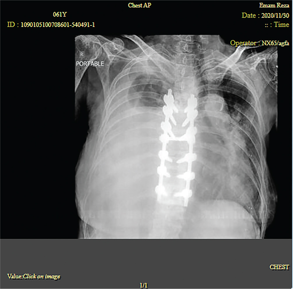
Figure 1.
Chest X-ray, right massive pleural effusion.
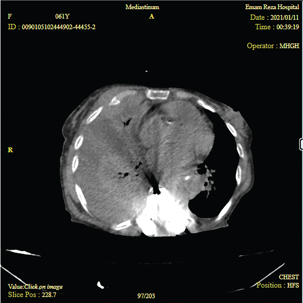
Figure 2.
CT of the thorax showed unilateral sided pleural effusions in the right side.
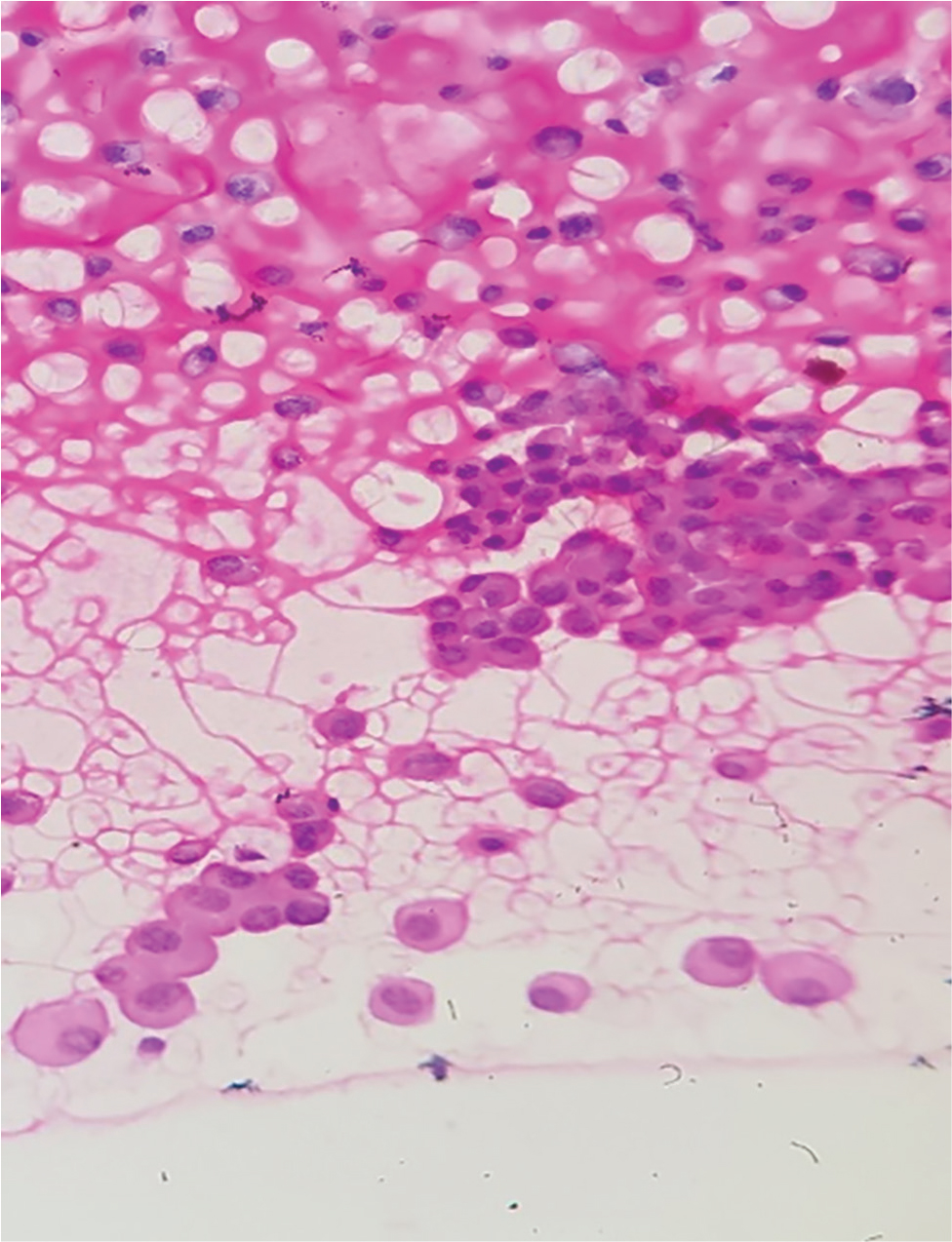
Figure 3.
Pleural fluid smear shows plasmacytoid cells with abundant cytoplasm and eccentric nuclei.there are some inflammatory cells in the background.
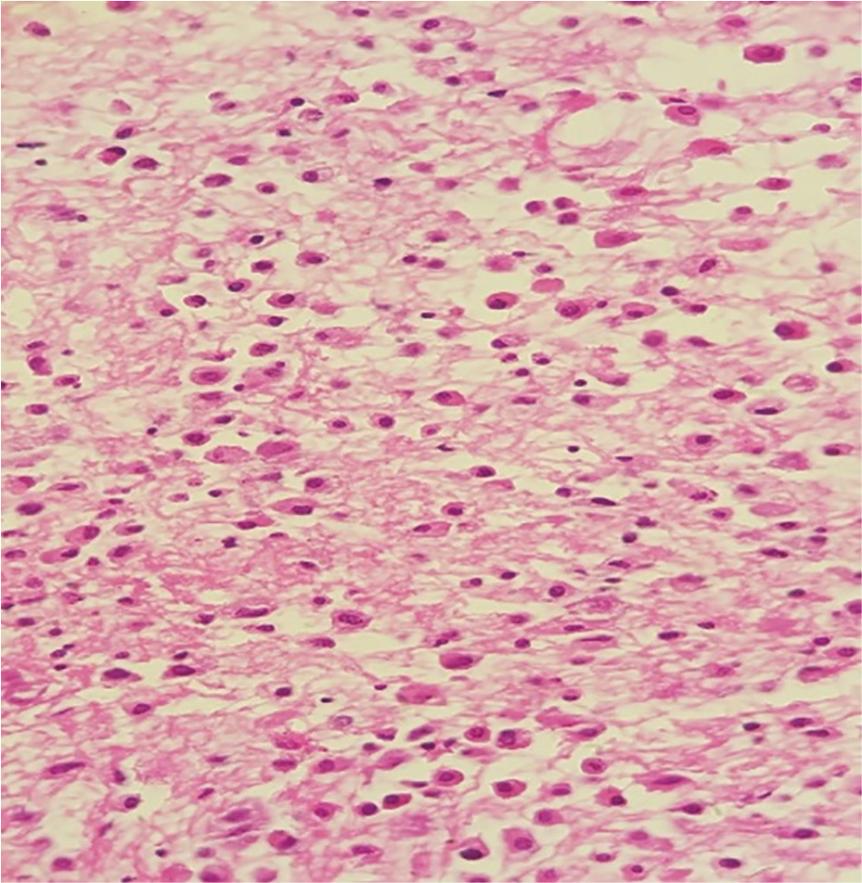
Figure 4.
Cell block sections reveal numerous plasmacytoid cells with abundant cytoplasm and eccentric nuclei.
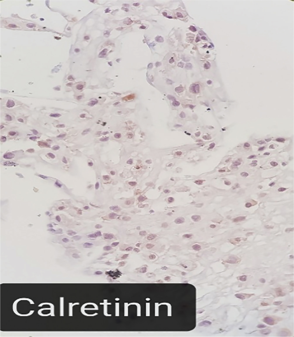
Figure 5.
Calretinin is negative in most plasmacytoid cells.
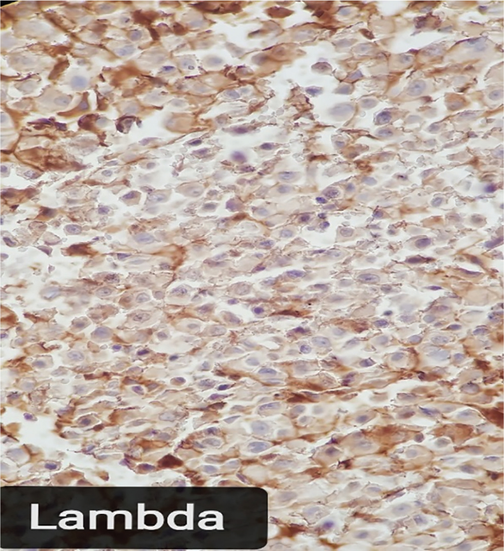
Figure 6.
Immunohistochemical staining is negative for lambda light chain in most plasma cells.
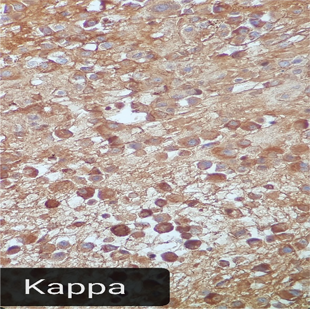
Figure 7.
Immunohistochemical staining shows diffuse cytoplasmic kappa light chain positivity in plasma cells.