
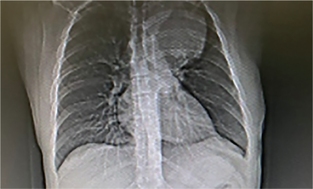
Figure 1.
Chest X-ray. A paramediastinal left tumor, clearly contured, 120x80 mm.
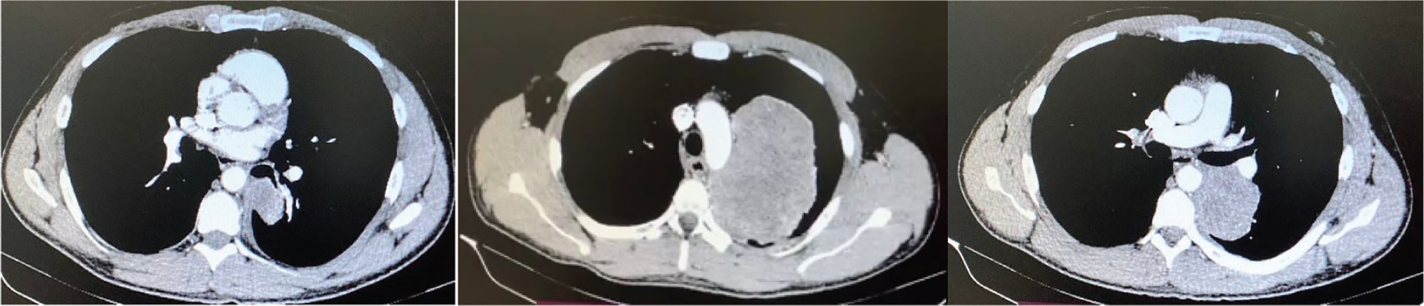
Figure 2.
Chest CT scan. A large mass in the left upper lobe which did not have a plane of clivage from arch and descending aorta, as well as from left the pulmonary artery.
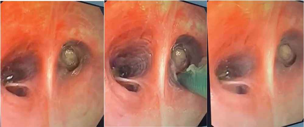
Figure 3.
Endobronchial aspect of the tumor.
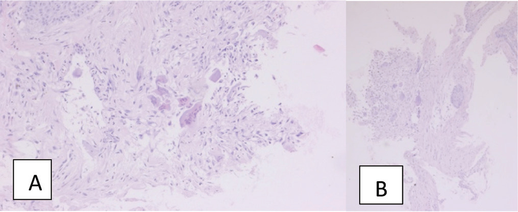
Figure 4.
(A,B). Histological aspect of the tumor. A- HE 20x. B- HE 10x.
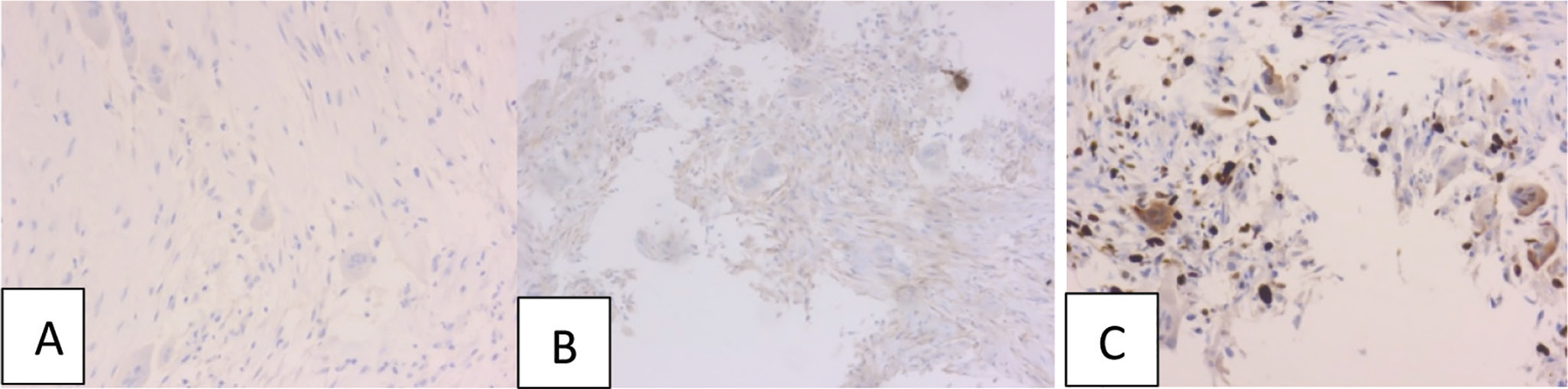
Figure 5.
Immunochemistry (IHC). A. IHC S100 20x. B. IHC CD99 20x. C. IHC Ki67 20x.
Table 1.
Subtypes of chondromas and sarcomas.
| Conventional chondrosarcoma | Fibrosarcoma | Malignant fibrous histiocytoma |
|---|---|---|
| Chondroma | Giant cell tumor osteosarcoma | Mesenchymal chondrosarcoma |
| Clear cell chondrosarcoma | Hemangiopericytoma | Neurofibroma of Bone (Schwangomul) |
| Classic osteosarcoma | High-grade surface osteosarcoma | Osteoclastoma |
| Ewing sarcoma | “Intraosseous” Osteosarcuma | Osteosarcoma in Paget’s Disease of Bone |
| Periosteal osteosarcoma | Peripheral neuroendocrine tumor | Small cell osteosarcoma |
| Periosteal condroma | Primitive neuroendocrine tumor | Telangiectatic osteosarcoma |