
Table 1
Histopathological diagnoses of 80 rare primary pulmonary neoplasms (Mayo Clinic, 1980–1990)
| Types of neoplasm | Patients no (%) |
|---|---|
| Lymphoma | 33 (41) |
| • Small cell | 22 |
| • Large cell | 6 |
| • Mixed cell | 5 |
| Carcinosarcoma | 16 (20) |
| Mucoepidermoid carcinoma | 12 (15) |
| Malignant fibrous histiocytoma | 4 (5) |
| Malignant melanoma | 3 (4) |
| Fibrosarcoma | 3 (4) |
| Leiomyosarcoma | 3 (4) |
| Angiosarcoma | 2 (2) |
| Haemangiopericytoma | 2 (2) |
| Osteosarcoma | 1 (1) |
| Blastoma | 1 (1) |
Table 2
Histopathological diagnosis of 32 rare primary pulmonary neoplasms (National Matsudo Hospital and National Cancer Hospital East – Japan)
| Types of malignant tumours (n = 20) | Patients no (%) |
| Epithelial tumours | |
| Adenocystic carcinoma | 1 (5) |
| Mucoepidermoid carcinoma | 2 (10) |
| Bronchial gland mixed tumour | 2 (10) |
| Myoepitelioma | 1 (5) |
| Soft tissue tumours | |
| Leiomyosarcoma | 1 (5) |
| Epitheloid haemangioendothelioma | 1 (5) |
| Hemangiopericytoma | 1 (5) |
| Miscellaneous tumours | |
| Carcinosarcoma | 3 (15) |
| Malignant melanoma | 1 (5) |
| Malignant lymphoma | 6 (30) |
| Coriocarcinoma | 1 (5) |
| Types of benign tumours (n = 12) | Patients no (%) |
| Epithelial tumours | |
| Papillomas | 3 (25) |
| Adenomas | 1 (8.3) |
| Soft tissue tumours | |
| Fibromas | 1 (8.3) |
| Lipoma | 3 (25) |
| Leiomyoma | 3 (25) |
| Miscellaneous tumours | |
| Meningioma | 1 (8.3) |
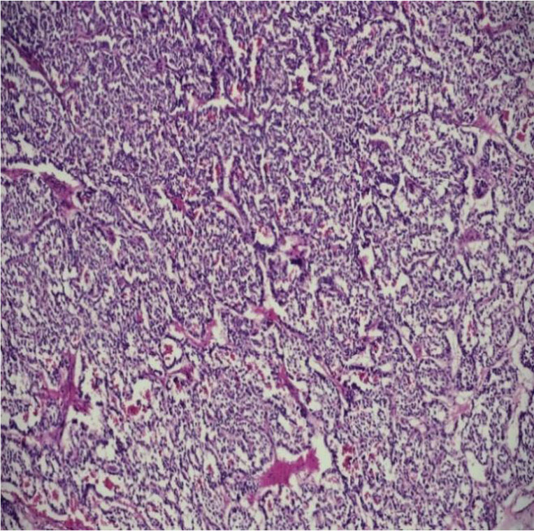
Figure 1
Interconnected trabeculae, separating from a vascular stroma - carcinoid tumor (H&E 10X).
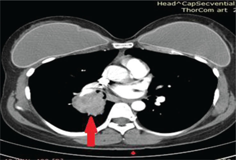
Figure 2
CT examination - Female pacient with a round hilar mass with irregular inferior edge - atypical carcinoid tumor.
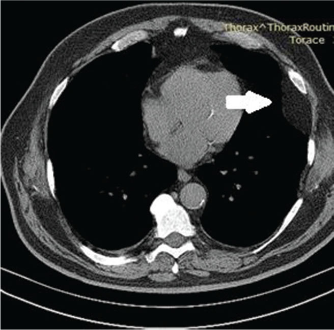
Figure 3
CT scan - Peripheral tumor with well defined border – pulmonary lipoma.
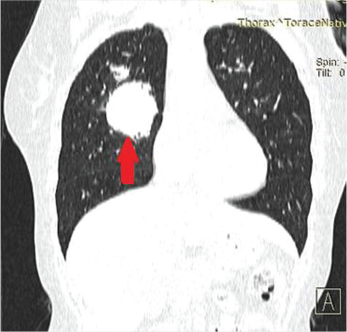
Figure 4
CT Scan - Large round mass with irregular edges localized in the right lung - Liposarcoma.
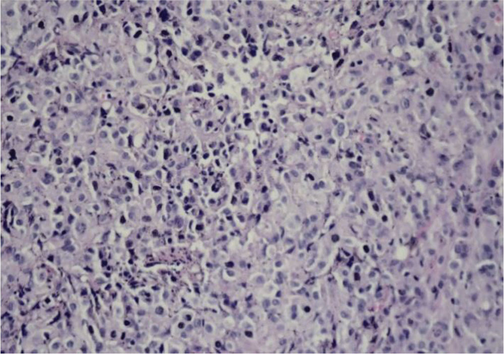
Figure 5
Multiple mesenchymal and epithelial elements without necrosis - Hamartoma (H&E 10X).
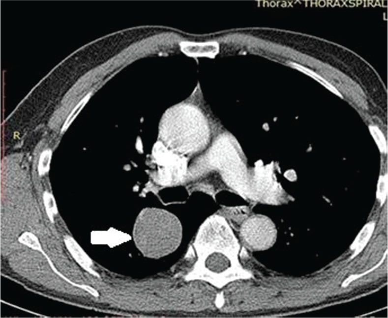
Figure 6
CT Scan - Well-defined lung mass, without necrosis but with slighty calcifications - Hamartoma.
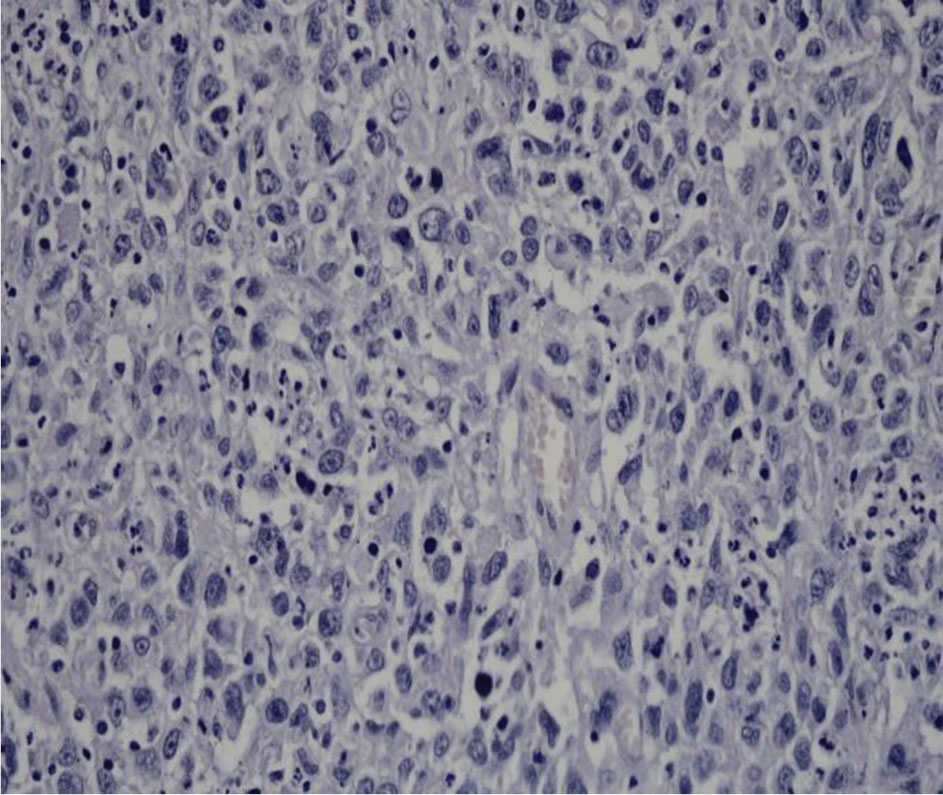
Figure 7
Cells with keratin with few atypia - Angiosarcoma (H&E 10X).
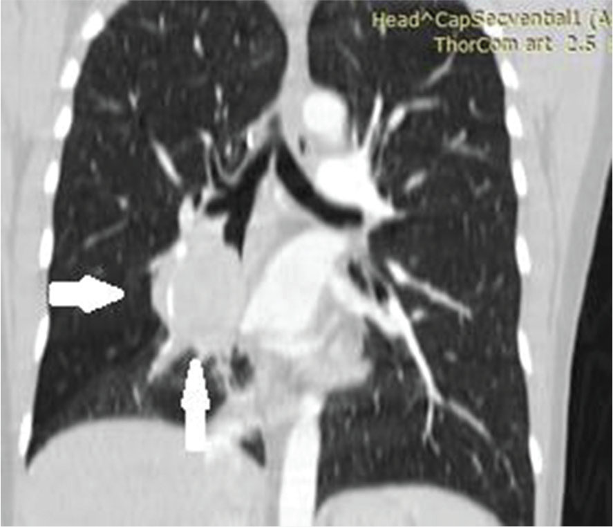
Figure 8
CT scan - Large central tumor situated in middle and lower lobe with spiculate margins - pulmonary angiosarcoma.
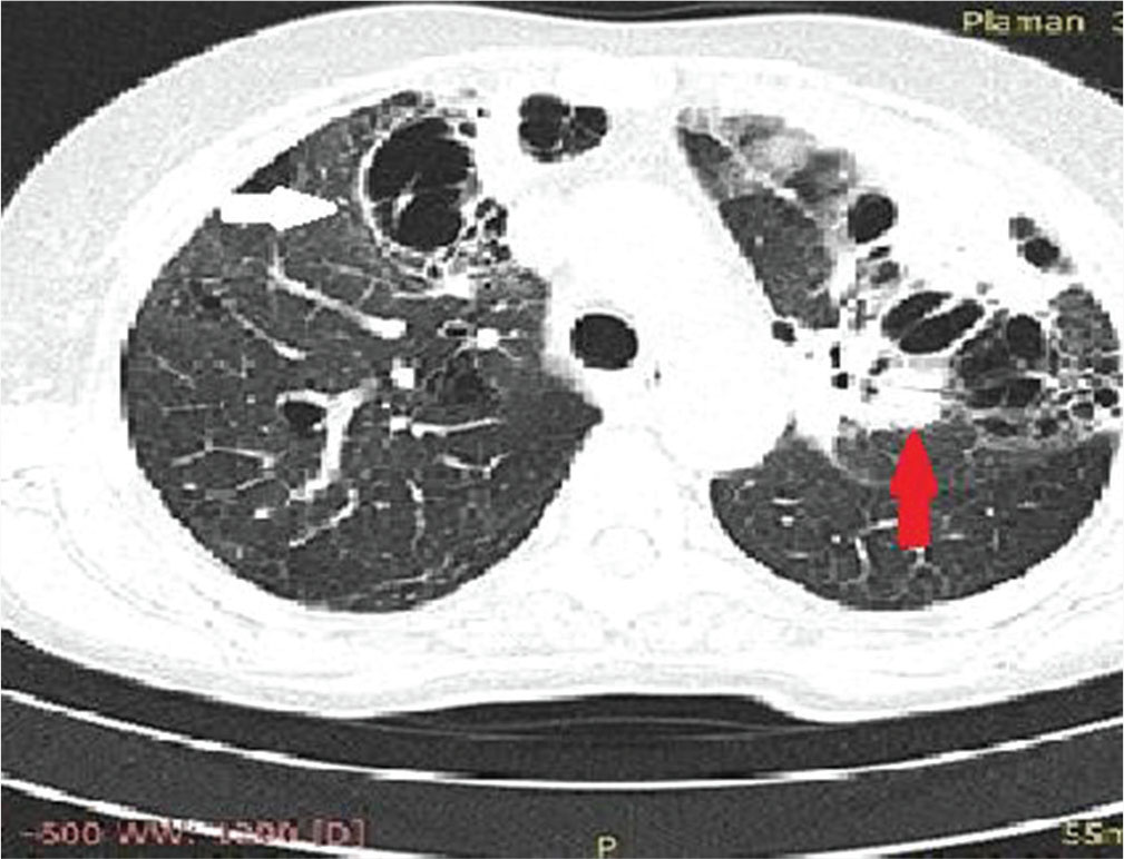
Figure 9
CT scan - Multicentric masses (red arrow) with central bronchiectasis and necrosis (white arrows) - pulmonary Angiocentric Lymphoma.