
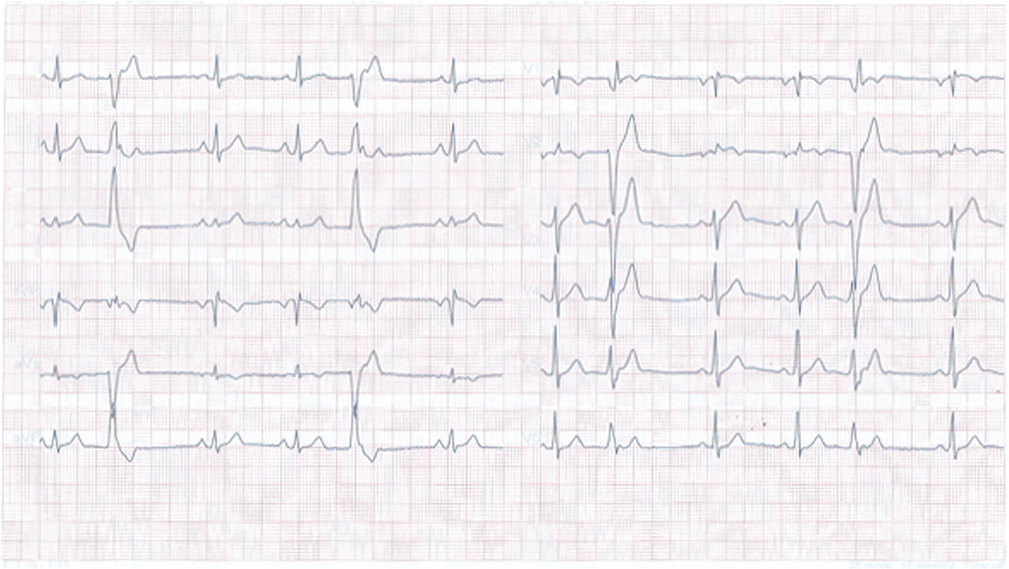
Figure 1
ECG recorded during an emergency presentation, showing frequent premature ventricular beats.
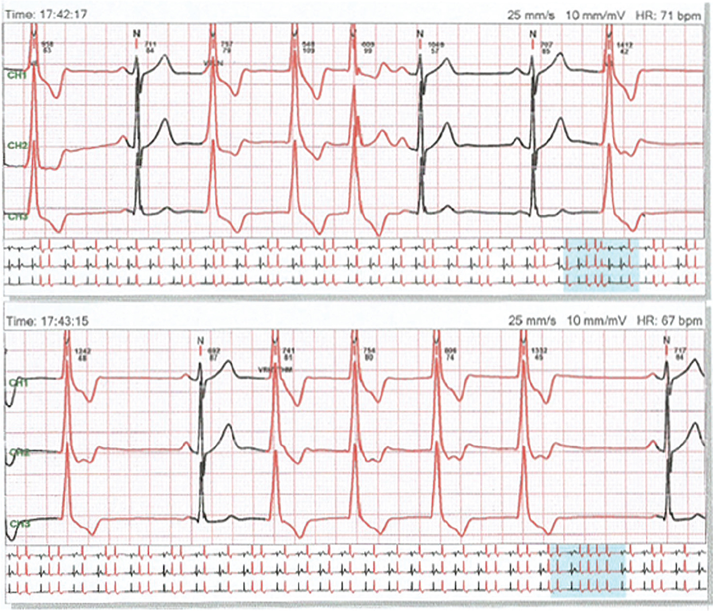
Figure 2
A 24-h recording of ECG showing frequent premature ventricular beats with unique morphology, mimicking a right bundle branch block.
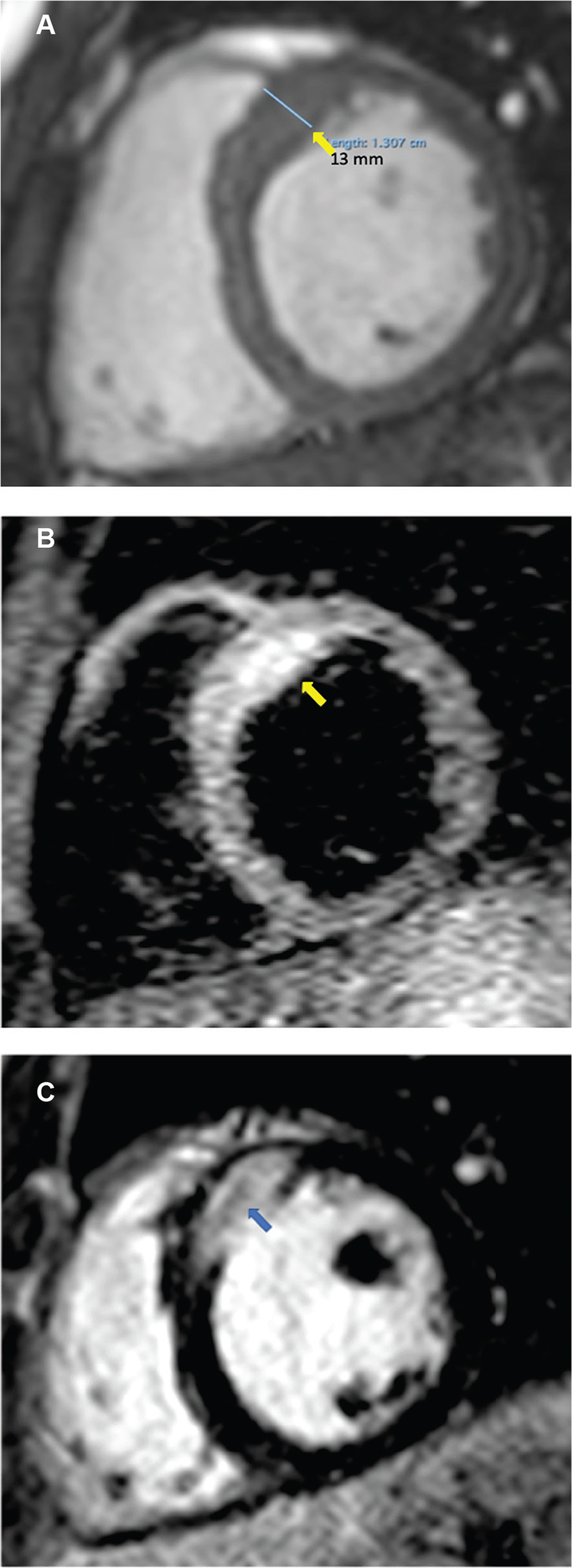
Figure 3
CMR. (A) Hypertrophy localised at the base of the septum. (B) T2 focal oedema at the base of the septum. (C) Late gadolinium enhancement: homogeneous late uptake of gadolinium in the granuloma.
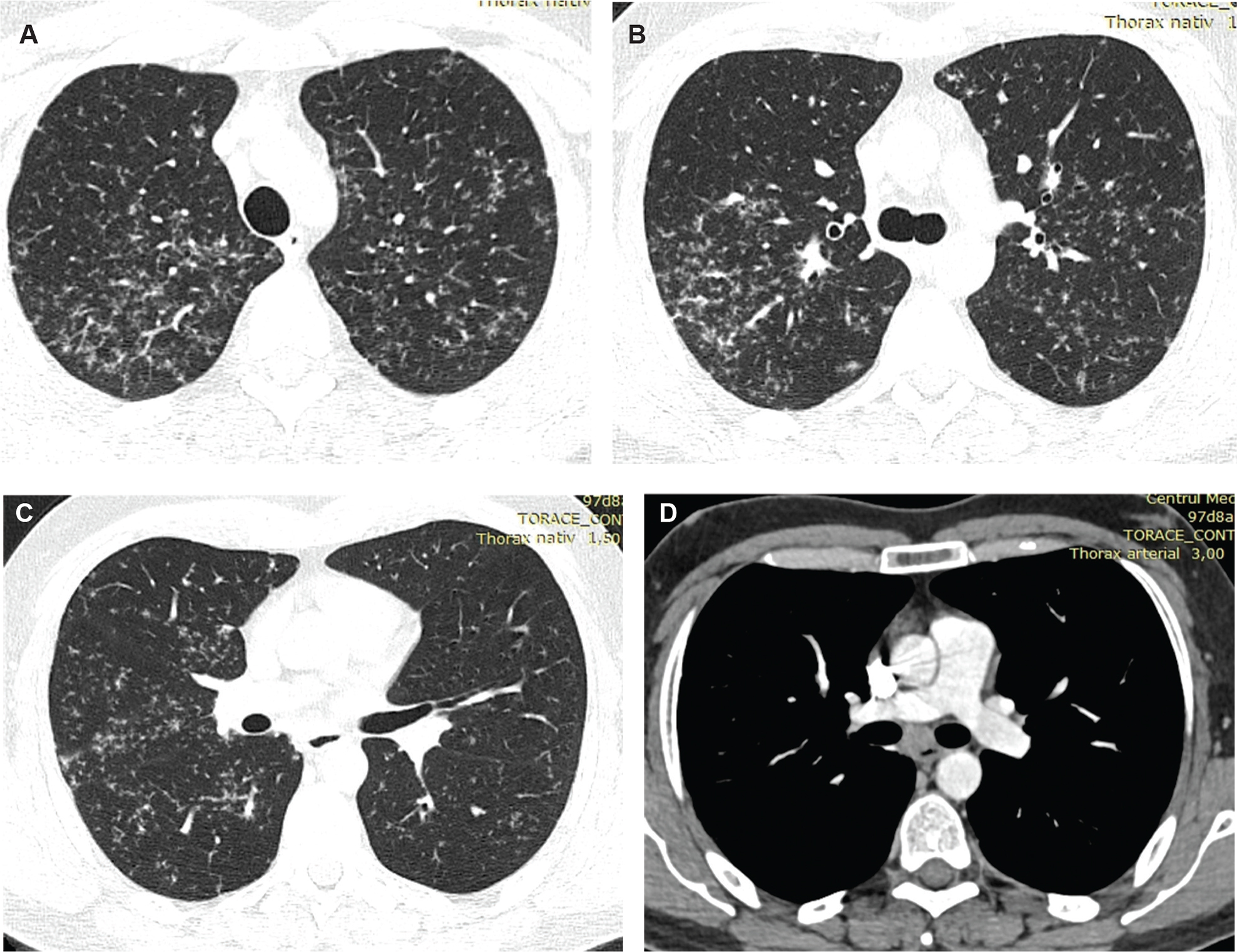
Figure 4
High-resolution CT of the lungs: multiple bilateral micronodules predominant in the upper lobes, increased infracarinal lymph nodes.