
Table 1
Histological diagnoses of samples obtained at thoracoscopy
| Diagnosis | N (%) (n=104) | |
|---|---|---|
| Pleural fibrosis | 8 (7.7) | |
| Nonspecific inflammation | 25 (24) | |
| Mesothelioma | Type not given | 2 (1.9) |
| Epithelioid | 27 (26) | |
| Sarcomatoid | 2 (1.9) | |
| Biphasic | 1 (1) | |
| Suspected | 5 (4.8) | |
| Adenocarcinoma | Lung | 4 (2.8) |
| Colon | 2 (1.9) | |
| Breast | 3 (2.9) | |
| Endometrial | 1 (1) | |
| Unknown primary | 11 (10.6) | |
| Papillary carcinoma | 1 (1) | |
| Lymphoma | 2 (1.9) | |
| Neuroendocrine tumour | 1 (1) | |
| No/limited biopsy | 9 (8.7) |
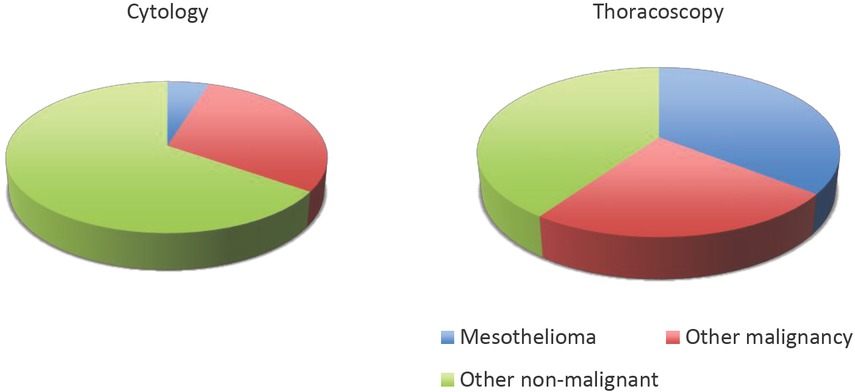
Figure 1
A comparison between the results obtained from pre-thoracoscopic cytology and the histology of thoracoscopy samples in 104 patients. The majority of cytological samples revealed only reactive lymphocytic cells giving a high false negative rate.