
1. Introduction
The rapid development of nanomedicine over the past two decades has highlighted mesoporous materials as a promising approach to overcome the limitations of conventional pharmacotherapy [1]. An effective drug delivery system should provide controlled drug loading and release while enabling targeted delivery to specific tissues or cells. Various nanomaterials, such as mesoporous carbon, mesoporous bioactive glass, and mesoporous metal–organic frameworks, are explored as drug delivery carriers due to their high surface area and tunable porosity [2]. Among them, mesoporous silica nanoparticles (MSNs) have gained particular attention due to their high specific surface area, large pore volume, and tunable pore structure [3]. Importantly, silica-based materials have been extensively studied and confirmed to exhibit good biocompatibility in both in vitro and in vivo systems [4]. These properties enable the efficient loading of a wide variety of therapeutic agents, including antibiotics, antivirals, and cardiovascular drugs [5]. In addition, designing MSNs with particle sizes below 100 nm is especially important, as it increases the surface-to-volume ratio and improves colloidal stability, which is crucial for their proper function in complex biological environments [6].
MSNs are particularly effective in addressing one of the major challenges in pharmacology: the poor water solubility of many drugs. Numerous nonsteroidal anti-inflammatory drugs (NSAIDs) exhibit low aqueous solubility, leading to reduced bioavailability and the need for higher doses to achieve a therapeutic effect. Consequently, the risk of adverse side effects increases [7]. Incorporating such drugs into a mesoporous silica matrix can significantly improve their dispersion and allow for controlled drug release [8,9]. Importantly, confinement of drug molecules within the nanopores often leads to their amorphization, which enhances dissolution rates and helps maintain stable therapeutic concentrations, ultimately reducing dose-dependent toxicity [10].
A representative example of such limitations is nimesulide (NMS), a selective cyclooxygenase-2 (COX-2) inhibitor [11]. Following oral administration, it exerts analgesic, antipyretic, and anti-inflammatory effects. However, its clinical application is significantly limited by its hydrophobic nature, resulting in poor aqueous solubility, suboptimal absorption, and consequently low bioavailability [12]. In addition, NMS is associated with a considerable risk of hepatotoxicity [13], which has led to regulatory restrictions in several countries. These limitations highlight the urgent need for innovative drug delivery strategies that can enhance its anti-inflammatory efficacy, improve solubility, and reduce systemic toxicity, particularly with respect to liver safety.
To address these limitations, the present study investigates the potential of a core–shell silica architecture as an advanced drug delivery system. This design enables additional control over drug loading and release by modifying the outer shell, which serves as a diffusion barrier and helps to reduce the initial “burst release” effect.
In this study, we present an in vitro evaluation of core–shell mesoporous silica nanocarriers designed to improve the delivery and safety of NMS. The aim was to determine whether structural modification of the silica carrier can improve drug loading, control release behaviour, and reduce the cytotoxicity associated with free NMS. We examined whether the engineered mesoporous shell could enhance NMS dissolution while providing a more controlled and safer release profile. To evaluate their biological performance, we performed cytotoxicity studies using the HepG2 cell line, a well-established in vitro model for assessing drug-induced hepatotoxicity [14]. HepG2 cells were selected due to their relevance in studies of hepatic metabolism and toxicological response, as NMS is primarily metabolized in the liver and is associated with hepatotoxic side effects [15]. This model allowed us to assess both the intrinsic biocompatibility of the developed silica carriers and their effect on the cellular response to NMS. The results showed that encapsulation of NMS in the mesoporous structure improved its physicochemical properties and reduced its toxic effect. These findings suggest that core–shell mesoporous silica nanocarriers are a promising strategy for improving NMS delivery and developing safer anti-inflammatory therapies.
2. Materials and methods
2.1. Synthesis of solid silica core nanoparticles
Silica nanospheres were prepared using a modified Stöber sol–gel method. Tetraethyl orthosilicate (TEOS, 9 mL) was added dropwise to a mixture of ethanol (150 mL) and aqueous ammonia solution (25%, 7.5 mL) under magnetic stirring. The reaction was carried out at room temperature for 24 h. After completion, the solvent was removed by evaporation, and the obtained product was collected and dried. The resulting silica nanoparticles exhibited a spherical morphology with an average diameter of approximately 70 nm, as confirmed by scanning electron microscopy (SEM) analysis.
2.2. Synthesis of core–shell MSNs
For the synthesis of core–shell nanoparticles, 100 mg of solid silica cores were dispersed in a mixture of distilled water (30 mL) and ethanol (12.6 mL) containing cetyltrimethylammonium bromide (CTAB, 160 mg). The suspension was sonicated for 20 min and subsequently stirred for 30 min. Then, aqueous ammonia solution (0.45 mL) and TEOS (0.28 mL) were added to the reaction mixture under continuous stirring.
The reaction was conducted at room temperature for 6 h (MSNs-6h) or 1 h (MSNs-1h). In an alternative approach (MSNs-6h ½ CTAB), the amount of CTAB was reduced to 80 mg while maintaining the reaction time of 6 h. After completion, the products were collected by centrifugation, washed several times with ethanol, and dried. Finally, the samples were calcined at 600°C for 4 h to remove the surfactant template and generate the mesoporous structure.
2.3. Preparation of the drug-loaded carrier
Drug loading was performed under light-protected conditions to prevent possible photodegradation of NMS. First, 50 mg of NMS was dissolved in 1 mL of acetone. The use of acetone as a solvent was introduced as a modification of the original procedure due to the high solubility of NMS in this medium and the reduced toxicity of the loading process. Next, 150 mg of synthesized silica nanoparticles was added to the drug solution. The resulting suspension was stirred continuously on a magnetic stirrer for 12 h. After incubation, the mixture was dried at 40°C until complete solvent evaporation. The obtained powder was washed twice with water and centrifuged at 12,000 rpm for 5 min. Finally, the material was dried again to obtain the drug-loaded carrier [9].
2.4. Drug loading capacity
To determine the amount of NMS successfully loaded into the silica nanoparticles, a total extraction procedure was applied. Briefly, 5 mg of the drug-loaded material was dispersed in 10 mL of ethanol. The suspension was then sonicated for 20 min in an ultrasonic bath to facilitate the release of the drug from the mesoporous structure. After extraction, the suspension was filtered through a 0.22 µm syringe filter to remove the silica matrix. The concentration of NMS in the filtrate was quantified by ultraviolet-visible (UV–Vis) spectroscopy. The drug loading capacity (%) was calculated according to the following equation:
2.5. Characterization techniques
Transmission electron microscopy (TEM, FEI Tecnai G2 F20 based at 200 kV accelerating voltage) was used to analyse the morphology and internal structure of the synthesized MSNs. The samples were dispersed in ethanol, sonicated to ensure proper separation, and then deposited onto carbon-coated copper grids. After solvent evaporation, the specimens were examined using a transmission electron microscope under appropriate accelerating voltage to visualize particle size, shape, and mesostructure ordering.
Nitrogen adsorption–desorption measurements were carried out for the synthesized mesoporous silica samples to evaluate their specific surface area and pore structure. The analyses were performed using a specific surface area analyser (Micrometrics ASAP 2460 system). Before the measurements, all samples were degassed under vacuum at 150°C to remove physically adsorbed moisture and other volatiles from the pore system, ensuring reliable determination of textural parameters.
The crystallographic structure of the synthesized nanomaterials was analysed using wide-angle X-ray diffraction (XRD, AERIS PANalytical X-ray diffractometer with Cu-Kα radiation). The measurements were performed to evaluate the phase composition and degree of crystallinity of the obtained samples. The diffraction patterns were recorded for the pristine core–shell structure before and after drug loading over a wide angular range (2θ).
The surface charge and colloidal stability of the silica nanoparticles and NMS were evaluated by zeta potential measurements using a zeta potential analyser (Zetasizer Nano-ZS ZEN3600). The samples were dispersed in a liquid medium, and their electrophoretic mobility was measured under an applied electric field.
2.6. Preparation of the NMS-loaded MSN solutions
The concentrations of NMS-loaded MSNs used in this study were established based on pharmacokinetic data provided in the drug safety data sheet. Following oral administration of a single 100 mg dose of NMS, its plasma concentration typically reaches 3–4 mg/L. This clinically relevant range was used as a reference point to design the in vitro exposure conditions.
Accordingly, a series of NMS-loaded MSN concentrations was prepared: 30, 15, 7.5, 3.75, and 1.875 mg/L (expressed as NMS equivalents). These concentrations correspond to MSN concentrations of 100, 50, 25, 12.5, and 6.25 mg/L, respectively. The conversion is based on the experimentally determined loading capacity of NMS in the MSNs, which was 30 mg of NMS per 100 mg of MSNs.
Both NMS-loaded MSNs and unloaded MSNs were dissolved in 10% ethanol before use. Before application, the NMS-loaded MSN solutions at varying concentrations were sonicated for 10 min at 80 W to prevent aggregation and ensure homogeneous dispersion.
This approach ensured that the tested concentrations reflect both pharmacologically relevant drug levels and the corresponding carrier doses, enabling a comprehensive evaluation of the cytotoxicity of the NMS-loaded delivery system.
2.7. Cell culture
The study was conducted on a human HepG2 cell line (HB-8065, ATCC), which is composed of epithelial-like cells derived from hepatocellular carcinoma. Adherent cell growth was achieved in an Eagle’s Minimum Essential Medium (30-2003, ATCC), supplemented with 10% fetal bovine serum (E5050, EURx) and 1% penicillin-streptomycin antibiotic (PCS-999-002, ATCC). The cells were maintained at 37°C in a humidified chamber with a 5% CO2 atmosphere.
2.8. Morphology analysis
After 48 h exposure to MSNs and NMS-loaded MSNs, HepG2 cells were imaged using a Keyence BZ-X810 fluorescence microscope at a magnification of 10× (Keyence, Neu-Isenburg, Germany).
2.9. Live/Dead® staining
For the visualization of live and dead HepG2 cells upon incubation with NMS-loaded MSNs and controls, a Live/Dead® staining was performed. Cells were seeded in a black polystyrene 96-well flat-bottom plate with a transparent bottom (Greiner Bio-One GmbH) at densities of 1 × 10⁴ cells/well. After 24 h, medium containing an increasing concentration of NMS-loaded MSNs was added to each well. Following 24 and 48 h exposure to the nanomaterials, cells were incubated with 2 μM calcein AM (L3224, Thermo Fisher Scientific), 2 μM ethidium homodimer-1 (L3224, Thermo Fisher Scientific), and 8 μM Hoechst 33342 (14533, Sigma-Aldrich) containing phosphate-buffered saline (PBS) (100 μL/well) for 30 min at 37°C. Finally, the cells were analysed with the Keyence BZ-X810 fluorescence microscope at 10× magnification (Keyence, Neu-Isenburg, Germany). Viable cells (green signal) were imaged using the green fluorescent protein (GFP) excitation/emission filter (475/525 nm), whereas for the dead cells (red signal), the tetramethylrhodamine isothiocyanate (TRITC) ex/em filter (545/605 nm) combination was applied. A 4′,6-diamidino-2-phenylindole (DAPI) ex/em filter (360/460 nm) was applied to detect the Hoechst 33342 blue signal (cell nuclei).
3. Results and discussion
3.1. Characterization of the carrier
The morphology and internal structure of the synthesized MSNs were examined by TEM, as shown in Figure 1a–c. All analysed samples (MSNs-1h, MSNs-6h, and MSNs-6h ½ CTAB) exhibited a well-defined core–shell architecture with a spherical morphology and high monodispersity. The particles remained non-aggregated and retained a regular shape regardless of the synthesis conditions.
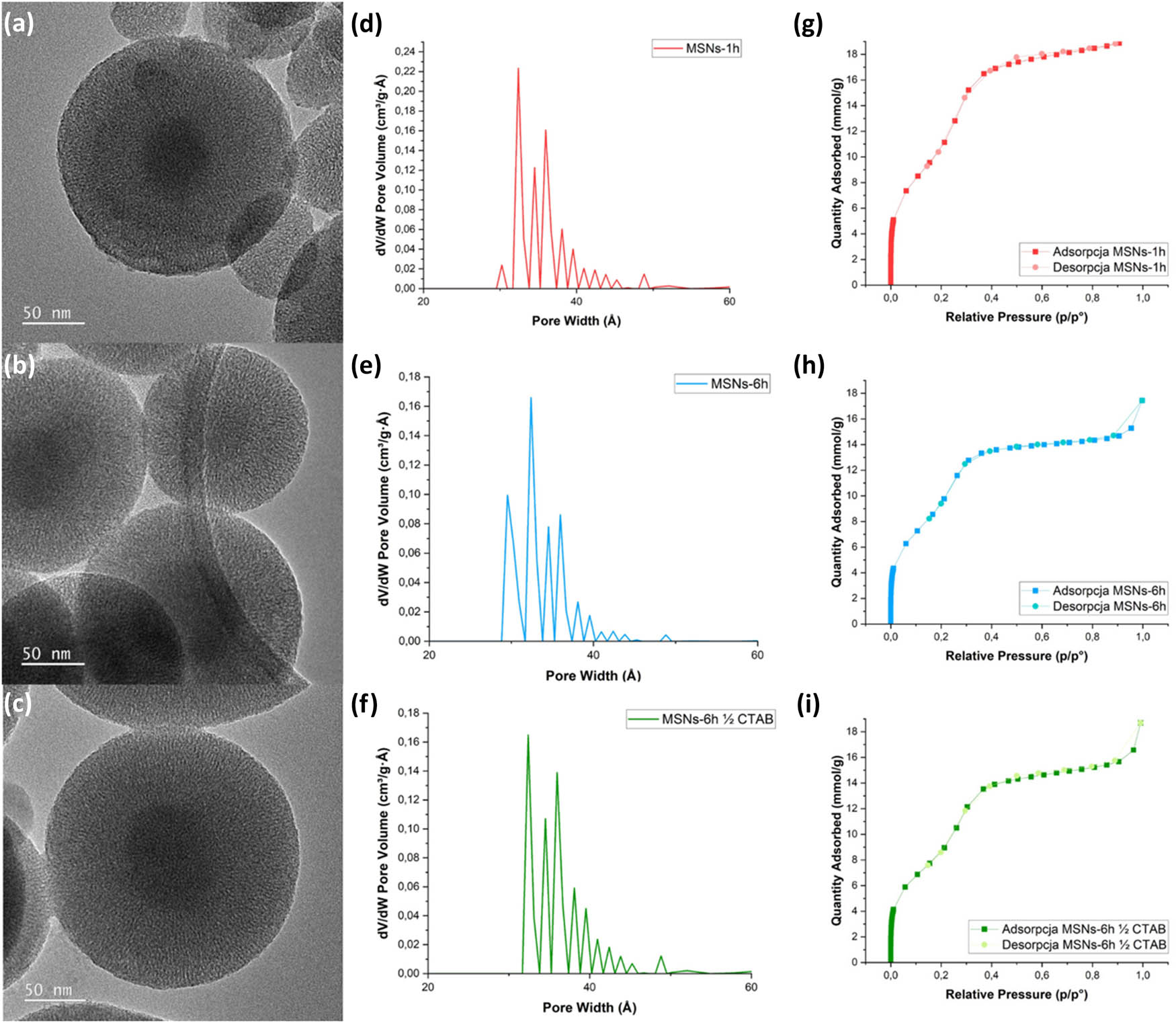
Figure 1
Structural characterization of the synthesized silica carriers: (a) MSNs-1h, (b) MSNs-6h, and (c) MSNs-6h ½ CTAB. TEM images showing core–shell morphology (scale bar = 50 nm); (d–f) pore size distribution curves derived from the Barrett-Joyner-Halenda (BJH) method; and (g–i) nitrogen adsorption–desorption isotherms.
A detailed analysis confirmed that all variants consist of a dense silica core with a uniform diameter of approximately 70 nm, surrounded by a highly ordered mesoporous shell. The shell is characterized by radially arranged pore channels extending from the core surface to the outer edge of the spheres.
To precisely evaluate the structural differences between the samples, particle size distribution sketches were determined via histogram analysis, as shown in Figure 2. The statistical data revealed that the total particle diameter is highly sensitive to the synthesis conditions. For MSNs-1h, the nanoparticles exhibited the largest dimensions, with diameters predominantly ranging between 280 and 360 nm (with a peak at 320–340 nm). Extending the reaction time to 6 h (MSNs-6h) resulted in a significant shift towards smaller particle sizes, yielding a narrower distribution centred around 180–200 nm. Interestingly, reduction of the surfactant concentration by 50% (MSNs-6h ½ CTAB) led to a moderate increase in size compared to MSNs-6h, with most particles falling within the 210–220 nm range. Consequently, the calculated average shell thickness varied from approximately 55–65 nm for MSNs-6h up to 125–135 nm for MSNs-1h.
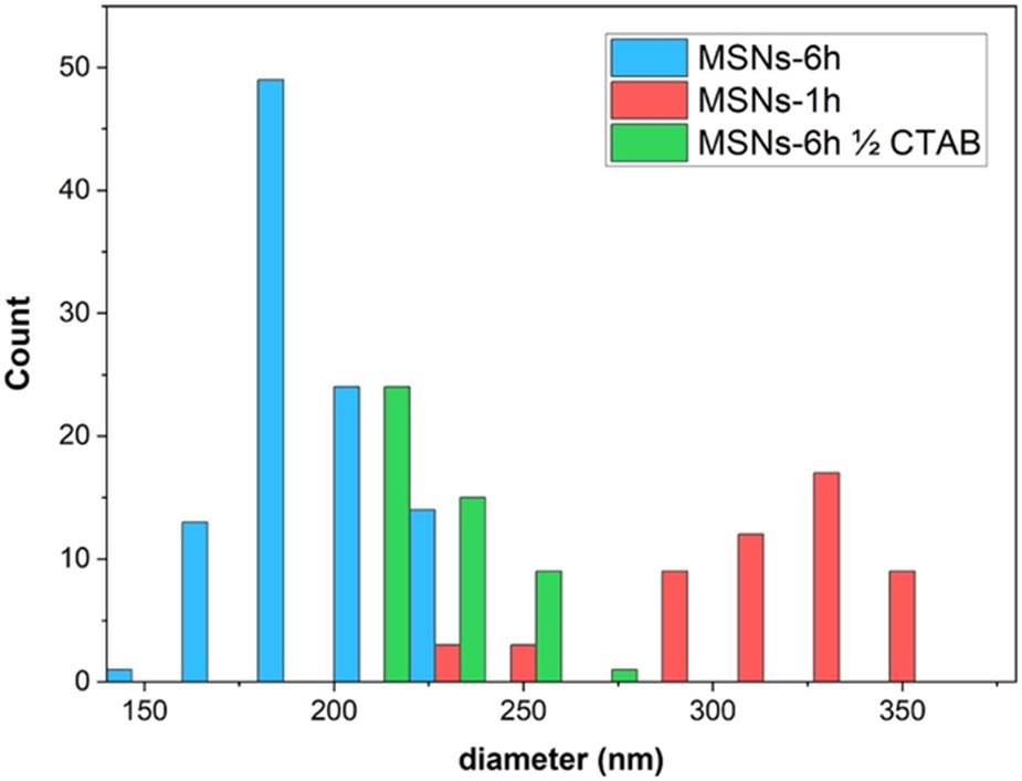
Figure 2
Particle size distribution histograms of the synthesized core–shell MSNs: (a) MSNs-1h, (b) MSNs-6h, and (c) MSNs-6h ½ CTAB, calculated from TEM image analysis.
The structural parameters of the synthesized silica nanoparticles were evaluated by nitrogen adsorption–desorption analysis in Figure 1g–i. All samples (MSNs-6h, MSNs-1h, and MSNs-6h ½ CTAB) exhibited type IV isotherms according to the International Union of Pure and Applied Chemistry (IUPAC) classification, confirming their mesoporous character [16]. A distinct increase in nitrogen uptake was observed in the relative pressure range of P/P₀ ≈ 0.2–0.4, corresponding to capillary condensation within mesopores. The narrow hysteresis loop, or its near absence, indicates the presence of uniform and well-ordered pores with a regular mesostructure. Pore size distribution analysis confirmed a high structural homogeneity of all materials, with pore diameters mainly in the range of 3–4 nm (Figure 1d–f). The largest pore diameter was observed for MSNs-1h, which can be attributed to the shorter reaction time and lower degree of silica framework condensation. Extending the synthesis time to 6 h (MSNs-6h) resulted in a slight narrowing of the mesoporous channels, whereas reducing the amount of CTAB in MSNs-6h ½ CTAB preserved the mesoporous structure with a similarly narrow pore size distribution.
Crucially, the obtained pore diameters are significantly larger than the molecular dimensions of typical small-molecule therapeutic agents, which usually do not exceed 1–2 nm. These dimensions are well-suited for accommodating molecules such as NMS (approximately 0.8–1.0 nm) [11], thereby ensuring efficient drug loading into the radial mesoporous channels. This favourable pore-to-molecular size ratio, combined with the high structural homogeneity of the materials, confirms that the synthesized core–shell matrices possess excellent potential as effective carriers for the controlled delivery of this hydrophobic active substance.
The zeta potential analysis in Table 1 demonstrated that synthesized MSNs and pristine NMS possess a negative surface charge. Since both components are negatively charged, the encapsulation of the drug within the silica matrix is not driven by electrostatic attraction. Instead, the drug loading mechanism is primarily associated with non-electrostatic interactions, particularly hydrogen bonding between the silanol groups present on the silica surface and the functional groups of NMS, as well as van der Waals forces. These interactions enable effective incorporation of the drug molecules into the porous structure of the carriers despite the electrostatic repulsion between the components. Furthermore, the obtained zeta potential values, ranging from −30 to +30 mV, indicate moderate colloidal stability of the developed systems [17].
Table 1
Zeta potential values of the synthesized silica carriers and pure NMS
| Zeta potential | MSNs-1h | MSNs-6h | MSNs-6h ½ CTAB | NMS |
| [mV] | −12.6 | −16.2 | −7.3 | −22.3 |
To evaluate potential interactions between NMS and the silica matrix, fourier-transform infrared spectroscopy (FTIS) spectroscopy was used. The spectra of pure silica, pure NMS, and the drug-loaded material show the characteristic bands of both components (Figure 3). No new absorption bands were observed after loading, indicating the absence of covalent bonding between NMS and silica. However, slight shifts and changes in the intensity of characteristic NMS bands, together with broadening of the silanol (Si–OH) region, were detected. These changes suggest weak non-covalent interactions, most likely hydrogen bonding between silanol groups and NMS functional groups, as well as van der Waals interactions within the mesoporous structure. Such interactions are typical for drug molecules confined in mesoporous silica systems and contribute to drug stabilization in a dispersed state [18].
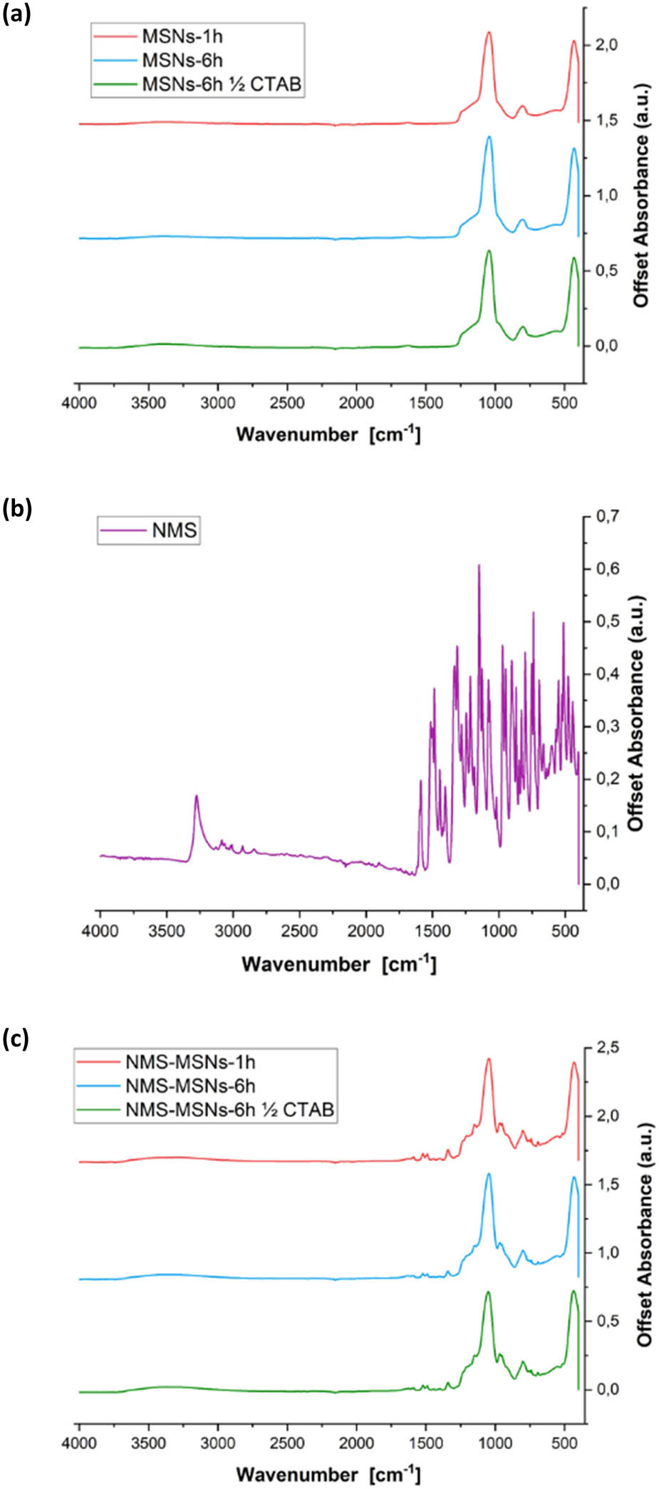
Figure 3
FTIR spectra of (a) the pristine silica carriers, (b) pure NMS, and (c) the NMS-loaded silica composite.
The diffraction patterns recorded for MSNs-1h, MSNs-6h, and MSNs-6h ½ CTAB showed nearly identical profiles, indicating no significant structural differences between the samples. In all cases, only one broad diffraction reflex centred at 2θ ≈ 22° was observed, with no sharp reflections detected at higher angles (Figure 4a). This pattern is characteristic of amorphous silica and confirms that the pore walls of all synthesized MSNs possess a fully amorphous structure [18]. The results indicate that neither the reaction time nor the reduced CTAB content induced crystallization of the silica framework. The diffractogram of pristine NMS shown in Figure 4b exhibits several sharp, high-intensity reflexes, typical of its crystalline nature [19]. For the NMS-MSNs-1h sample, as shown in Figure 4c, the crystalline reflections of NMS disappeared completely, resulting in a perfectly smooth, amorphous profile. The larger pore diameter of the 1-h variant (∼4 nm, as shown by BET) provided spatial confinement, successfully ensuring that NMS was entirely incorporated inside the mesoporous channels.
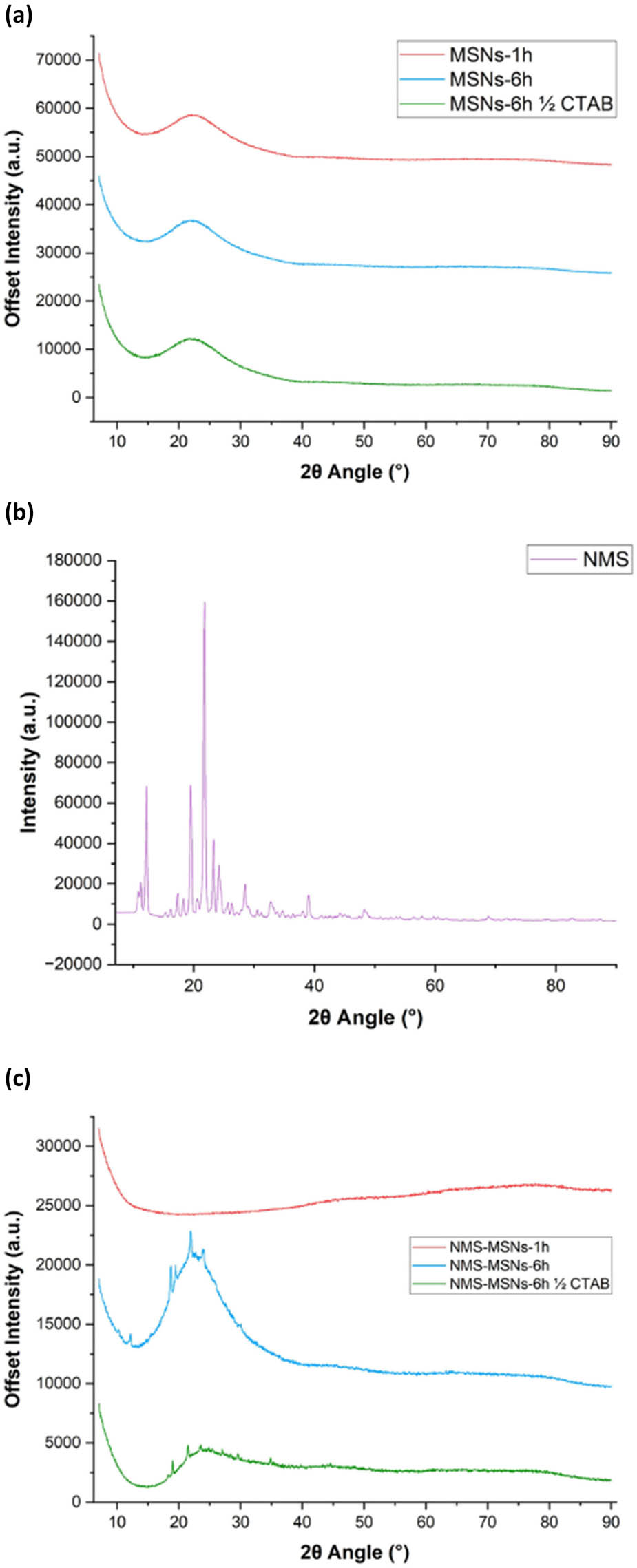
Figure 4
XRD patterns of (a) empty silica carriers, (b) pure NMS, and (c) drug-loaded systems (NMS-MSNs).
In contrast, the XRD patterns for both NMS-MSNs-6h and NMS-MSNs-6h ½ CTAB retained low-intensity, sharp crystalline reflections superimposed on the broad silica halo. This suggests that in the 6-h samples, which had narrower pores (∼3 nm), a small amount of NMS did not fully enter the porous structure and instead crystallized on the surface of the nanoparticles.
TEM analysis of the NMS-loaded carriers (Figure 5a–f) confirmed the successful incorporation of the drug into the MSNs. Compared with the pristine materials, the loaded samples exhibited reduced pore contrast and partial filling of the mesoporous channels, indicating the presence of NMS within the pore system. Importantly, the spherical morphology and well-defined mesostructure of the carriers were preserved after the loading process, demonstrating that drug incorporation did not change the structural integrity of the silica framework. In addition, part of the drug is present on the outer surface of the silica. These observations provide direct morphological evidence of successful NMS encapsulation within the mesoporous silica matrix.
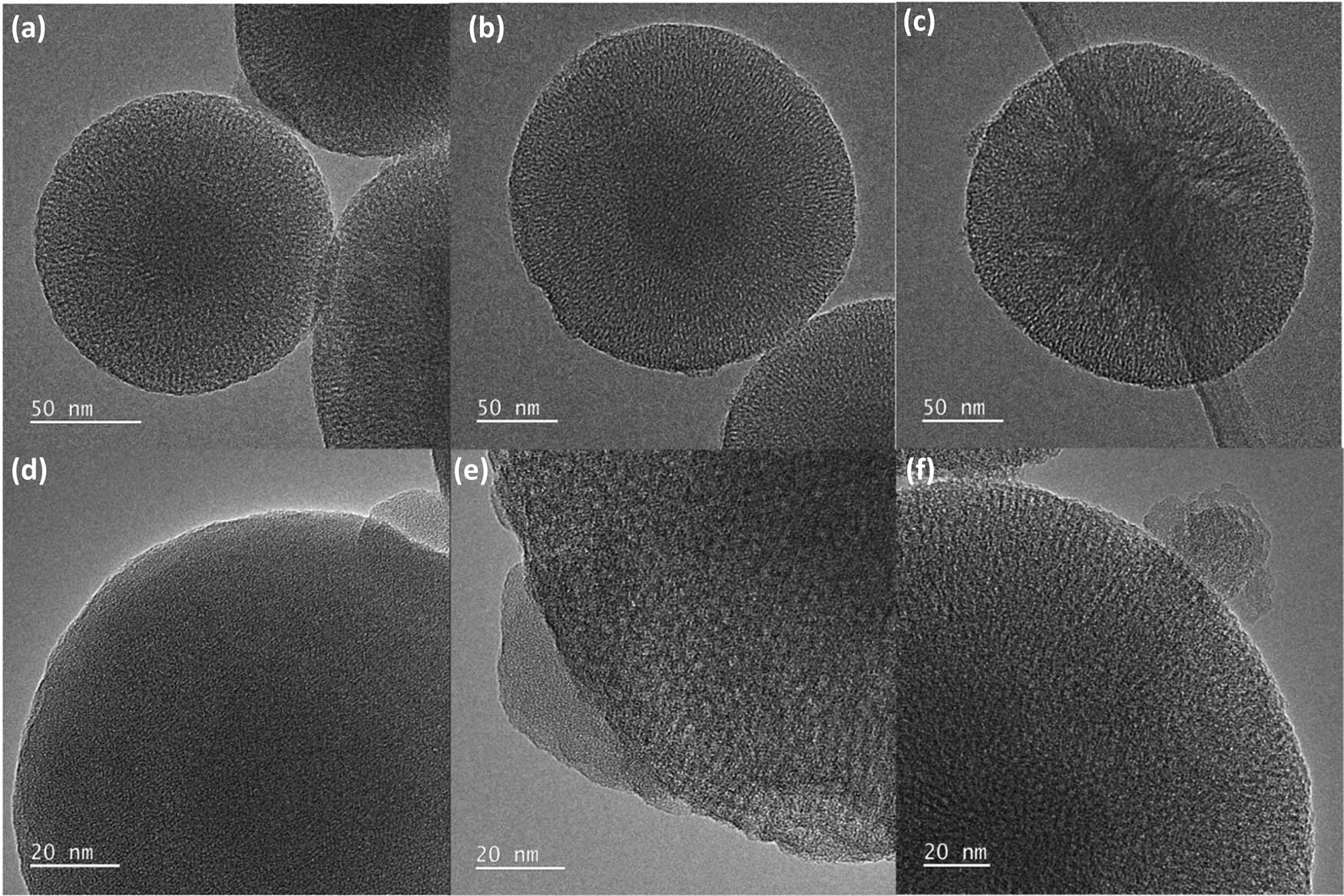
Figure 5
TEM micrographs of MSNs loaded with NMS: (a) 1 h, (b) 6 h, and (c) 6 h ½ CTAB; (d) 1 h, (e) 6 h, and (f) 6 h ½ CTAB.
The drug loading capacity of the synthesized core–shell carriers was evaluated to determine the effect of structural parameters on the encapsulation efficiency of NMS. The results obtained using the total extraction method showed clear differences between the tested materials. The loading capacity reached 17 wt% for MSNs-6h, 18 wt% for MSNs-6h ½ CTAB, and 23.5 wt% for MSNs-1h, indicating that the synthesis conditions influenced the amount of drug incorporated into the mesoporous structure.
These results are consistent with the textural properties determined by nitrogen adsorption–desorption analysis. The highest loading capacity observed for MSNs-1h can be attributed to its larger pore diameter, which likely promoted more effective diffusion of NMS molecules into the mesoporous network and increased the available space for drug incorporation. In comparison, MSNs-6h and MSNs-6h ½ CTAB exhibited similar loading capacities, which corresponds well with their comparable pore characteristics. Both samples showed narrower pore channels than MSNs-1h, which can potentially limit the extent of drug diffusion.
The obtained results indicate that pore architecture, particularly pore diameter, plays a key role in determining the drug loading capacity of the developed core–shell mesoporous silica carriers.
3.2. Morphological analysis of HepG2 cells
After treatment with NMS-loaded MSNs and control MSNs, HepG2 cells were examined to assess potential morphological alterations. The analysis revealed no observable changes in cell morphology following exposure to either NMS-loaded MSNs or control MSNs across all tested concentrations. Cells maintained their typical epithelial-like morphology, including normal cell shape, adherence, and confluency, comparable to untreated control cells (Figure 6). No signs of cellular stress, such as shrinkage, detachment, or membrane disruption, were detected. These findings indicate that both the MSNs and the NMS-loaded formulation did not induce detectable morphological alterations in HepG2 cells under the experimental conditions. In contrast, exposure to pristine NMS at a high concentration of 30 mg/L induced visible cytotoxic effects, characterized by a distinct cellular shrinkage and partial detachment from the surface.
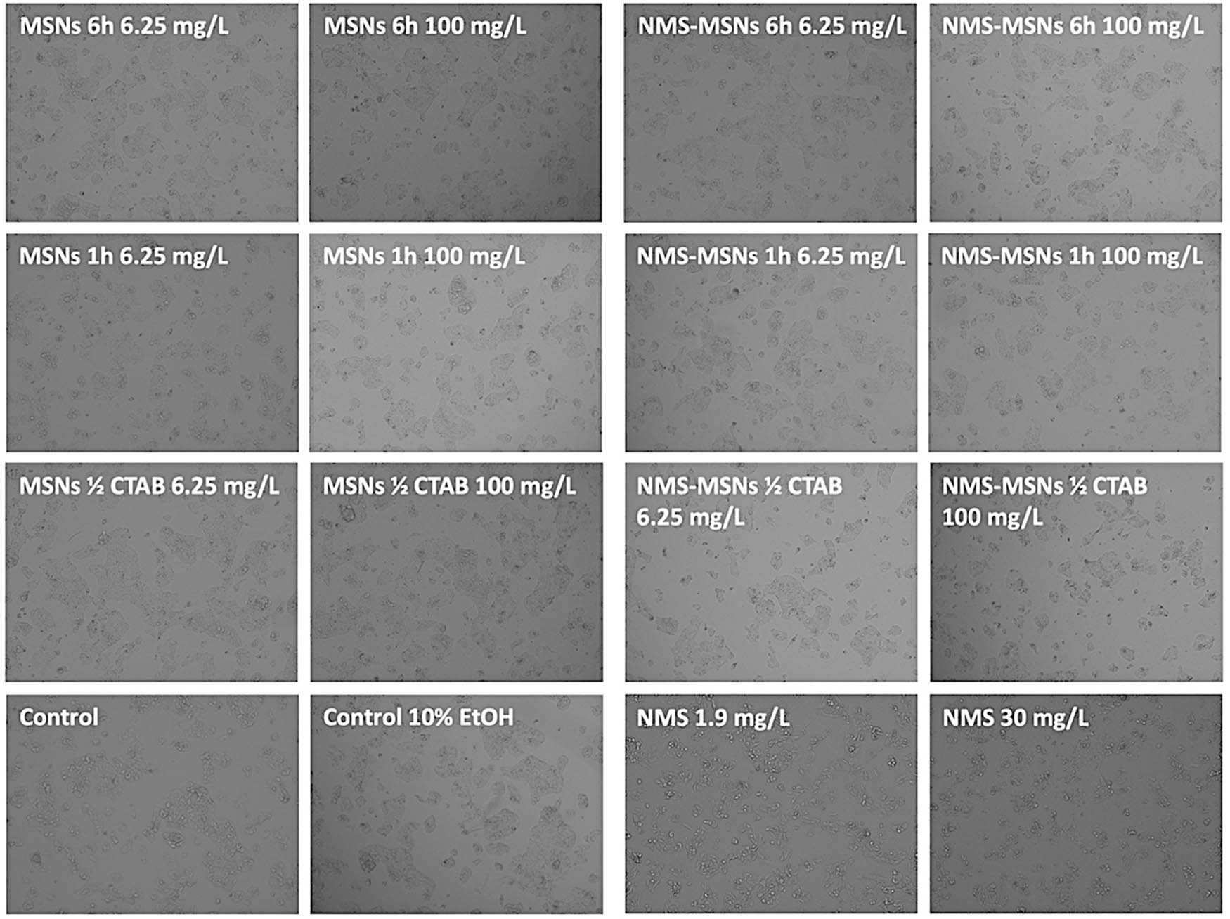
Figure 6
Morphological assessment of HepG2 cells after treatment with MSNs, NMS-loaded MSNs (NMS-MSNs), and free NMS.
3.3. Live/dead staining analysis
Cell viability was further assessed using live/dead staining to visualize the proportion of viable and non-viable cells after exposure to MSNs and NMS-loaded MSNs under different conditions. Representative fluorescence images show predominantly green-stained cells (live) with only a minor fraction of red-stained cells (dead) across all tested variants, including different concentrations (6.25–100 mg/L) after 48 h of incubation. No substantial differences in the ratio of live to dead cells were observed between cells treated with MSNs and those treated with NMS-loaded MSNs. In addition, the results were comparable to both untreated controls and the 10% ethanol control, indicating that neither the carrier system nor the drug-loaded formulation induced pronounced acute cytotoxic effects under the tested conditions. Even at the highest concentration (100 mg/L), only a slight increase in dead cells was visible, which remained minimal and consistent with the quantitative viability results. In contrast, exposure to pristine NMS at 30 mg/L led to a significant decrease in green fluorescence along with a noticeable accumulation of red-stained dead cells, confirming the direct hepatotoxic effect of the free drug at higher doses. This distinct increase in dead cells observed exclusively for free NMS suggests that encapsulation within the MSNs successfully reduces the direct cytotoxic effects of NMS on HepG2 cells. Overall, the live/dead staining, as shown in Figure 7, confirms that both MSNs and NMS-loaded MSNs exhibit good biocompatibility towards HepG2 cells, with no evident disruption of cell membrane integrity or significant induction of cell death.
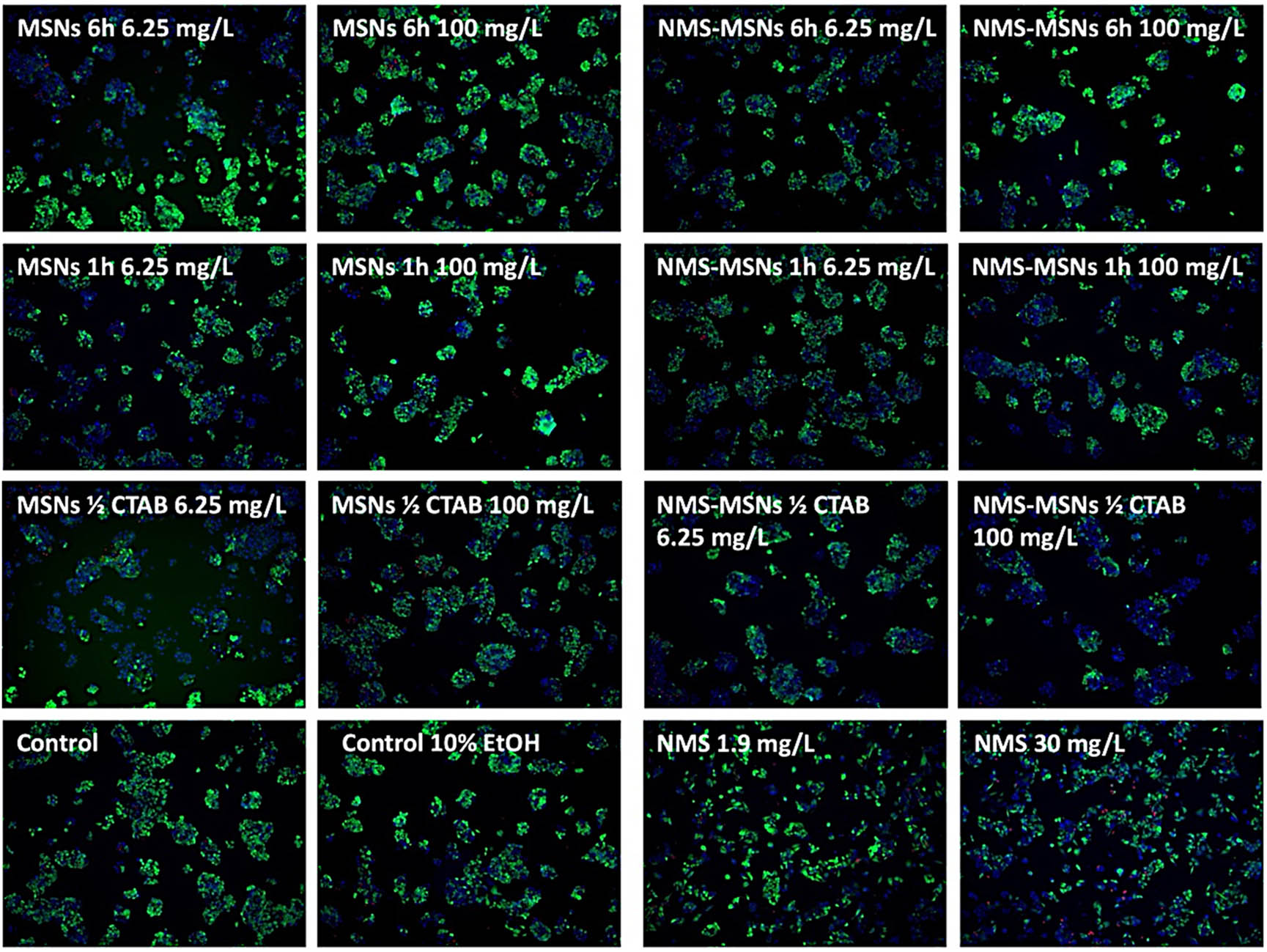
Figure 7
Live/dead staining of HepG2 cells after exposure to MSNs, NMS-loaded MSNs, and free NMS.
4. Discussion
The results clearly show that even small changes in the synthesis conditions have a strong effect on the final properties of the core–shell MSNs. Shortening the condensation time to 1 h limits the further densification of the silica network, which helps to preserve a more open pore structure. As a result, the material keeps wider mesopores compared to samples synthesized for 6 h, where a more compact silica framework is formed. This difference is important because it directly affects how easily drug molecules can enter and be stored inside the pores.
The increase in pore size to around 4 nm in MSNs-1h improves the accessibility of the porous network for NMS. In mesoporous silica systems, drug loading is often limited not only by surface area but also, and mainly, by how easily molecules can diffuse into the pores. This explains why the MSNs-1h sample shows a higher loading capacity compared to the 6 h variants, and this behaviour is in line with general trends reported for ordered mesoporous materials [9]. Compared to traditional mesoporous matrices like conventional MCM-41, which typically have smaller pore sizes (approximately 2 nm) that limit the encapsulation of larger NSAID molecules [20], the optimized MSNs-1h platform offers improved structural accessibility. Furthermore, while larger-pore materials like Santa Barbara Amorphous-15 (SBA-15) often show long diffusion pathways within elongated channels [21], the short-channel core–shell structure designed in this study avoids these transport limitations, leading to an increased loading capacity of 23.5 wt%. This performance compares favourably with alternative nanostructured formulations for NMS, such as polymeric nanoparticles or solid lipid carriers, which often show lower drug loading and pronounced burst-release behaviour [22,23]. Consequently, the designed core–shell silica framework represents a highly efficient and stable alternative for delivering poorly water-soluble anti-inflammatory drugs.
The biological studies performed on HepG2 cells indicate that both the empty carriers and the drug-loaded systems are well tolerated by the cells under the tested conditions. The lack of clear toxicity is consistent with previous reports showing that amorphous silica nanoparticles can be considered biologically safe at moderate concentrations [4]. At the same time, the results suggest that encapsulating NMS inside the mesoporous structure may reduce its direct negative effect on cells by controlling how quickly and in what amount the drug is released.
Overall, the study shows that controlling the synthesis time is a simple but effective way to adjust pore structure, which then influences how much drug can be loaded and how the system behaves in a biological environment.
5. Conclusions
Mesoporous core–shell silica nanoparticles were successfully synthesized and evaluated as carriers for NMS. We obtained regular MSNs with spherical morphology, a stable particle size of approximately 180–360 nm, and a well-developed mesoporous structure. Our study demonstrated that tailoring the synthesis conditions directly influenced the structural properties of the nanoparticles and their drug loading performance. The key factor determining the carrier properties was the condensation time – shortening the synthesis time from 6 to 1 h enabled a controlled increase in pore diameter from 3 to 4 nm. This structural modification directly improved the application potential of the material, resulting in an increase in NMS loading capacity from 17 to 23.5 wt%.
Preliminary biological evaluation showed that both pure silica nanoparticles and NMS-loaded systems exhibited good biocompatibility towards HepG2 cells. Within the tested concentration range, no significant cytotoxicity was observed, and the cells maintained normal morphology. The obtained results confirm that mesoporous core–shell silica nanoparticles can serve as a safe and effective carrier for NMS. Appropriate control of synthesis parameters allows tuning of the material properties, creating opportunities for the further development of this type of system in modern drug delivery applications. Further functional studies, including cell viability assays (alamarBlue assay and water-soluble tetrazolium-1 (2-(4-iodophenyl)-3-(4-nitrophenyl)-5-(2,4-disulfophenyl)-2H-tetrazolium, monosodium salt) (WST-1) assay), reactive oxygen species (ROS) generation analysis, and migration assays, are currently in progress. In addition, pharmacokinetic studies evaluating the release profile of NMS (drug dissolution) from mesoporous silica formulations are being conducted to determine the release kinetics and assess the influence of the silica carrier on drug availability. Pharmacodynamic investigations focusing on COX inhibition, including COX-1 and COX-2 assays, are also underway to evaluate whether encapsulation of NMS within MSNs affects its anti-inflammatory activity and selectivity towards COX isoforms. To complement these studies, comprehensive long-term stability and degradability investigations of the carriers under physiological storage conditions are also planned.
Acknowledgements
The authors are grateful to Bartosz Sroda for technical support.
Funding information
Authors state no funding involved.
Author contributions
Sofiia Volosiuk: investigation, methodology, formal analysis; Sara Wasilewska: investigation, methodology, formal analysis; Elżbieta Czarniewska: investigation, biological analysis; Dariusz Wawrzyniak: investigation, biological analysis, data curation; Karolina Wenelska: formal analysis, data curation; Ewa Mijowska: formal analysis, supervision, conceptualization.
Conflict of interest statement
The authors declare no conflict of interest.
Data availability statement
The data that support the findings of this study are available from the corresponding author upon reasonable request.