
Fig. 1.
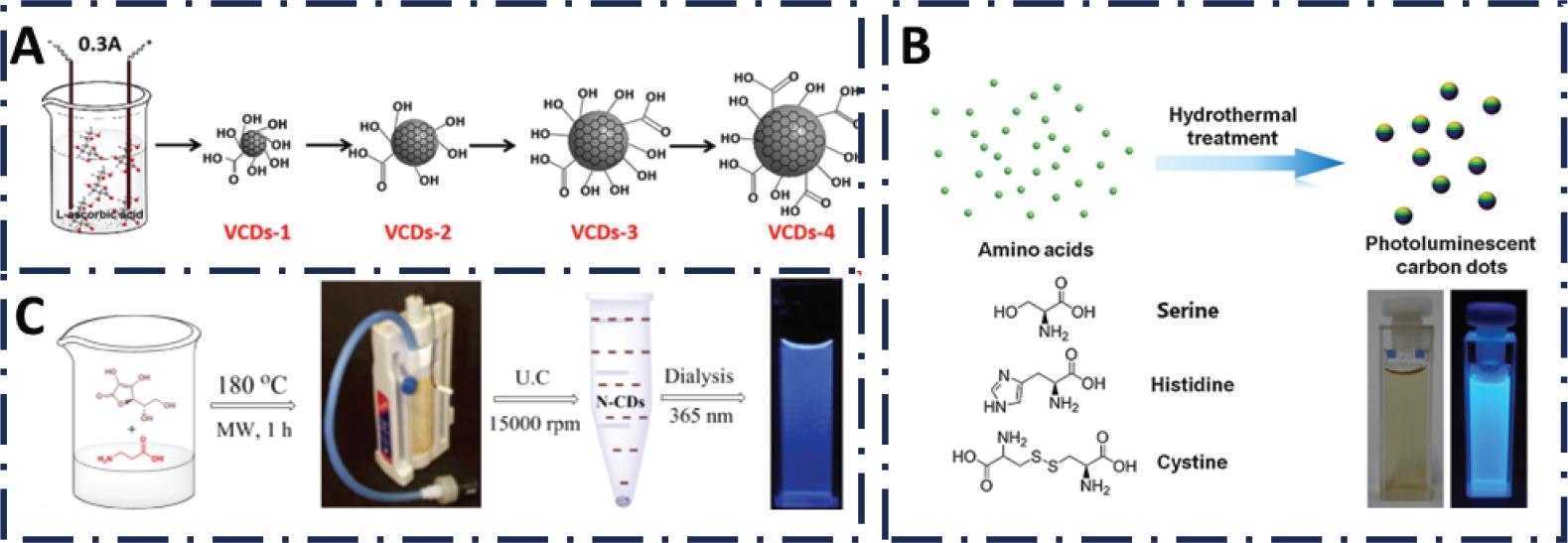
Fig. 2.
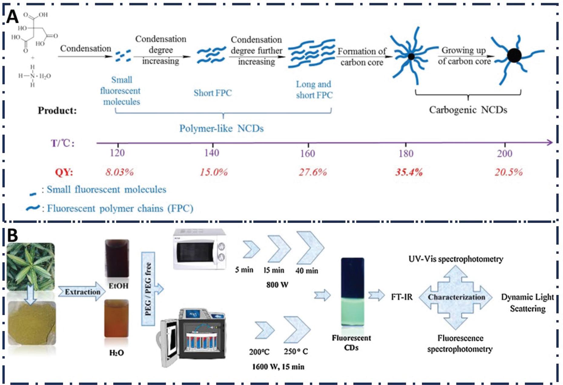
Fig. 3.
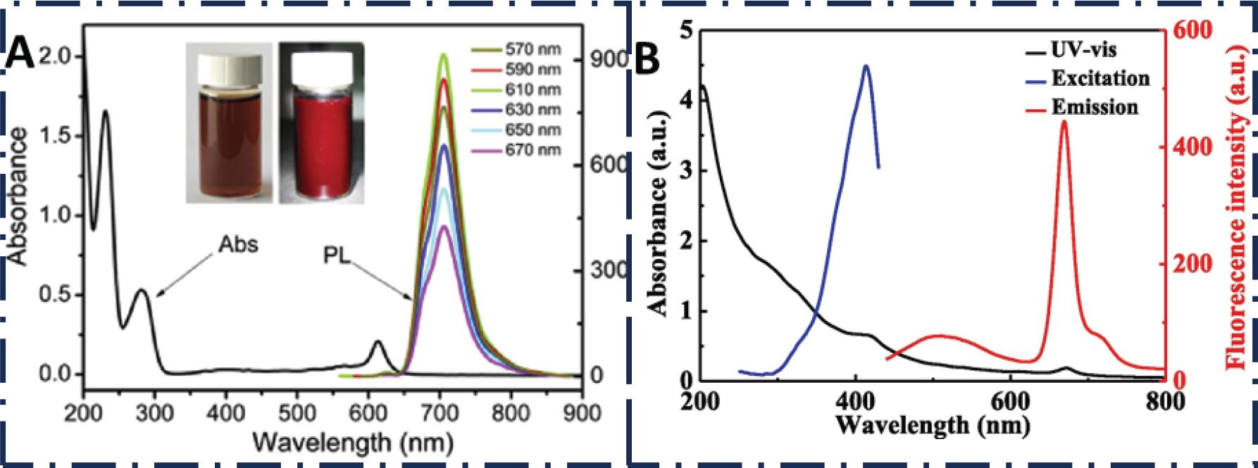
Fig. 4.
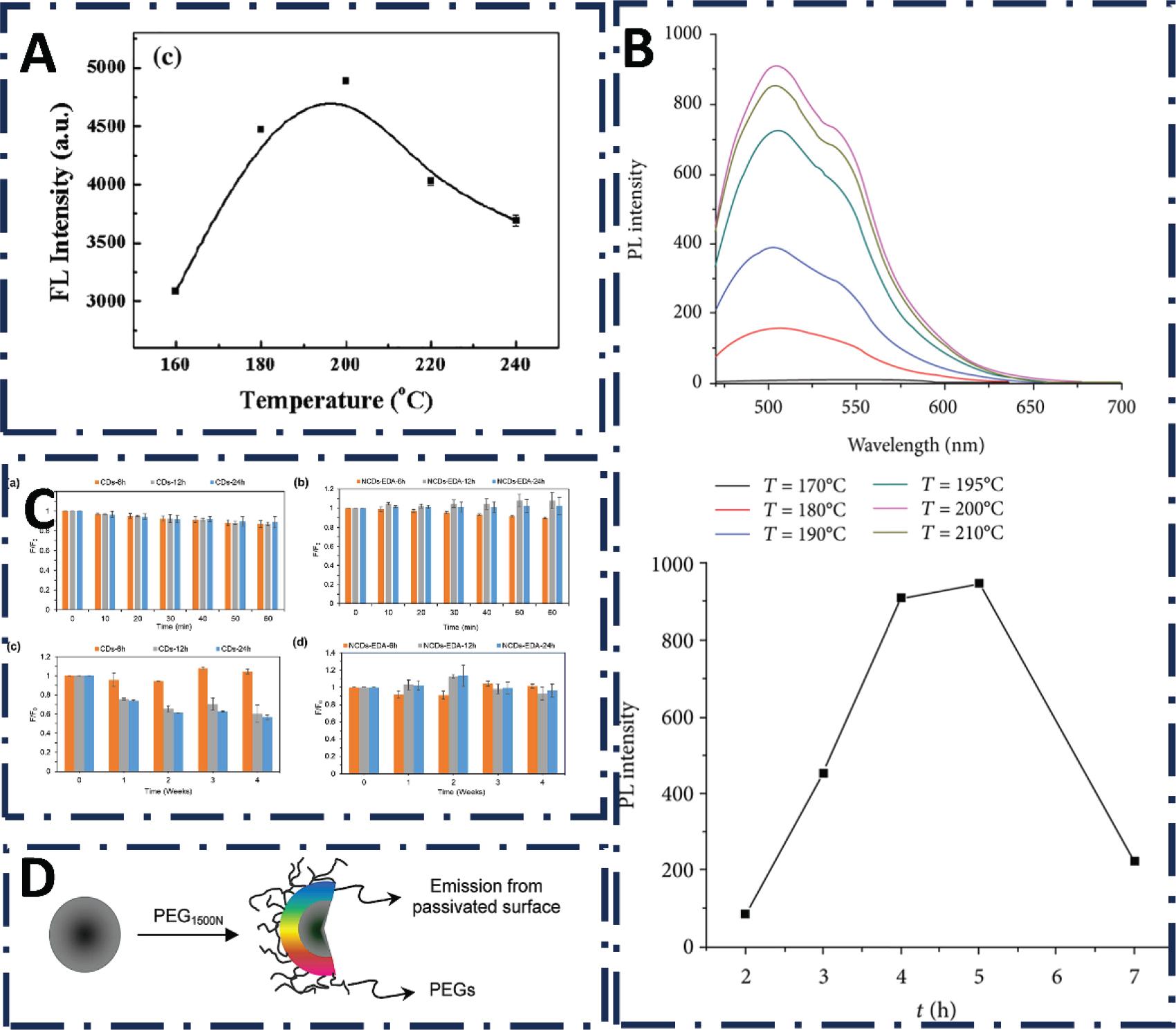
Fig. 5.
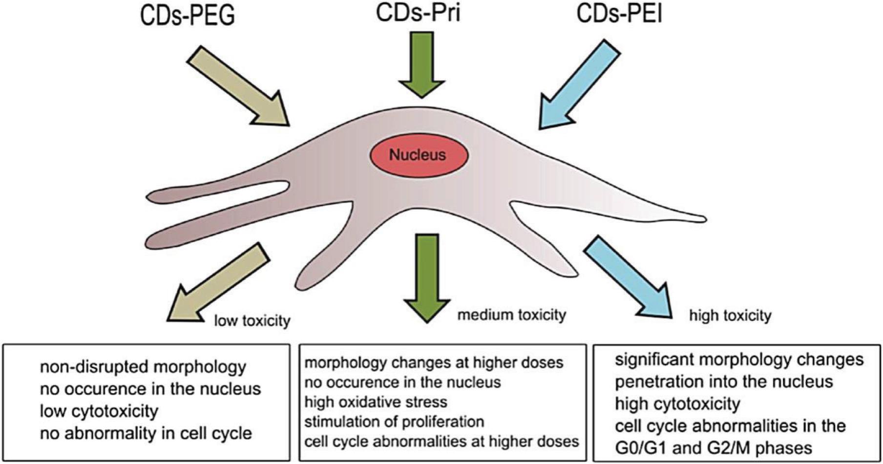
Fig. 6.
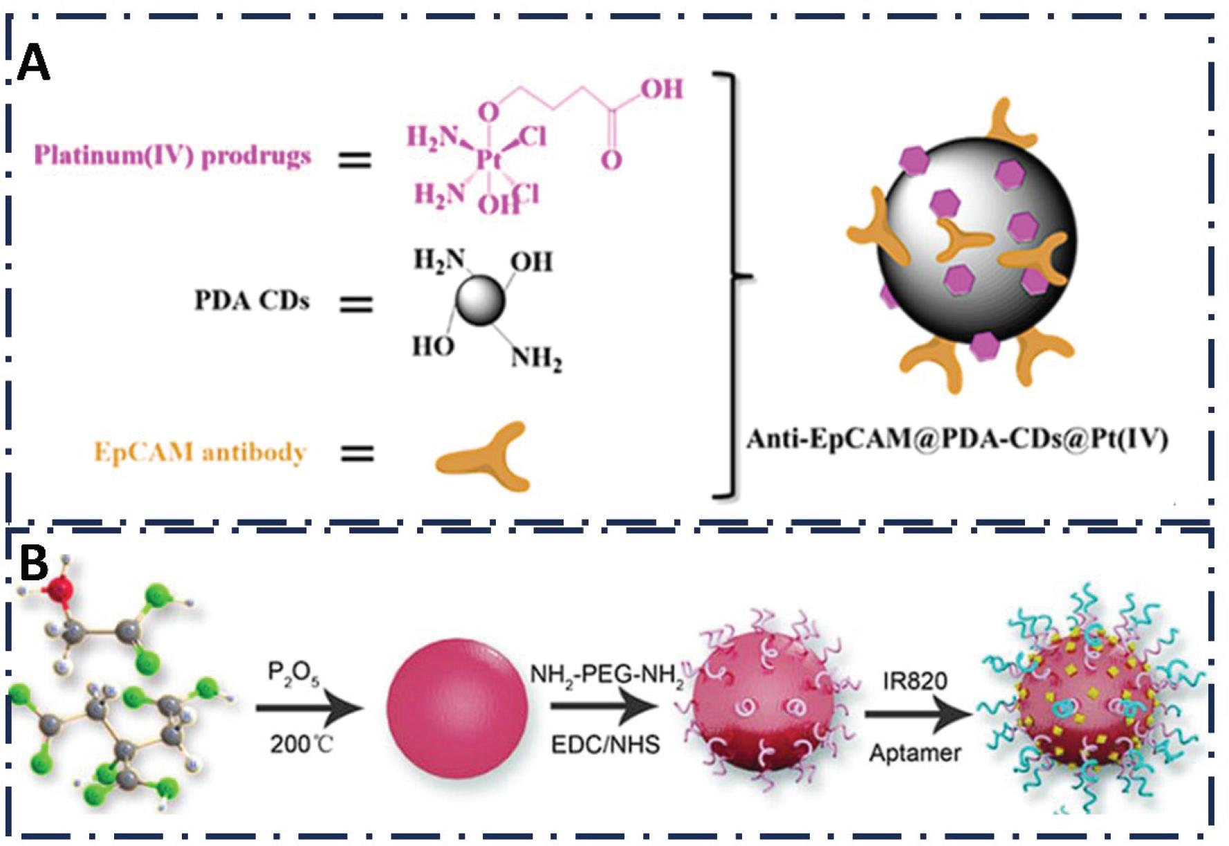
Fig. 7.
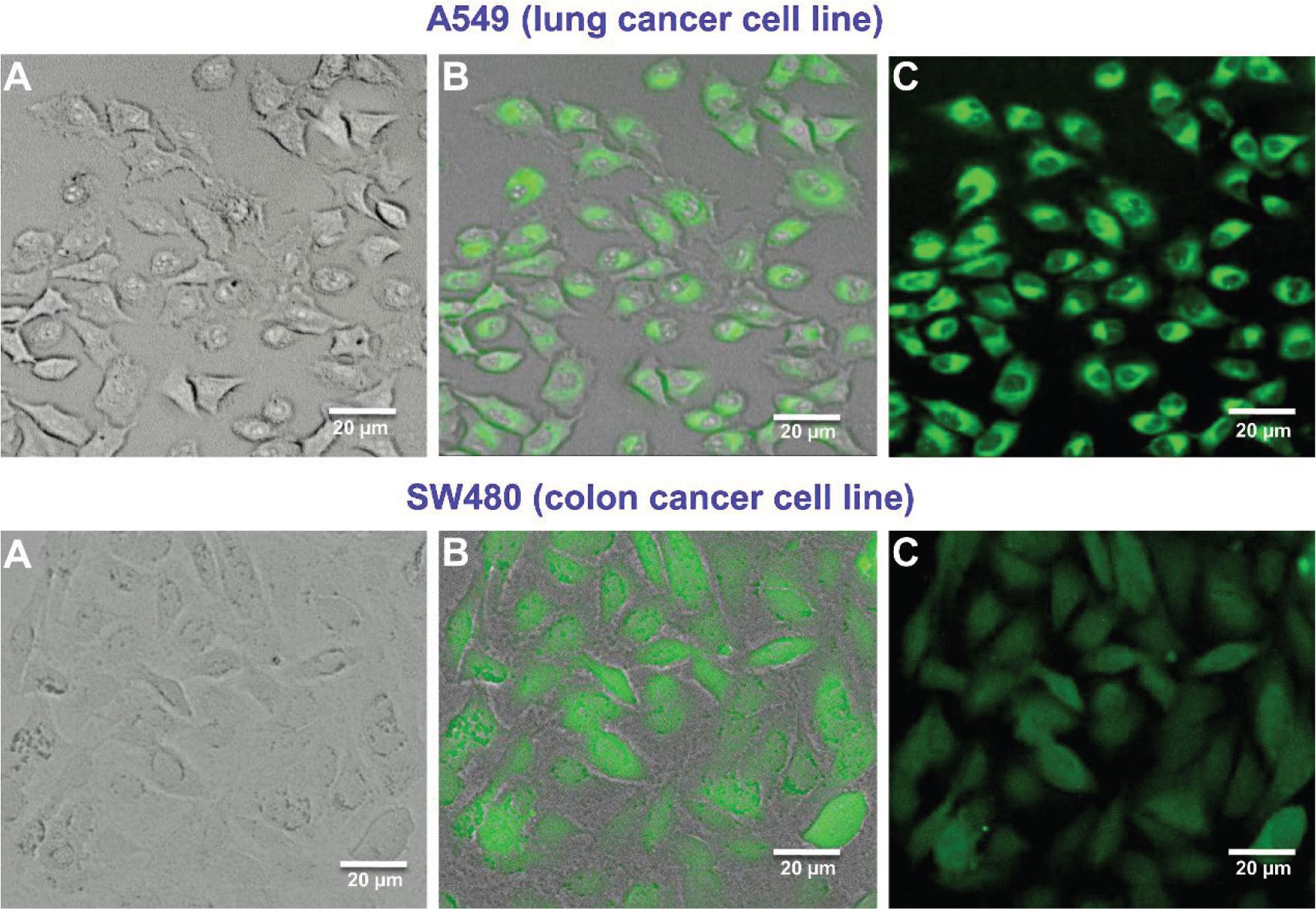
Fig. 8.
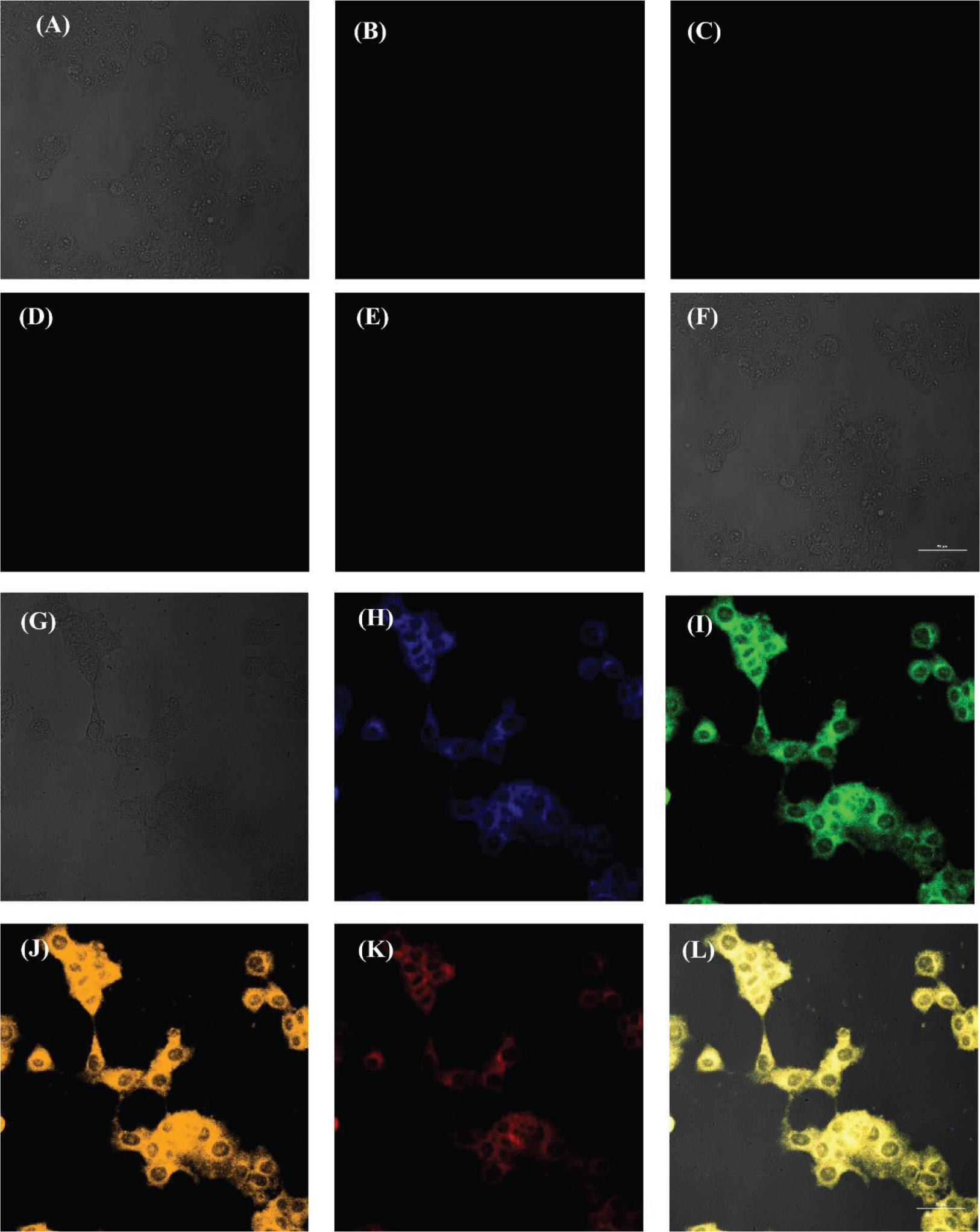
Fig. 9.
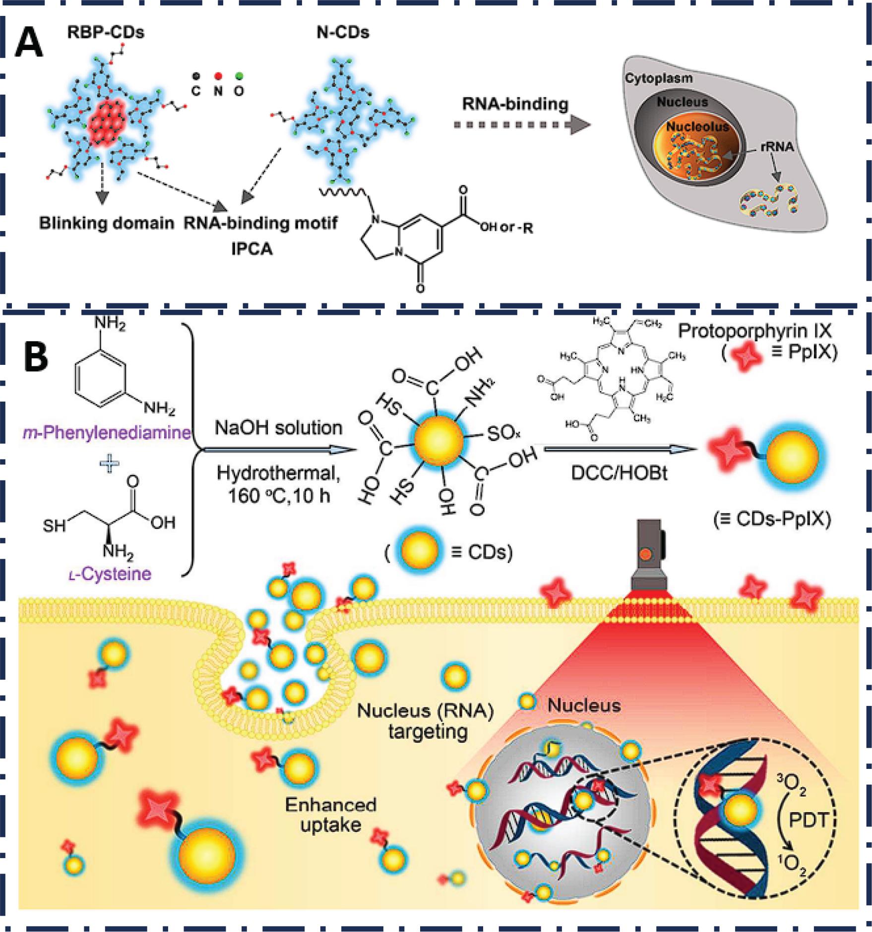
Fig. 10.
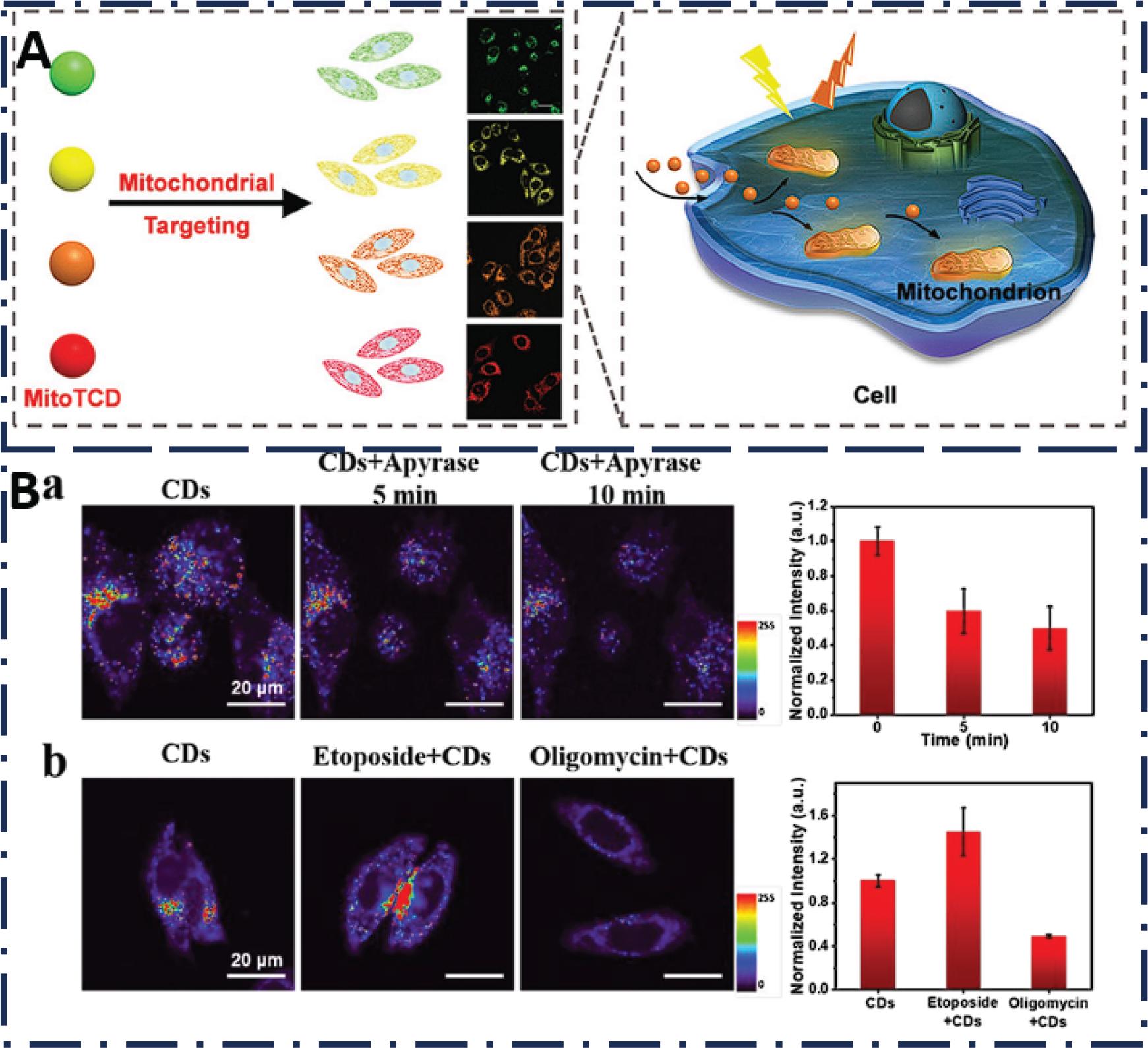
Fig. 11.
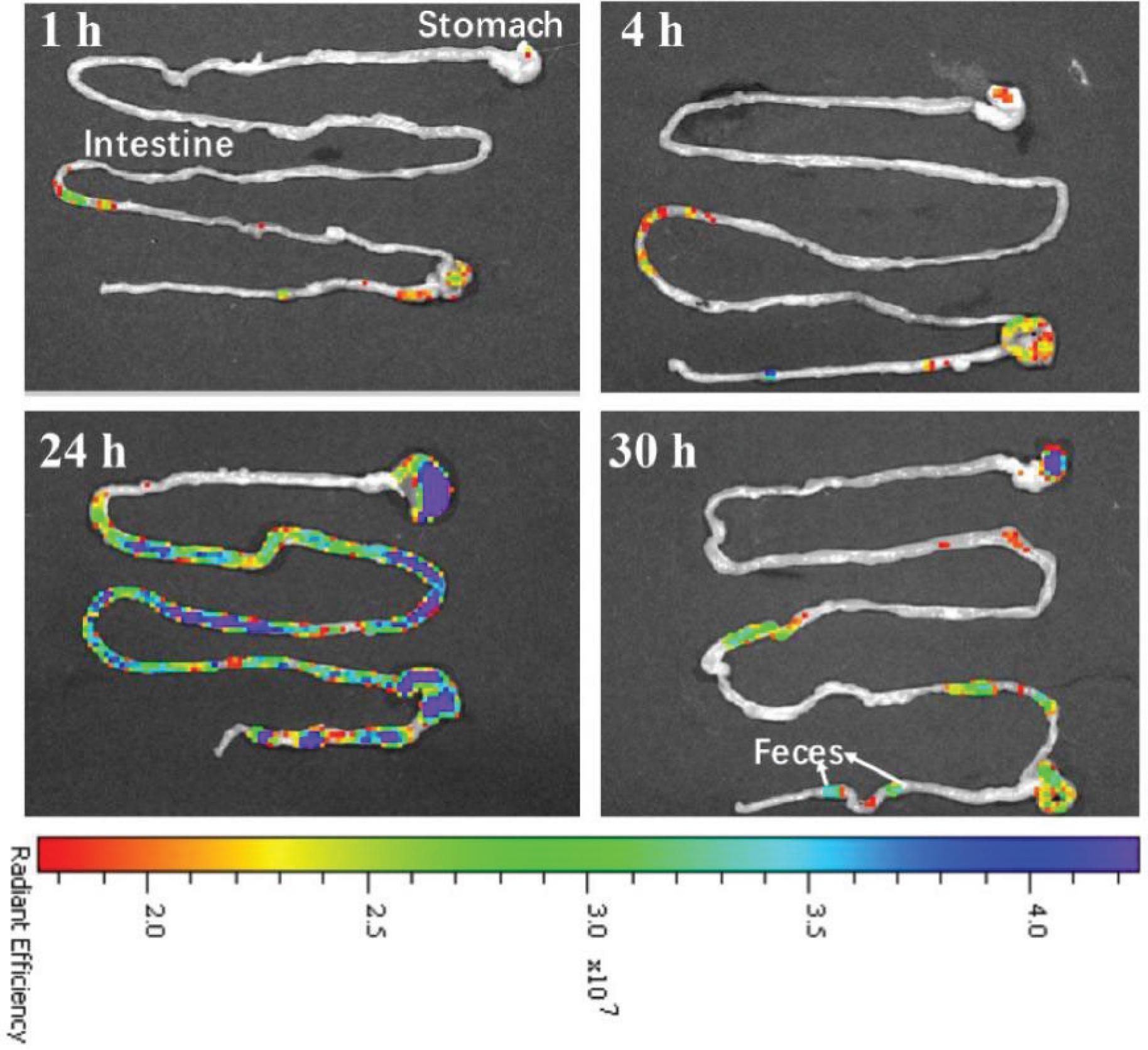
Fig. 12.
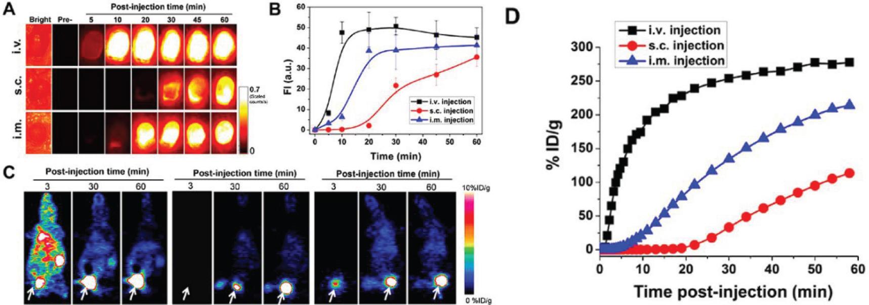
Fig. 13.
Fig. 14.
Common source for CDs synthesis
| Source | Ref. | Source | Ref. |
|---|---|---|---|
| Spinach | (39,136–139) | Onion | (40,140–143) |
| Apple | (41,144,145) | Orange | (42,146,147) |
| Trapa bispinosa | (148) | Cannabis sativa | (149) |
| Neem | (43,150) | Cocoons | (44,151) |
| Coffee | (45,152–154) | Oils | (46,155,156) |
| Milk | (47,157,158) | Egg white | (48,159,160) |
| Honey | (49,161,162) | Yeast extract | (163) |