
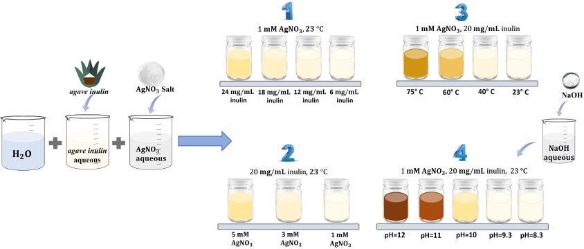
Fig. 1.
Experimental setup for the synthesis of AgNPs. Variables were (1) agave inulin; (2) AgNO3; (3) temperature; (4) pH
Table 1.
Parameters and values used in the preparation of samples to study agave inulin’s effect on the synthesis of AgNPs
| Sample | A | B | C | D |
|---|---|---|---|---|
| S1A | 6 | 1 | 23 | 6.5 |
| S2A | 12 | 1 | 23 | 6.5 |
| S3A | 18 | 1 | 23 | 6.5 |
| S4A | 24 | 1 | 23 | 6.5 |
Table 2.
Parameters and values used in the preparation of samples to study the AgNO3 effect on the synthesis of AgNPs
| Sample | A | B | C | D |
|---|---|---|---|---|
| S1B | 20 | 1 | 23 | 6.5 |
| S2B | 20 | 3 | 23 | 6.5 |
| S3B | 20 | 5 | 23 | 6.5 |
Table 3.
Parameters and values used in the preparation of samples to study temperature effect on the synthesis of AgNPs
| Sample | A | B | C | D |
|---|---|---|---|---|
| S1C | 20 | 1 | 23 | 6.5 |
| S2C | 20 | 1 | 40 | 6.5 |
| S3C | 20 | 1 | 60 | 6.5 |
| S4C | 20 | 1 | 75 | 6.5 |
Table 4.
Parameters and values used in the preparation of samples to study pH effect on the synthesis of AgNPs
| Sample | A | B | C | D |
|---|---|---|---|---|
| S1D | 20 | 1 | 23 | 12 |
| S2D | 20 | 1 | 23 | 11 |
| S3D | 20 | 1 | 23 | 10 |
| S4D | 20 | 1 | 23 | 9.3 |
| S5D | 20 | 1 | 23 | 8.3 |
Table 5.
Contents of the test tubes to study antibacterial activity
| Tube | LB (mL) | E. coli (μL) | Inulin (μL) | Sample (μL) |
|---|---|---|---|---|
| 1 | 5 | – | – | – |
| 2 | 5 | 100 | – | – |
| 3 | 5 | 100 | 100 | – |
| 4 | 5 | 100 | – | S1bac (100) |
| 5 | 5 | 100 | – | S2bac (100) |
| 6 | 5 | 100 | 100 | – |
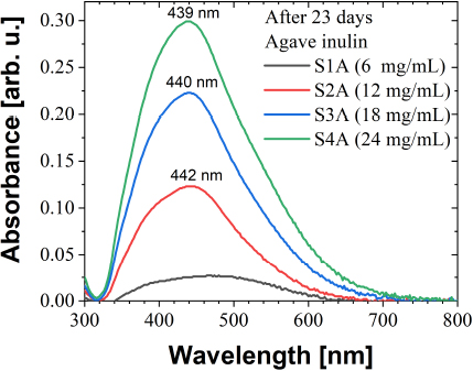
Fig. 2.
UV-Vis absorbance of nanoparticles obtained using different agave inulin concentrations
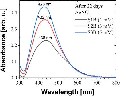
Fig. 3.
UV-Vis absorbance of nanoparticles obtained using different AgNO3 concentration
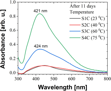
Fig. 4.
UV-Vis absorbance of nanoparticles obtained using different temperatures
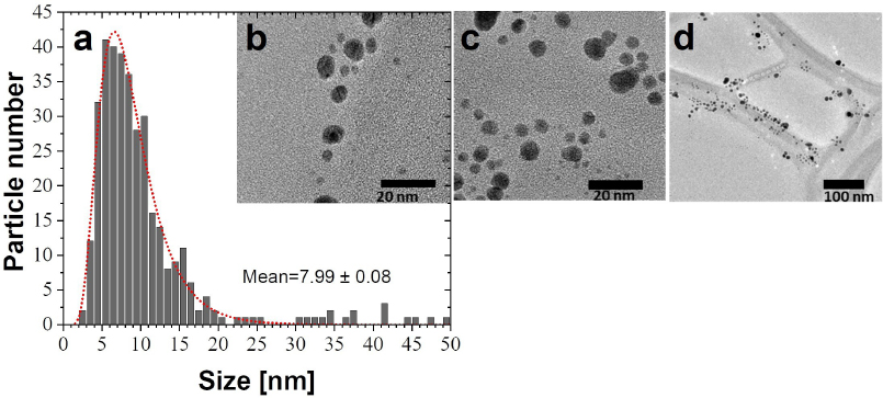
Fig. 5.
(a) Size distribution, fitted with a Log-normal model (dotted line) from several micrographs (for instance b, c, and d) of AgNPs obtained using a temperature of 75°C
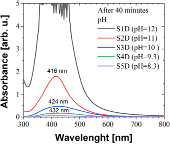
Fig. 6.
UV-Vis absorbance of nanoparticles obtained using different pH values
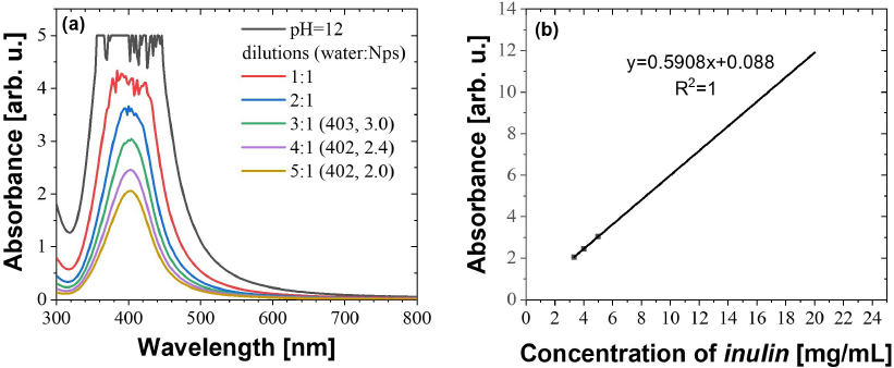
Fig. 7.
(a) UV-Vis spectra of nanoparticles obtained with a pH of 12 using different solutions in water. (b) Line calibration of the maximum absorbance versus the agave inulin concentration (mg/mL)
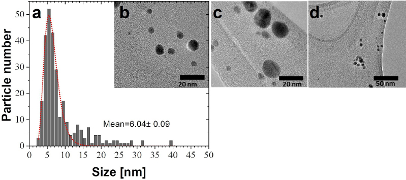
Fig. 8.
(a) Size distribution, fitted with a Log-normal model (dotted line) from several micrographs (for instance b, c, and d) of AgNPs obtained using pH 11
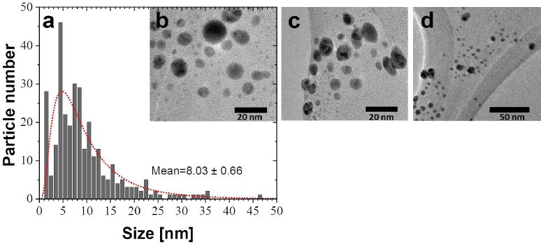
Fig. 9.
(a) Size distribution, fitted with a Log-normal model (dotted line) from several micrographs (for instance b, c, and d) of AgNPs obtained using pH 10
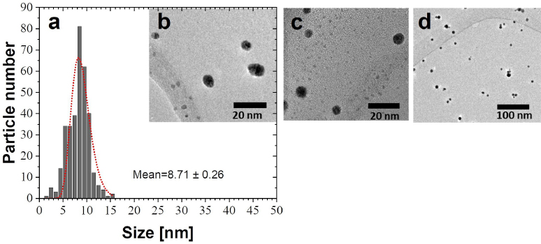
Fig. 10.
(a) Size distribution, fitted with a Log-normal model (dotted line) from several micrographs (for instance b, c, and d) of AgNPs obtained using a pH of 12
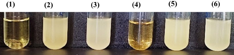
Fig. 11.
Bactericidal effect. LB medium was used to determine the bactericidal effect. (1) Positive control: LB medium; (2) Negative control: LB medium and 100 μL E. coli strain; (3) LB medium, 100 μL E. coli strain, and 100 μL agave inulin; (4) LB medium, 100 μL of E. coli strain, and 100 μL of AgNPs (S1bac); (5) LB medium, 100 μL of E. coli strain, and 100 μL of AgNPs (S2bac); (6) LB medium, 100 μL E. Coli strain, and 100 μL agave inulin