
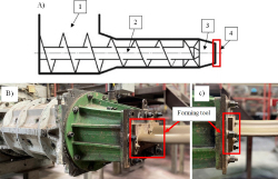
Fig. 1.
Ceramic roof tile extrusion process – A) schematic diagram of construction of extruder barrels: 1 - vacuum zone, 2 - screw, 3 - pressure head, 4 - forming tool [25], B) main view of extruder, C) process of band forming
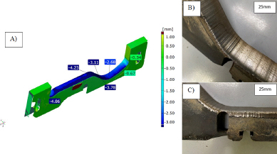
Fig. 2.
Example of worn tool - A) results of 3D scanning, B) scratches, C) crack
Table 1.
Chemical composition of X153CrMoV12 steel
| Steel X153CrMoV12 (NC11LV) – chemical composition [%] | |||||||||||
|---|---|---|---|---|---|---|---|---|---|---|---|
| C | Si | Mn | P | S | Cr | Mo | Ni | Al | Cu | V | W |
| 1.59 | 0.25 | 0.38 | 0.012 | 0.008 | 11.72 | 0.88 | 0.20 | 0.03 | 0.06 | 0.76 | 0.05 |
Table 2.
Parameters of the test samples’ heat treatment
| Sample no. | Austenitizing temperature [°C] | Tempering temperature [°C] | Tempering time [h] |
|---|---|---|---|
| 960/450 | 960 | 450 | 2 |
| 1060/450 | 1060 | 450 | 2 |
| 1020/200 | 1020 | 200 | 2 |
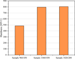
Fig. 3.
Results of hardness measurements of steel after the performed heat treatment variants
Table 3.
Results of the impact energy of samples with a V-notch
| Material | Sample no. | Test temperature | Energy |
|---|---|---|---|
| °C | J | ||
| NC11LV | 960/450 | 50 | 3.8 |
| 1060/450 | 50 | 3.7 | |
| 1020/200 | 50 | 2.8 | |
| 960/450 | 200 | 4.9 | |
| 1060/450 | 200 | 4.6 | |
| 1020/200 | 200 | 3.1 | |
| Hardox 600 | 1 | 50 | 21.6 |
| 2 | 200 | 24.3 |
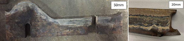
Fig. 4.
Examples of damage in the form of cracks to the NC11LV tools forming the clay band
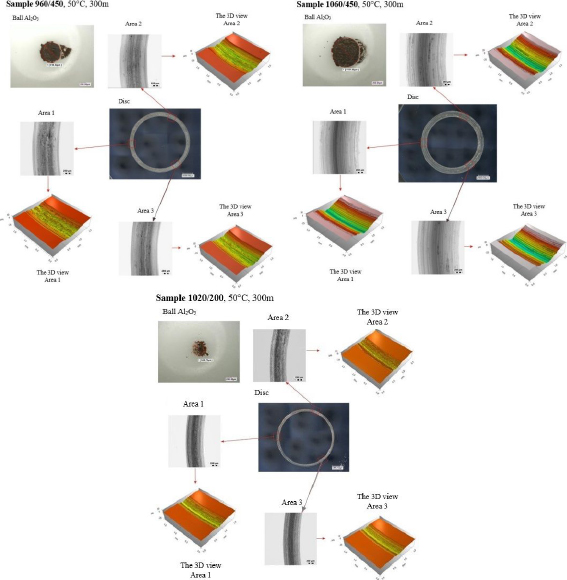
Fig. 5.
Results of the ball-on-disc tests for the analyzed materials
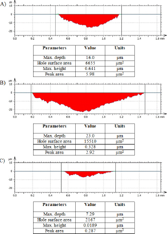
Fig. 6.
Track profiles for the ball-on-disc test: A) sample 960/450, B) sample 1060/450, C) sample 1020/200
Table 4.
Results of the measurement of the track volume in the ball-on-disc test
| Parameters | Sample 960/450 | Sample 1060/450 | Sample 1020/200 |
|---|---|---|---|
| Track volume | 9622503 | 20987030 | 3397096 |
| Max. track depth | 17.0 | 21.7 | 7.46 |
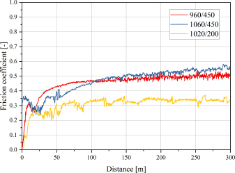
Fig. 7.
Friction coefficients in the ball-on-disc test for both analyzed materials
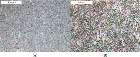
Fig. 8.
View of (A) microstructure of steel NC11. sample 960/450 (B) magnified fragment of the area. Light microscopy, etched state
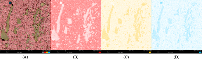
Fig. 9.
Microscopic SEM image, sample 960/450 (A) with the distribution of: iron (B), chromium (C) and vanadium (D). SEM/EDS
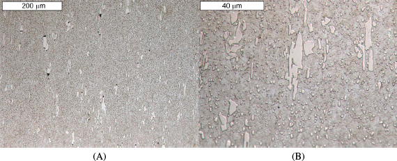
Fig. 10.
View of (A) microstructure of steel NC11LV – sample 1060/450 (B) magnified fragment of the area. Light microscopy, etched state
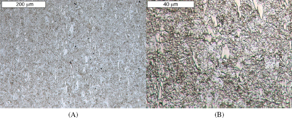
Fig. 11.
View of (A) microstructure of steel NC11LV – sample 1020/200, (B) magnified, fragment of the area. Light microscopy, etched state
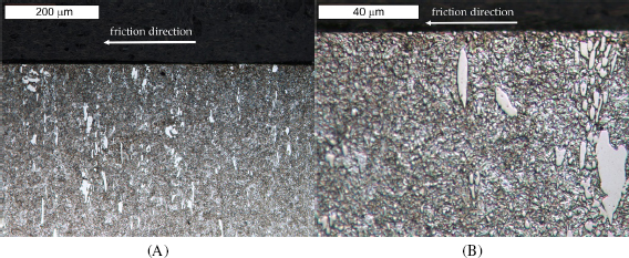
Fig. 12.
View of (A) microstructure of steel NC11LV in the sub-surface area in sample 960/450, (B) magnified fragment of the area. Light microscopy, etched state
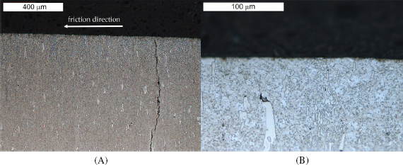
Fig. 13.
View of (A) microscopy of steel NC11LV in the sub-surface area, sample 1060/450, (B) magnified fragment of the area. Light microscopy, etched state
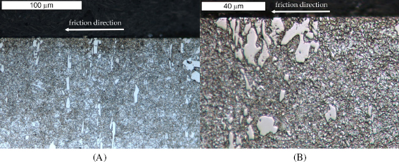
Fig. 14.
View of (A) microstructure of steel NC11LV in the sub-surface area of sample 1020/200, (B) magnified fragment of the area. Light microscopy, etched state
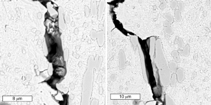
Fig. 15.
Course of a crack developing on the surface. Etched state, SEM – sample 1060/450
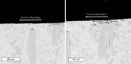
Fig. 16.
Spalling of carbides observed in the sub-surface area of steel in state 2. Etched state, SEM, sample 1060/450
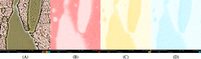
Fig. 17.
A microscopic SEM image, sample 1060/450 (A) together with the distribution of: iron (A), chromium (B) and vanadium (D)
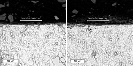
Fig. 18.
Surface changes of steel NC11LV. Etched state, SEM – sample 1020/200