
Table 1
Chemical compositions of S355JR substrate material and Metcoclad 625 powder
| Material | C | Mn | Si | P | S | Cr |
|---|---|---|---|---|---|---|
| wt.% | ||||||
| S355JR | 0.2 | 1.5 | 0.2–0.5 | Max 0.04 | Max 0.04 | Max 0.3 |
| Oerlikon Metcoclad 625 | – | – | – | – | – | 20.0–23.0 |
| Material | Ni | Mo | Nb | Al | Cu | Fe |
|---|---|---|---|---|---|---|
| wt.% | ||||||
| S355JR | Max 0.3 | – | – | Max 0.02 | Max 0.03 | Balance |
| Oerlikon Metcoclad 625 | 58.0–63.0 | 8.0–10.0 | 3.0–5.0 | – | – | Max 5.0 |
Table 2
Technical specifications of TRUMPF Trudisk 3302 laser
| Property | Value |
|---|---|
| Wavelength, μm | 1.3 |
| Maximum output power, W | 3300 |
| Laser beam divergence, mm/rad | <8.0 |
| Fiber core diameter, μm | 200 |
| Collimator focal length, mm | 200 |
| Focusing lens focal length, mm | 200 |
| Beam spot diameter, μm | 200 |
| Fiber length, m | 20 |
Table 3
Laser-cladding parameters
| Designation | Powder mixture | Laser power, W | Speed, mm/min |
|---|---|---|---|
| P1-1 | P1 | 2,000 | 240 |
| P1-2 | P1 | 2,150 | 258 |
| P1-3 | P1 | 2,300 | 276 |
| P2-1 | P2 | 2,000 | 240 |
| P2-2 | P2 | 2,150 | 258 |
| P2-3 | P2 | 2,300 | 276 |
| P3-1 | Metcoclad 625 | 2,000 | 240 |
| P3-2 | Metcoclad 625 | 2,150 | 258 |
| P3-3 | Metcoclad 625 | 2,300 | 276 |
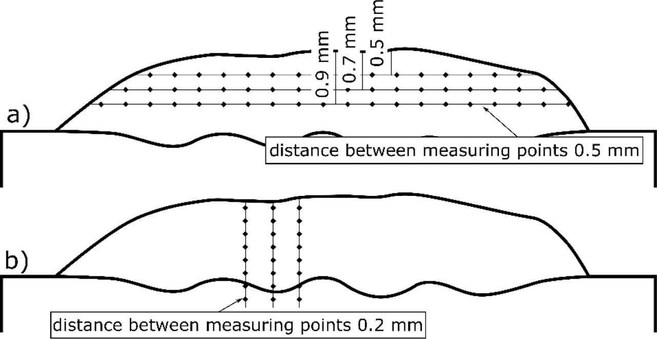
Fig. 1
Vickers hardness lines scheme. (A) Measurements across the multi-pass coating. (B) Measurements from the coating surface toward the substrate material
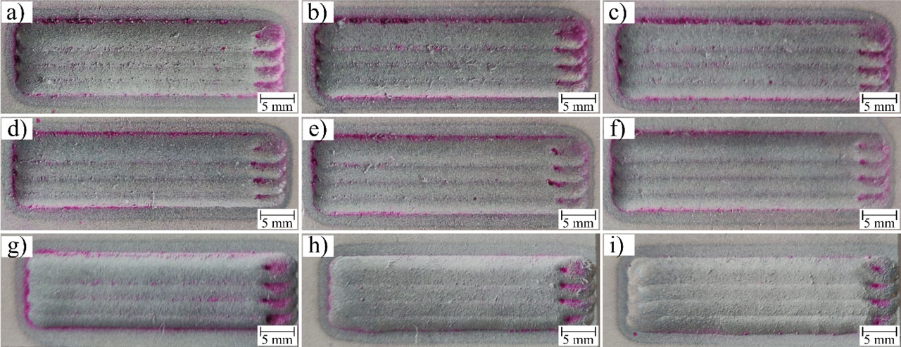
Fig. 2
Penetrant test results of laser-cladded coatings: (A) P1-1; (B) P1-2; (C) P1-3; (D) P2-1; (E) P2-2; (F) P2-3; (G) P3-1; (H) P3-2; and (I) P3-3. Note: Designations are according to Table 3
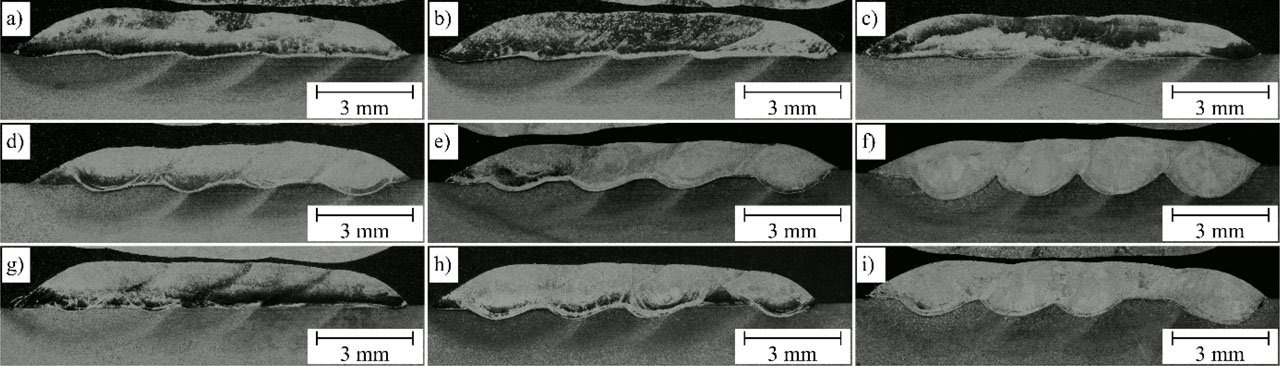
Fig. 3
Macrographs of laser-cladded coatings: (A) P1-1; (B) P1-2; (C) P1-3; (D) P2-1; (E) P2-2; (F) P2-3; (G) P3-1; (H) P3-2; and (I) P3-3. Note: Designations are according to Table 3
Table 4
The laser-cladded coating thickness, dilution, and reinforcing particles volume fraction
| Designation | Coatings thickness, mm | Dilution, % | Reinforcing particles volume fraction, vol.% |
|---|---|---|---|
| P1-1 | 1.43 ± 0.2 | 7.4 ± 0.2 | 3.0 ± 0.3 |
| P1-2 | 1.35 ± 0.2 | 8.5 ± 0.3 | 2.6 ± 0.5 |
| P1-3 | 1.22 ± 0.3 | 8.8 ± 0.4 | 2.7 ± 0.1 |
| P2-1 | 1.23 ± 0.2 | 13.3 ± 0.3 | 6.4 ± 0.9 |
| P2-2 | 1.04 ± 0.1 | 20.0 ± 0.5 | 5.5 ± 0.2 |
| P2-3 | 0.95 ± 0.1 | 30.8 ± 0.3 | 4.5 ± 0.6 |
| P3-1 | 1.42 ± 0.3 | 5.6 ± 0.1 | – |
| P3-2 | 1.37 ± 0.2 | 13.3 ± 0.5 | – |
| P3-3 | 1.16 ± 0.1 | 22.3 ± 0.3 | – |
[i] Note: Designations are according to Table 3
Table 5
The average EDS chemical composition of laser-cladded coatings
| Designation | Ni | Cr | Mo | Nb | Fe | Ti |
|---|---|---|---|---|---|---|
| wt.% | ||||||
| P1-1 | 56.5 ± 1.8 | 18.7 ± 0.4 | 10.7 ± 0.7 | 4.9 ± 0.6 | 6.9 ± 1.7 | 2.4 ± 0.1 |
| P1-2 | 55.4 ± 2.7 | 18.3 ± 0.7 | 10.3 ± 1.0 | 4.3 ± 0.8 | 8.9 ± 3.5 | 2.8 ± 0.2 |
| P1-3 | 54.4 ± 1.2 | 18.0 ± 0.4 | 11.0 ± 0.3 | 4.7 ± 0.5 | 9.1 ± 1.3 | 2.8 ± 0.1 |
| P2-1 | 48.6 ± 2.5 | 15.6 ± 0.6 | 10.2 ± 1.1 | 5.0 ± 0.8 | 16.3 ± 5.1 | 4.1 ± 0.3 |
| P2-2 | 43.2 ± 5.1 | 14.5 ± 1.9 | 9.1 ± 1.0 | 4.3 ± 0.6 | 24.8 ± 8.4 | 4.0 ± 0.5 |
| P2-3 | 33.3 ± 2.3 | 11.2 ± 0.6 | 7.8 ± 0.4 | 4.0 ± 0.5 | 40.1 ± 2.7 | 3.3 ± 0.3 |
| P3-1 | 60.2 ± 1.6 | 19.5 ± 0.8 | 10.5 ± 0.8 | 4.0 ± 1.5 | 5.7 ± 0.5 | – |
| P3-2 | 54.0 ± 1.1 | 17.7 ± 0.3 | 9.0 ± 0.5 | 4.3 ± 0.3 | 15.0 ± 2.1 | – |
| P3-3 | 46.1 ± 1.1 | 15.3 ± 0.5 | 7.5 ± 0.4 | 3.8 ± 0.5 | 27.2 ± 2.0 | – |
[i] EDS, energy dispersive spectrometer.
[ii] Note: Designations are according to Table 3
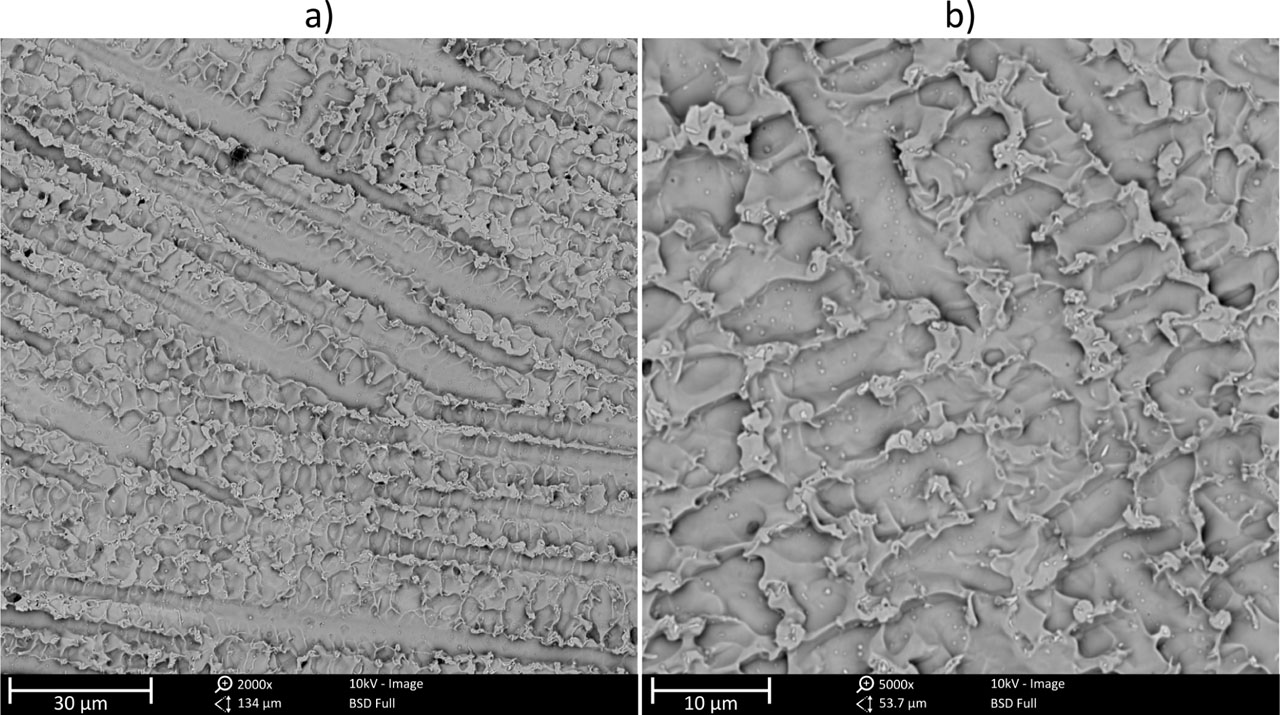
Fig. 4
SEM microstructure of the representative metallic Inconel 625 laser-cladded coating, magnification. (A) Lower magnification. (B) Higher magnification. SEM, scanning electron microscopes
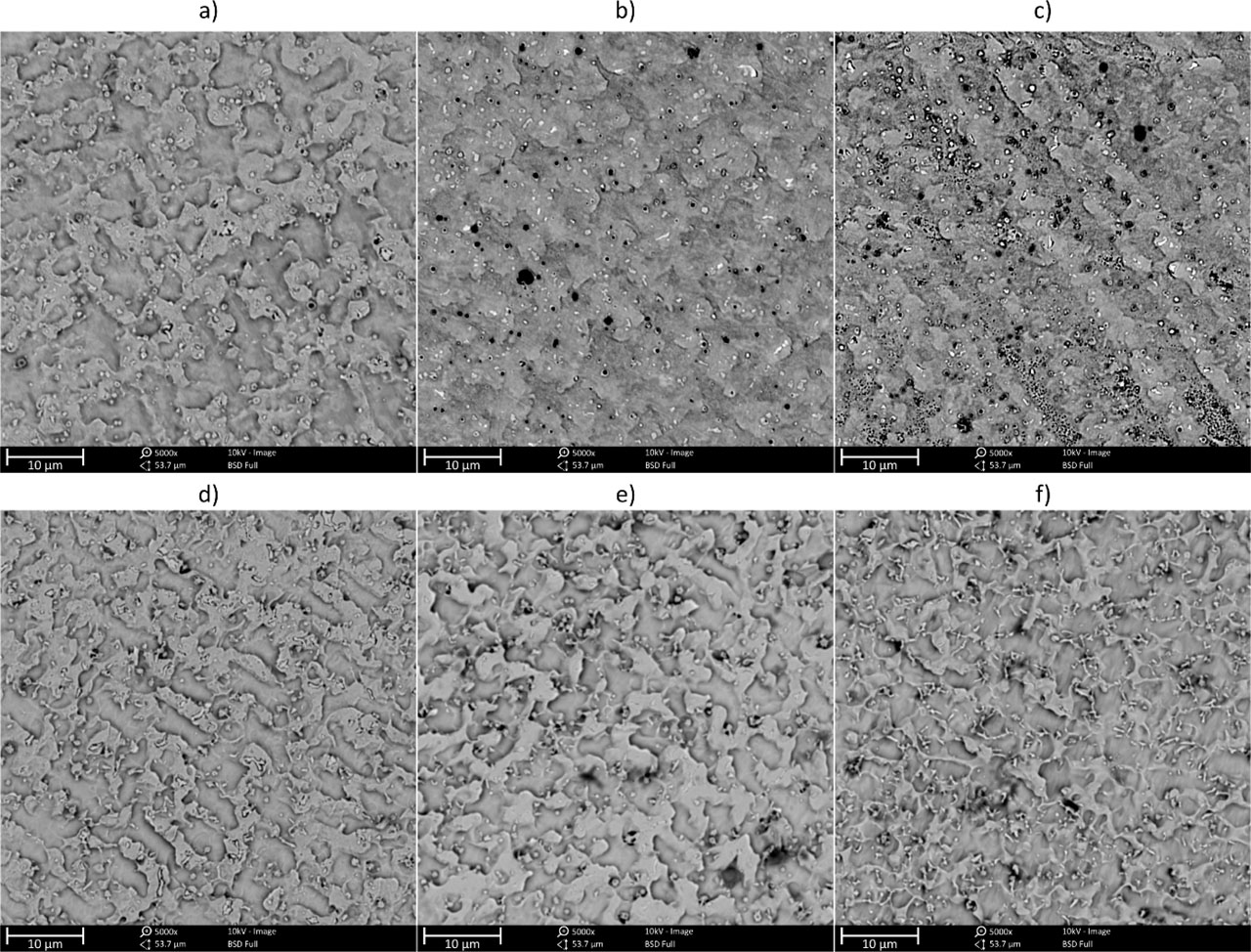
Fig. 5
SEM microstructure of the laser-cladded in situ composite coatings, magnification 5000×. (A) P1-1; (B) P1-2; (C) P1-3; (D) P2-1; (E) P2-2; and (F) P2-3. Note: Designations are according to Table 3. SEM, scanning electron microscopes
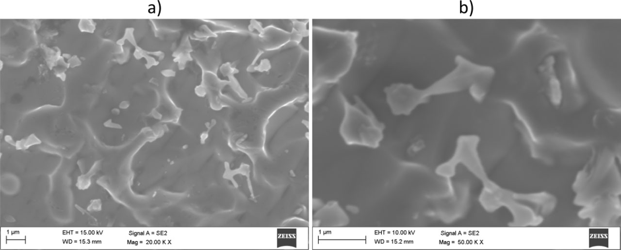
Fig. 6
SEM microstructure of the representative laser-cladded in situ composite coating, magnification. (A) Lower magnification. (B) Higher magnification. SEM, scanning electron microscopes
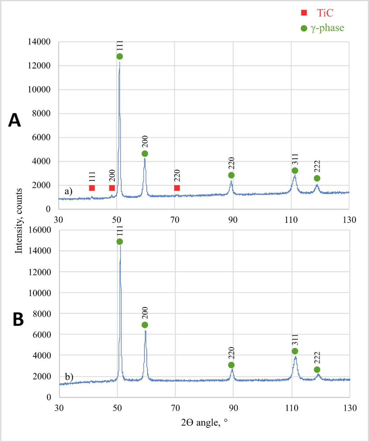
Fig. 7
XRD patterns of (A) representative in situ composite coating and (B) metallic Inconel 625 coating. XRD, X-ray diffraction
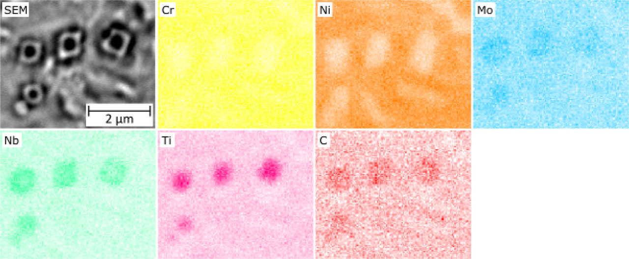
Fig. 8
EDS maps of in situ formed primary reinforcing particles. EDS, energy dispersive spectrometer
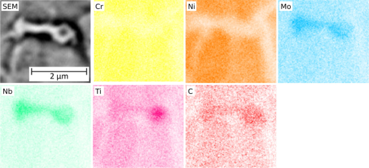
Fig. 9
EDS maps of in situ formed eutectic reinforcing particles. EDS, energy dispersive spectrometer
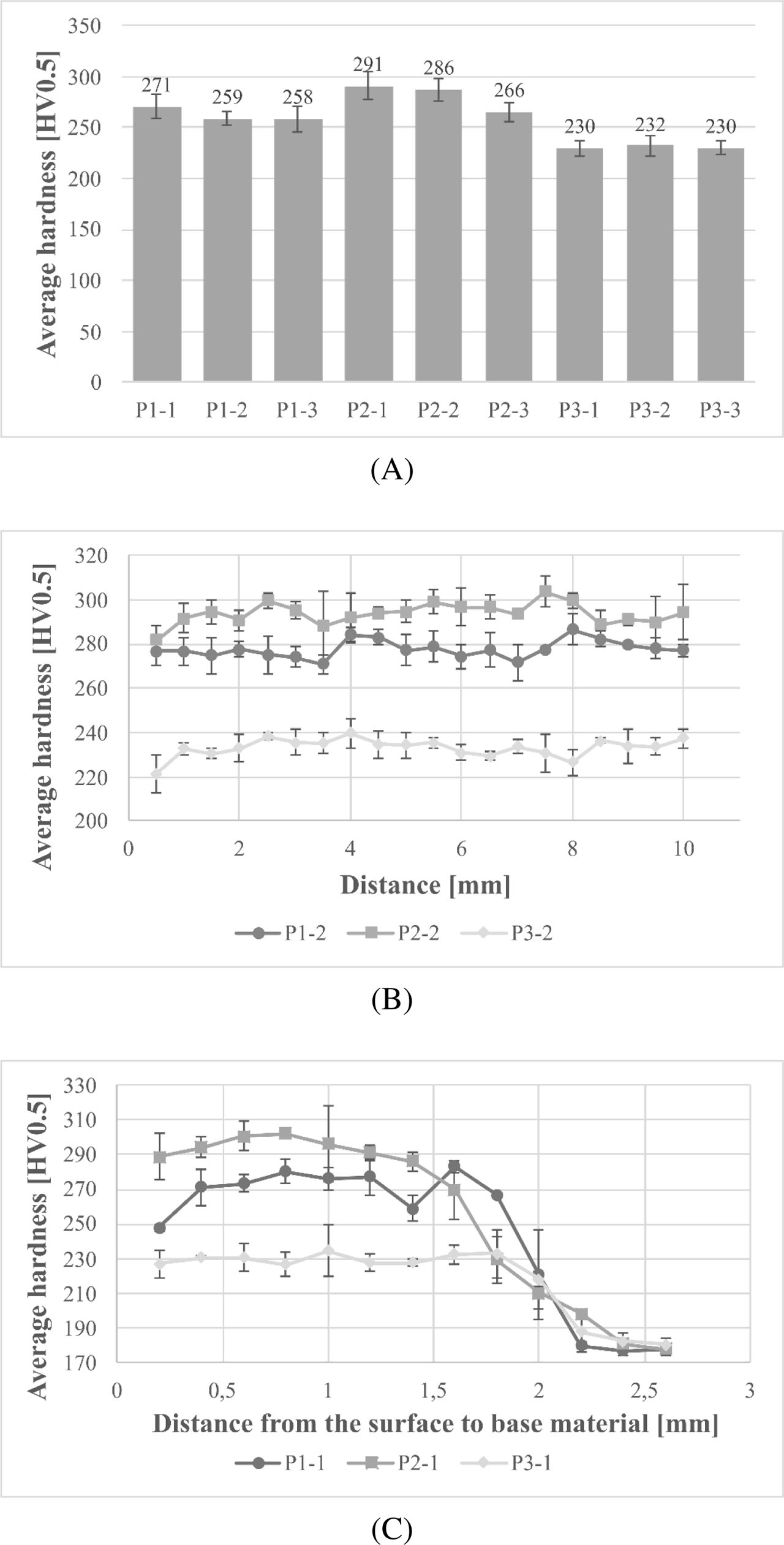
Fig. 10
Hardness results of laser-cladded coatings with standard deviation. (A) Average hardness. (B) Hardness distribution across the multi-pass coatings according to Figure 1A. (C) Hardness distribution from the surface to substrate material according to Figure 1B. Note: Designations are according to Table 3
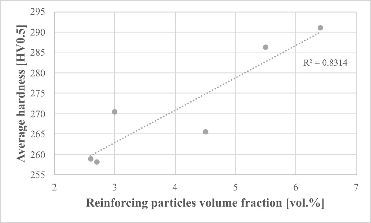
Fig. 11
Influence of reinforcing particles volume fraction on the average hardness of laser-cladded composite coatings
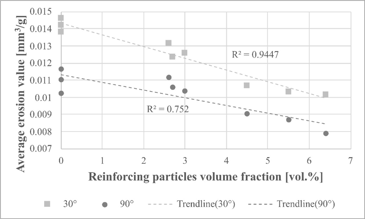
Fig. 12
Influence of reinforcing particles volume fraction on the erosion values of laser-cladded coatings
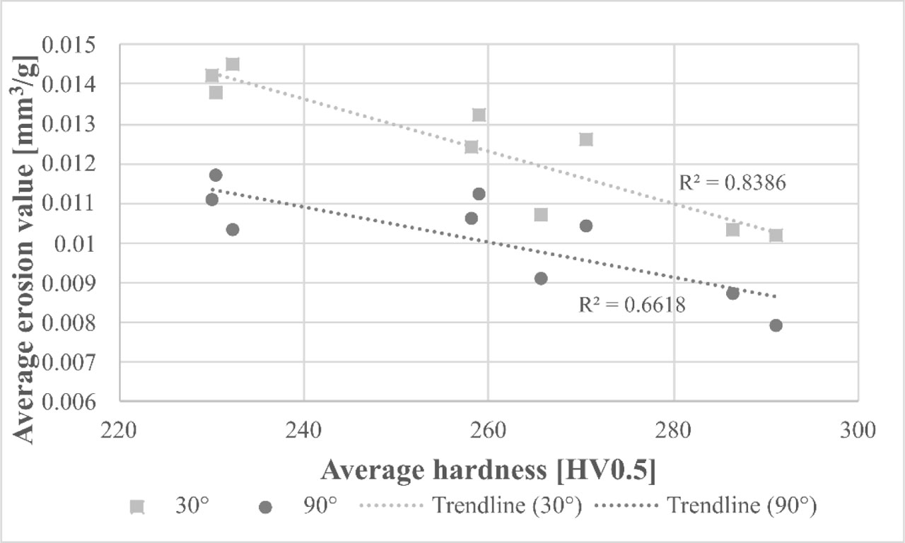
Fig. 13
Influence of average hardness on the erosion values of laser-cladded coatings
Table 6
Average results of erosion rates and erosion values of laser-cladded coatings
| Designation | Erosion rate, mg/min | Erosion value, mm3/g | ||
|---|---|---|---|---|
| 30° | 90° | 30° | 90° | |
| P1-1 | 0.21 ± 0.05 | 0.17 ± 0.02 | 0.0126 ± 0.0032 | 0.0104 ± 0.0012 |
| P1-2 | 0.22 ± 0.04 | 0.19 ± 0.02 | 0.0132 ± 0.0024 | 0.0112 ± 0.0013 |
| P1-3 | 0.21 ± 0.04 | 0.18 ± 0.02 | 0.0124 ± 0.0021 | 0.0106 ± 0.0009 |
| P2-1 | 0.17 ± 0.03 | 0.13 ± 0.04 | 0.0102 ± 0.0018 | 0.0079 ± 0.0022 |
| P2-2 | 0.17 ± 0.03 | 0.14 ± 0.01 | 0.0103 ± 0.0018 | 0.0087 ± 0.0007 |
| P2-3 | 0.18 ± 0.04 | 0.15 ± 0.03 | 0.0107 ± 0.0021 | 0.0091 ± 0.0016 |
| P3-1 | 0.24 ± 0.04 | 0.19 ± 0.04 | 0.0142 ± 0.0021 | 0.0111 ± 0.0022 |
| P3-2 | 0.25 ± 0.03 | 0.21 ± 0.03 | 0.0146 ± 0.0015 | 0.0103 ± 0.0015 |
| P3-3 | 0.23 ± 0.03 | 0.23 ±0.02 | 0.0138 ± 0.0018 | 0.0117 ± 0.0012 |
[i] Note: Designations are according to Table 3
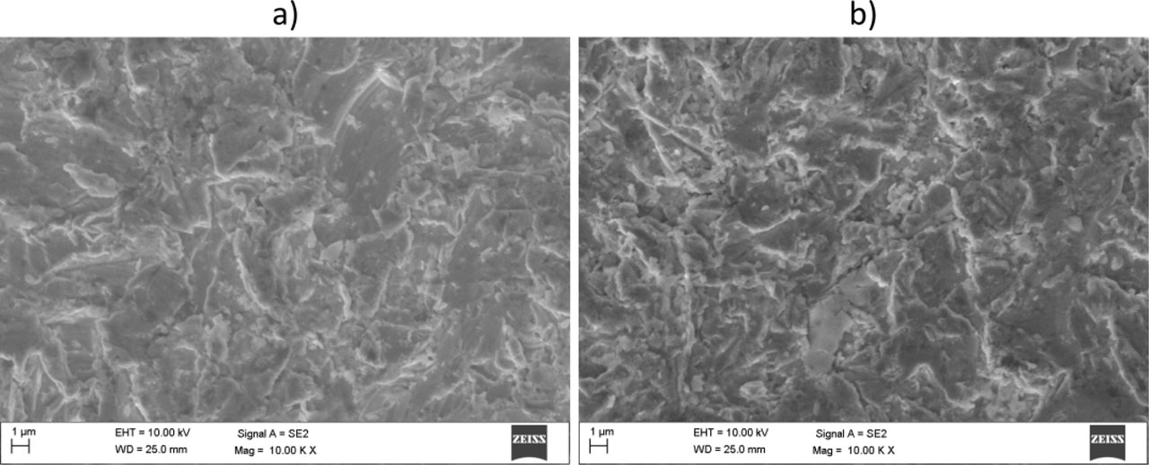
Fig. 14
SEM micrographs of the craters of the representative metallic Inconel 625 coating after solid particle erosion tests. Impingement angles (A) 30° and (B) 90°. SEM, scanning electron microscopes
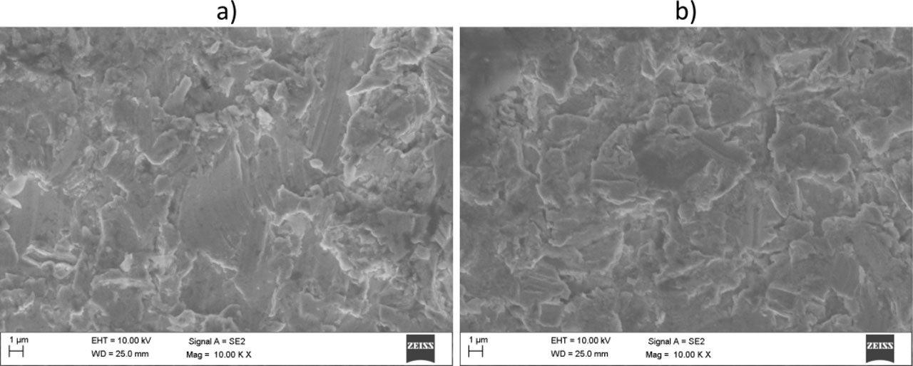
Fig. 15
SEM micrographs of the craters of the representative composite coating after solid particle erosion tests. Impingement angles (A) 30° and (B) 90°. SEM, scanning electron microscopes