
Introduction
Equine herpesviruses (EHV) are important pathogens of horses worldwide, causing respiratory disease, abortion and neurological disorders, which result in substantial economic losses in the equine industry (13, 29). Globally, EHV-1 and EHV-4 are highly prevalent, with reported seroprevalence rates ranging from 30% to over 80% in different horse populations (1, 6, 10). Cases of EHV-1-associated abortion and outbreaks of neurological disease with this cause have been documented in Europe, North America, and Asia (5, 7, 14, 21, 25, 26). Primarily, EHV-1 causes respiratory illness, reproductive failure and neurological complications, whereas EHV-4 predominantly induces respiratory infections, particularly in young horses (20, 23). Both viruses are highly contagious and can be transmitted through direct contact between horses or via contaminated fomites. Surveillance of these pathogens is therefore critical to understand their epidemiology, guide control strategies and prevent outbreaks.
Mongolia has a continental climate characterised by long, cold winters and short summers, and its livestock sector is largely based on a nomadic system. Horses are a key component of the livestock economy, and numbered approximately 4.9 million in 2025 (16). In recent years, the development of equestrian sports has led to the importation of high-performance stallions and mares from abroad, increasing the risk of introducing infectious agents into local horse populations. Consequently, several viral, bacterial and parasitic diseases have been reported in Mongolian horses, including tick-borne encephalitis (12), equine infectious anaemia (24), equine glanders (11), and equine piroplasmosis (15, 17). Extensive grazing systems also allow frequent contact between horses and other domestic or wild animals, elevating the risk of cross-species transmission (18, 19).
Previous serological surveys in Mongolia have detected antibodies against multiple equine viral pathogens, including EHV-1, EHV-3, equine arteritis virus, and equine influenza virus, indicating widespread exposure (19). However, comprehensive nationwide data on the prevalence of EHV-1 and EHV-4 are limited. To address this knowledge gap, the present study conducted serological surveillance of horses across 12 Mongolian provinces. The study aimed to determine antibody prevalence and investigate the effects of age, sex, and geographic distribution on exposure.
Material and Methods
Serum samples
In 2025, blood samples were randomly collected from 1,222 apparently healthy horses across 12 Mongolian provinces (Fig. 1 and Table 1). Approximately 2 mL of blood was drawn from the jugular vein into vacutainer tubes without anticoagulant (Zhejiang Gongdong Medical Technology Co., Taizhou, China). Serum was separated by centrifugation at 2,000 × g for 10 min and stored at −20°C until analysis. All procedures were approved by the Scientific Committee of the Institute of Veterinary Medicine, Mongolian University of Life Sciences (Approval No. 2025002). The study population included horses managed under different production systems, including racing horses, breeding animals, and traditionally managed horses maintained under extensive grazing conditions. Some animals had a history of participation in horse racing events, interprovincial movement or importation from abroad.
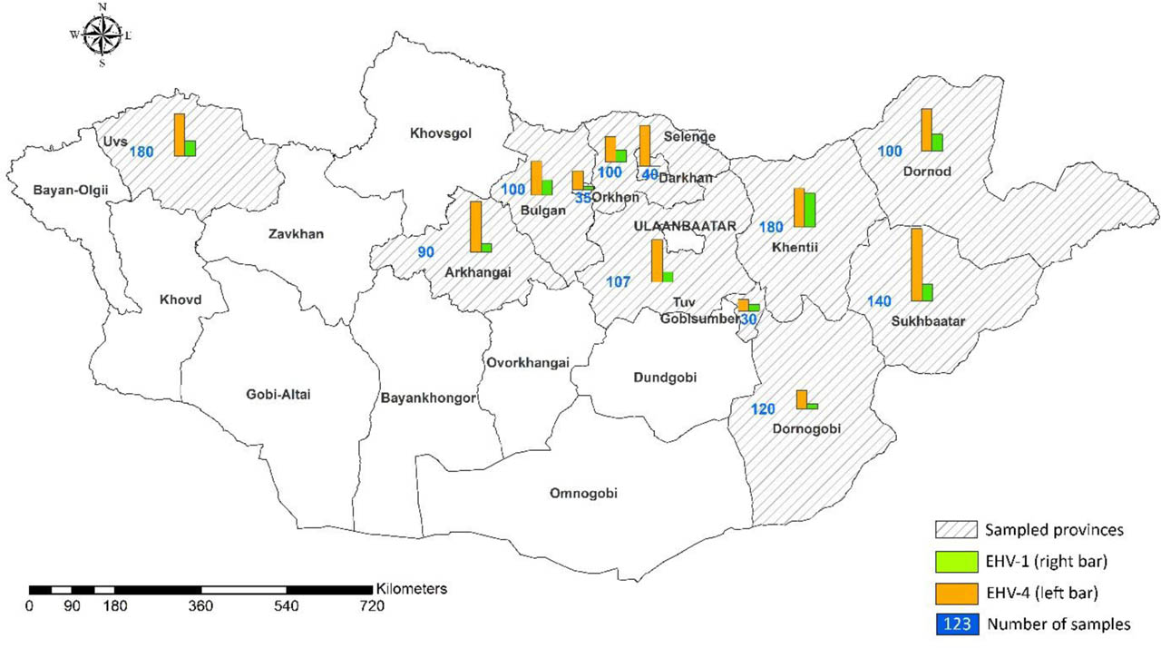
Fig. 1.
Map showing the Mongolian provinces where horses were surveyed for equine herpesvirus type 1 (EHV-1) and type 4 (EHV-4) and the provinces' prevalence rates. The map was created using ArcGIS v. 10.1 (Environmental Systems Research Institute, Redlands, CA, USA)
Table.1.
ELISA detection of equine herpesvirus infections in horses from Mongolia
| Province | n | Number positive for EHV-1 | % (95% CI) | Number positive for EHV-4 | % (95% CI) | Number positive for both | % (95% CI) |
|---|---|---|---|---|---|---|---|
| Bulgan | 100 | 7 | 7.0 (3.4–13.7) | 18 | 18.0 (11.7–26.7) | 2 | 2.0 (0.6–7.0) |
| Dornod | 100 | 6 | 6.0 (2.8–12.5) | 20 | 20.0 (13.3–28.9) | 4 | 4.0 (1.6–9.8) |
| Tuv | 107 | 6 | 5.6 (2.6–11.7) | 25 | 23.4 (16.4–32.2) | 5 | 4.7 (2.0–10.5) |
| Selenge | 100 | 7 | 7.0 (3.4–13.7) | 15 | 15.0 (9.3–23.3) | 3 | 3.0 (1.0–8.5) |
| Sukhbaatar | 140 | 10 | 7.1 (3.9–12.6) | 42 | 30.0 (23.0–38.0) | 8 | 5.7 (2.9–10.9) |
| Khentii | 180 | 20 | 11.1 (7.3–16.5) | 24 | 13.3 (9.1–19.1) | 2 | 1.1 (0.3–4.0) |
| Dornogovi | 120 | 3 | 2.5 (0.9–7.1) | 11 | 9.2 (5.2–15.7) | 1 | 0.8 (0.1–4.6) |
| Uvs | 180 | 8 | 4.4 (2.3–8.5) | 25 | 13.9 (9.6–19.7) | 1 | 0.6 (0.1–3.1) |
| Arkhangai | 90 | 9 | 10.0 (5.4–17.9) | 31 | 34.4 (25.4–44.7) | 5 | 5.6 (2.4–12.4) |
| Darkhan | 40 | 0 | 0.0 (0–8.8) | 24 | 60.0 (44.6–73.7) | 1 | 2.5 (0.4–12.9) |
| Orkhon | 35 | 2 | 5.7 (1.6–18.6) | 10 | 28.6 (16.3–45.1) | 2 | 5.7 (1.6–18.6) |
| Govisumber | 30 | 4 | 13.3 (5.3–29.7) | 8 | 26.7 (14.2–44.4) | 0 | 0.0 (0–11.4) |
| Total | 1,222 | 82 | 6.7% (5.4–8.2) | 253 | 20.7% (18.5–23.1) | 34 | 2.8% (2.0–3.9) |
ELISAs
All serum samples were screened for antibodies against equine herpesvirus type 1 (EHV-1) and equine herpesvirus type 4 (EHV-4) using ELISAs as previously described (2, 3), with minor modifications. Briefly, synthetic peptides gE1 (KKPPKQPQPRLRVKTPPPVT) for EHV-1 and gG4 (MKNNPIYSEGSLMLNVQHDDSIHT) for EHV-4, commercially synthesised by Rigen Biotech Co., Ltd, China, were used as antigens. Antigen stock solutions were diluted in 0.05 M carbonate–bicarbonate buffer (pH 9.6) to a final concentration of 5 μg/mL. Each 96-well microplate was divided into three sections: wells coated with gE1 peptide (EHV-1), wells without antigen as negative controls, and wells coated with gG4 peptide (EHV-4). For each serum sample, parallel measurements were performed in these three types of well. Plates were incubated at 37°C for 2 h. After removal of unbound antigen, the wells were blocked with 100 μL of 10% bovine serum in PBS at 37°C for 30 min. The plates were then washed twice with 250 μL of PBS containing 0.05% Tween-20 (wash buffer). Subsequently, 50 μL of horse serum diluted 1 : 20 in blocking solution was added to each well and incubated at 37°C for 30 min. After the wells had been washed three times with wash buffer, 50 μL of rabbit anti-horse IgG (whole molecule) conjugated with horseradish peroxidase (Sigma-Aldrich, St. Louis, MO, USA) and diluted 1 : 5,000 in blocking solution was added to each well and the plates were incubated at 37°C for 30 min. After three additional washes, 50 μL of tetramethylbenzidine substrate solution (Sigma-Aldrich) was added, and the plates were again incubated at 37°C for 30 min. The reaction was stopped by adding 50 μL of stop solution (Sigma-Aldrich), and OD values were measured at 450 nm using a microplate reader.
Determination of antibody titres
Final OD values were calculated by subtracting the OD values of antigen-free control wells from those of the corresponding antigen-coated wells (EHV-1 or EHV-4). Negative control sera were obtained from a commercial equine herpesvirus ELISA kit (INgezim Rinoneumonitis 14.HVE.K1; Gold Standard Diagnostics, Budapest, Hungary), and positive control sera for both EHV-1 and EHV-4 were provided by the Japan Racing Association (Shimotsuke, Japan). The assay cut-off value was calculated using the following formula:
where M is the mean of negative controls and SD is the standard deviation of negative controls.Samples with corrected OD values ≥ 0.2 were considered positive. The peptide-based ELISA used in this study was previously validated for the detection of antibodies against EHV-1 and EHV-4, demonstrating high sensitivity and specificity (2, 3).
Statistical analysis
The 95% CIs for prevalence rates of EHV-1 and EHV-4 were calculated using the OpenEpi online software tool (8) based on the Wilson score interval (28). Prevalence was compared between groups stratified by province, sex and, age category. The P-values to determine the statistical significance of differences between the positive rates were calculated using a chi-squared test with N-1 correction (4, 22) (Medcalc Software, Ostend, Belgium). Differences between the positive rates were considered statistically significant if P-values were <0.05.
Results
Testing by ELISA detected antibodies against equine herpesvirus type 1 (EHV-1) and type 4 (EHV-4) in 1,222 horses across 12 provinces of Mongolia (Table 1). Overall, 82 horses (6.7%) were positive for EHV-1, 253 horses (20.7%) for EHV-4, and 34 horses (2.8%) showed dual seropositivity. In total, 301 horses (24.6%) were seropositive for at least one of the two viruses. The overall EHV-4 prevalence was significantly higher than EHV-1 (P-value < 0.0001). At the provincial level, EHV-1 positivity ranged from 0.0% to 13.3%, and EHV-4 positivity from 9.2% to 60.0%. The prevalence of EHV-4 consistently exceeded that of EHV-1 in all provinces (Table 1). These findings should be interpreted cautiously because the sample size was relatively small.
Geographically (Fig. 1), high EHV-4 positivity (≥30%) was observed in Darkhan, Arkhangai and Sukhbaatar provinces, and lower positivity emerged in Dornogovi and Khentii. Rates of ≥10% for EHV-1 positivity were found in Khentii, Arkhangai and Govisumber provinces, but a 0.0% rate was noted in Darkhan. Govisumber province was the only one in which no simultaneous positivity for both EHV-1 and EHV-4 was discovered. While higher EHV-1 positivity generally correlated with increased EHV-4 positivity in within-province patterns, exceptions were observed (Table 1). Darkhan province, for example, exhibited a high EHV-4 positive rate (60.0%) despite no detected EHV-1 infection.
Positive rates were compared across sexes (564 males and 658 females) and age groups (1-to-3-year-olds, n = 215; >3-year-olds, n = 1,007). The prevalence of EHV-1 was comparable between males (7.6%) and females (5.9%). However, that of EHV-4 was significantly higher (P-value = 0.003) in female animals (23.9%) than in male animals (17.0%) (Table 2).
Table 2.
Rates of equine herpesvirus infections by sex in Mongolian horses
| Virus genotype | Number of positive males (n = 564) | % (95% CI) | Number of positive females (n = 658) | % (95% CI) | P-value |
|---|---|---|---|---|---|
| Equine herpesvirus 1 | 43 | 7.6 (5.6–10.1) | 39 | 5.9 (4.2–8.0) | 0.23 |
| Equine herpesvirus 4 | 96 | 17.0 (14.0–20.4) | 157 | 23.9 (20.7–27.3) | 0.003 |
A strong association was observed between age and EHV-4 prevalence, with 1–3-year-old horses exhibiting a significantly higher positive rate (28.4%) than horses older than 3 years (19.1%) (P-value = 0.0023) (Table 3). However, no significant age-related differences were found for EHV-1, with prevalence rates of 9.3% and 6.2% in the respective age groups. These findings indicate that sex and age significantly impact EHV-4 infection rates in Mongolian horses.
Table 3.
Rates of equine herpesvirus infections by age group in Mongolian horses
| Virus genotype | Number of positive 1–3-yr olds (n = 215) | % (95% CI) | Number of positive >3-yr-olds (n = 1,007) | % (95% CI) | P-value |
|---|---|---|---|---|---|
| Equine herpesvirus 1 | 20 | 9.3 (6.1–13.8) | 62 | 6.2 (4.8–7.8) | 0.099 |
| Equine herpesvirus 4 | 61 | 28.4 (22.5–34.9) | 192 | 19.1 (16.7–21.6) | 0.0023 |
Discussion
This study provides the first large-scale nationwide serological assessment of EHV-1 and EHV-4 in horses in Mongolia, and helps address an important knowledge gap in the epidemiology of these pathogens. Our findings reveal a substantial circulation of both viruses, with EHV-4 being significantly more prevalent than EHV-1, and highlight significant variations in prevalence based on geographical location, sex and age.
The seroprevalence observed in the present study was lower than that reported in several other countries. In Spain, antibodies against EHV-1 and EHV-4 were detected in a substantially larger proportion of horses, reflecting the widespread circulation of these viruses in intensively managed equine populations (6). Similarly, a study conducted in Israel reported high seroprevalence rates for both EHV-1 and EHV-4, particularly among horses involved in organised equestrian activities (1). In New Zealand, serological investigations also demonstrated extensive exposure to EHV-1 among horses from different management systems (9). In Morocco, considerably higher prevalence rates of both EHV-1 and EHV-4 than those observed in Mongolia were reported (10). These differences may reflect variations in horse density, animal movement, management practices, vaccination programmes, and environmental conditions. Despite lower overall prevalence in Mongolia compared with many international reports, the detection of antibodies across all surveyed provinces indicates that both EHV-1 and EHV-4 are endemic in the country's horses. Globally, EHV seroprevalence rates often range from 30% to over 80% in various horse populations (27, 29, 30). Although the overall prevalence observed in this study is at the lower end of this range or falls below it, particularly for EHV-1, the widespread geographical distribution supports the endemic circulation of these viruses in Mongolia. The significantly higher prevalence of EHV-4 than of EHV-1 aligns with general epidemiological patterns, where EHV-4 is often more commonly detected in respiratory outbreaks, particularly among younger horses, and can have a higher seroprevalence than EHV-1 in some populations (27).
The distinct epidemiological patterns observed for EHV-1 and EHV-4 in Mongolia are noteworthy. The prevalence of EHV-4 was consistently higher than that of EHV-1 in all sampled provinces. This virus type showed high positivity (≥30%) in Darkhan, Arkhangai and Sukhbaatar provinces, suggesting localised high-transmission zones. Conversely, EHV-1-positive cases (≥10%) were concentrated in Khentii, Arkhangai and Govisumber. An interesting finding was the complete absence of EHV-1 antibodies in Darkhan province, despite the high EHV-4 prevalence there of 60.0%. Only in Khentii Province were the EHV-4 (13.3%) and EHV-1 (11.1%) seroprevalence rates relatively similar. In contrast, considerably greater differences between the two viruses were observed in the other provinces. This finding may suggest that these viruses circulate independently of each other or that different regional factors may influence their respective transmission dynamics. Such regional disparities could be influenced by local horse management practices, population densities, movement patterns, and environmental factors. The nomadic and extensive grazing systems prevalent in Mongolia (19) might facilitate broader dissemination of infectious agents, but localised factors could still create specific hotspots or protect certain areas. The caution regarding the small sample sizes in some provinces (e.g. Darkhan, Orkhon, and Govisumber) means that these provincial-level interpretations should be made carefully.
Analysis of demographic factors revealed significant differences for EHV-4. Female horses had a significantly higher (P-value = 0.003) EHV-4-positive rate (23.9%) than males (17.0%). Similarly, 1–3-year-old horses exhibited a significantly higher (P-value = 0.0023) EHV-4 prevalence (28.4%) than horses older than 3 years (19.1%). This pattern for EHV-4 is consistent with its known epidemiology, as young horses are generally considered more susceptible to primary respiratory infection (2, 3). The higher seroprevalence observed in female horses may be associated with differences in management, breeding practices or exposure opportunities. However, these factors were not analysed in detail in the present study, and, therefore, the reasons for the observed sex-associated difference remain speculative. In contrast, no significant differences were observed for EHV-1 prevalence across sexes or age groups. This finding suggests that EHV-1 exposure in Mongolia might not be as directly influenced by typical age- or sex-related management practices as EHV-4 exposure, or that the immune response and duration of antibodies differ between the two viruses. The EHV-1 prevalence being uncorrelated with age could indicate that exposure is widespread and occurs at various stages of life, or that transient immunity or intermittent shedding patterns obscure a clear age-related trend in seroprevalence. Although demographic factors were not significantly associated with EHV-1 seropositivity, the widespread occurrence of EHV-1 remains epidemiologically significant because of its impact on equine health and reproduction.
The economic significance of EHV infections, particularly EHV-1 causing abortion and neurological disorders, and EHV-4 causing respiratory disease (27, 29), underscores the importance of these findings. The importance of EHV-1 surveillance is further emphasised by reports of abortion outbreaks on breeding farms. Abortion storms associated with EHV-1 have been documented in several countries, including the United Kingdom (14), Poland (25) and France (5), highlighting the substantial reproductive losses that may occur following virus introduction into susceptible horse populations. Mongolia's growing equestrian sports sector and the importation of high-performance horses further increase the risk of infectious disease introduction and spread (11). Our study provides crucial baseline epidemiological data essential for developing targeted, risk-based surveillance programmes, and control strategies. Identifying high-prevalence areas and age and sex groups susceptible to EHV-4 infection may facilitate more efficient allocation of resources for surveillance, biosecurity measures, and future disease-control programmes.
Despite the comprehensive nature of this study, several limitations should be acknowledged. Seropositivity reflects past EHV-1/4 exposure, but seronegativity does not exclude the possibility of previous infection, because antibody levels may decline over time. Comparing seropositivity and seronegativity rates potentially underestimates the true prevalence. Furthermore, seroprevalence only indicates previous exposure rather than active infection or clinical disease at the time of sampling. Although the overall sample size was large, the number of samples collected from some provinces was relatively small, requiring cautious interpretation of province-level estimates. An important limitation when comparing the findings of the present study with those reported in other countries is the difference in vaccination practices among the study populations. This difference arises because, unlike the horse population included in our study, horses investigated in other countries may have been vaccinated against these viruses. Since vaccination against EHV-1 and EHV-4 is not routinely practiced in Mongolia, very few horses in the study population had received vaccination. In contrast, routine vaccination may have been implemented in the horse populations investigated in other countries. Consequently, antibodies detected in seroprevalence studies conducted elsewhere may reflect not only natural exposure to the viruses but also vaccine-induced immunity. In the Mongolian horse population, however, the detected antibody responses most likely resulted exclusively from natural exposure to these viruses.
In addition, information on management practices, movement history, and reproductive status was not analysed in detail, which may have influenced the observed seroprevalence patterns. Future studies should incorporate direct viral detection methods (e.g. PCR) to determine active infection rates and circulating strains. Further research is also warranted to investigate specific risk factors associated with herd management practices, horse movement, and interactions with other animal species within the unique nomadic environment of Mongolia. Longitudinal studies could provide valuable insights into infection dynamics, duration of immunity, and the true economic impact of the diseases caused by EHV-1 and EHV-4.
Conclusion
This study provides the first large-scale, nationwide serological data on EHV-1 and EHV-4 prevalence in Mongolian horses. The high prevalence of EHV-4, particularly in young and female horses, and significant regional variations for both viruses, emphasise the need for targeted surveillance and control programmes. These findings lay a critical foundation for protecting the health and economic viability of Mongolia's important equine industry.
Acknowledgements
We would like to thank the local veterinarians for their assistance in collecting serum samples.
Notes
[2] Conflicts of interest Conflict of Interests Statement: The authors declare that there is no conflict of interests regarding the publication of this article.
[3] Financial disclosure Financial Disclosure Statement: This study was supported by the Mongolian Foundation for Science and Technology (Project No. Шyy3 2024/03).
[4] Animal Rights Statement: All procedures were approved by the Scientific Committee of the Institute of Veterinary Medicine, Mongolian University of Life Sciences (Approval No. 2025002).
[5] CRediT Authorship Contribution Statement: Purevtseren Dulam: research concept and design, collection and/or assembly of data, data analysis and interpretation, writing the article. Myagmarsuren Odonchimeg: collection and/or assembly of data, data analysis and interpretation, critical revision of the article. Davaajav Otgonsuren: data analysis and interpretation, critical revision of the article. Batsuren Nanzaddorj: collection and/or assembly of data. Davaanyam Nyamtseren: collection and/or assembly of data. Ochirvaani Bumantsetseg: collection and/or assembly of data. Yondonjamts Enkhmandakh: collection and/or assembly of data. Enkhbaatar Batmagnai: collection and/or assembly of data. Hiroshi Bannai: collection and/or assembly of data. Badgar Battsetseg: collection and/or assembly of data. Punsantsogvoo Myagmarsuren: research concept and design, collection and/or assembly of data, final approval of the article. Dashzevge Erdenechimeg: research concept and design, collection and/or assembly of data, data analysis and interpretation, writing the article, critical revision of the article, final approval of the article.