
Table 1.
Chick embryo weight parameters and heart volumes at different periods (Mean±SD)
| Parameter | Incubation day 10 | Incubation day 13 | Incubation day 16 | Incubation day 21 |
|---|---|---|---|---|
| Embryo weight (g) | 3.11 ± 0.21a | 8.91 ± 0.46b | 22.74 ± 2.72c | 41.18 ± 2.26d |
| Heart weight (g) | 0.021 ± 0.002a | 0.071 ± 0.007b | 0.182 ± 0.031c | 0.237 ± 0.062d |
| Relative organ weight (g) | 0.686 ± 0.038a | 0.793 ± 0.058b | 0.804 ± 0.152b | 0.577 ± 0.151a |
| Heart volume (mm3) | 0.371 ± 0.05a | 1.279 ± 0.11b | 1.922 ± 0.43c | 3.239 ± 0.57d |
Table 2.
Chick embryo heart diameters at different periods (Mean±SD)
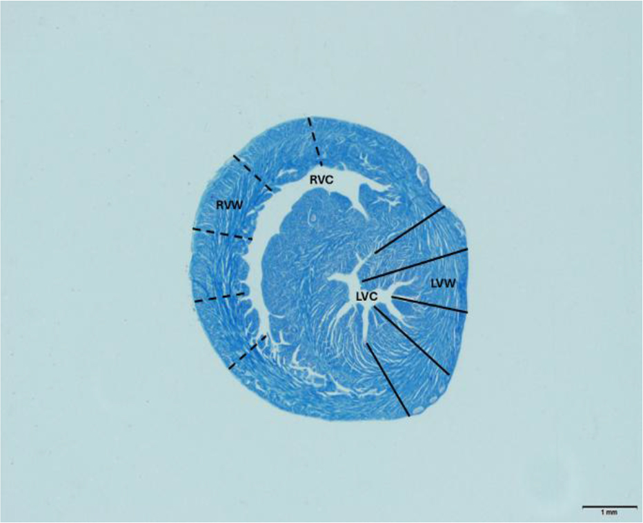
Fig. 1.
Morphometric measurement of the ventricular wall of heart. RVW – right ventricular wall; RVC – right ventricular cavity; LVW – left ventricular wall; LVC – left ventricular cavity. Crossmon’s trichrome staining. Scale bar – 1 mm
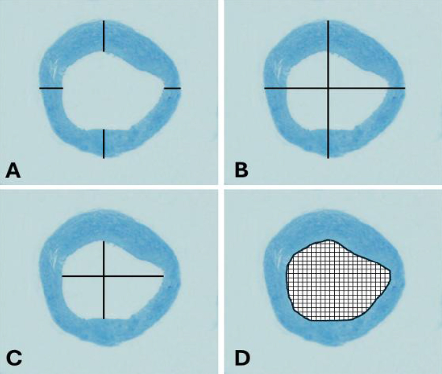
Fig. 2.
Morphometric measurement of sections of a chick embryo aorta. A – aortic wall thickness; B – aortic diameter; C – aortic lumen diameter; D – aortic lumen area
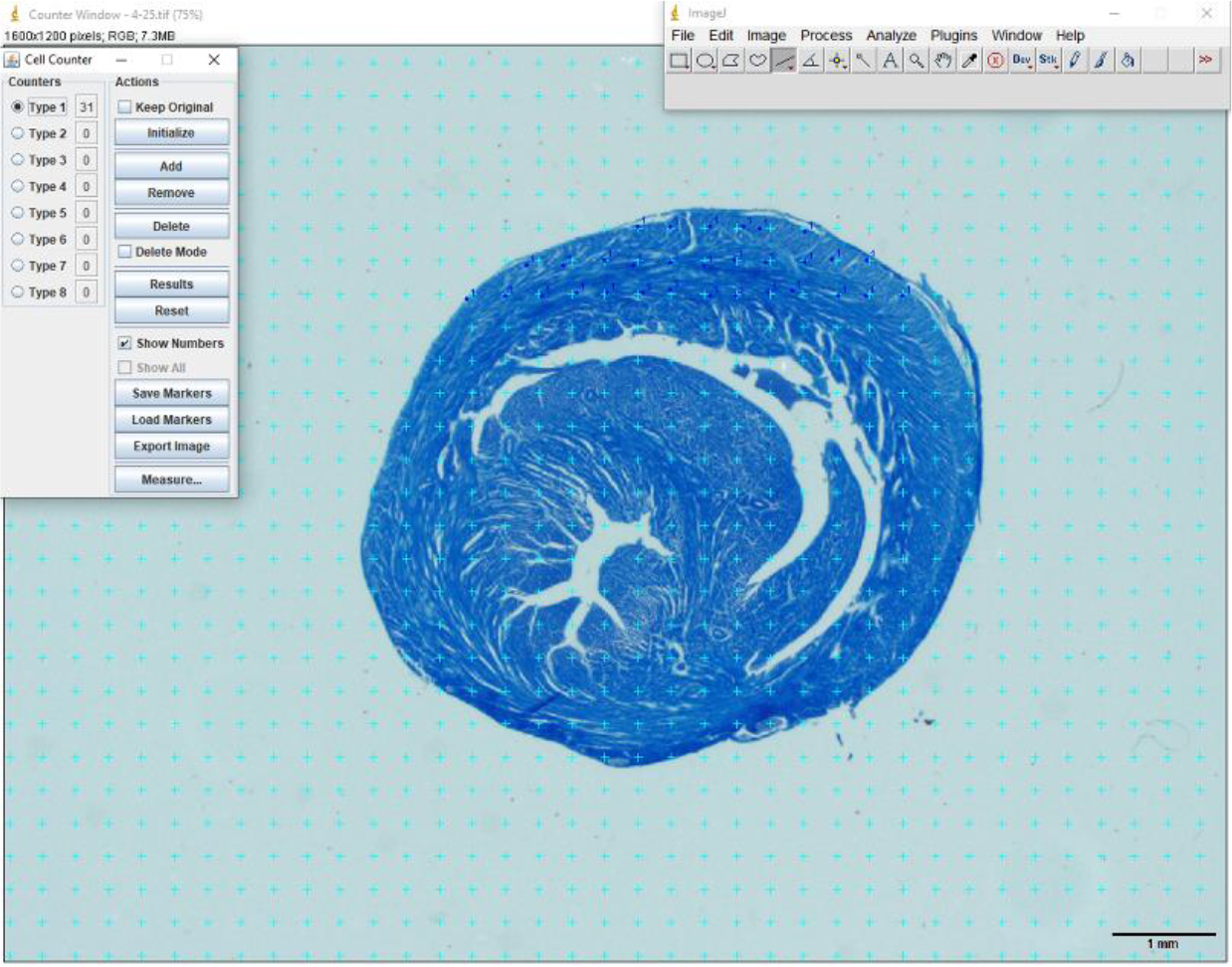
Fig. 3.
Application of the point counting grid for chick embryo heart sections
Table 3.
Morphometric measurements of chick embryo heart walls at different periods (Mean±SD)
| Parameter (thickness unless stated) | Incubation day 10 | Incubation day 13 | Incubation day 16 | Incubation day 21 |
|---|---|---|---|---|
| Left atrial wall (μm) | 8.87 ± 0.88a | 12.67 ± 2.08ab | 15.75 ± 5.25b | 16.40 ± 2.13b |
| Right atrial wall (μm) | 11.47 ± 3.87 | 13.50 ± 2.80 | 13.45 ± 2.35 | 15.53 ± 4.97 |
| Left/right atrium ratio | 0.89 ± 0.44 | 0.95 ± 0.084 | 1.14 ± 0.18 | 1.16 ± 0.43 |
| Left ventricular wall (μm) | 60.53 ± 17.08a | 98.56 ± 13.83b | 115.22 ± 21.58b | 156.28 ± 26.07c |
| Right ventricular wall (μm) | 52.73 ± 6.32a | 79.72 ± 12.68a | 89.06 ± 30.19a | 109.83 ± 30.41b |
| Left/right ventricle ratio | 1.15 ± 0.31 | 1.22 ± 0.39 | 1.47 ± 0.35 | 1.48 ± 0.37 |
| Interatrial septum (μm) | 6.27 ± 1.79a | 6.67 ± 1.25a | 16.78 ± 6.78b | 16.53 ± 5.09b |
| Interventricular septum (μm) | 69.07 ± 8.13a | 87.28 ± 20.13a | 118.83 ± 15.52b | 139.50 ± 15.74b |
Table 4.
Chick embryo heart muscle layer thickness in the ventricular wall at different periods (Mean±SD)
| Layer | Incubation day 10 | Incubation day 13 | Incubation day 16 | Incubation day 21 |
|---|---|---|---|---|
| Left epicardium (μm) | 3.90 ± 0.33 | 5.49 ± 0.57 | 7.55 ± 1.16 | 14.24 ± 0.68 |
| Right epicardium (μm) | 5.01 ± 0.63 | 8.08 ± 1.46 | 8.42 ± 1.28 | 26.73 ± 2.48 |
| Left myocardium (μm) | 36.29 ± 5.14 | 52.26 ± 2.82 | 71.31 ± 7.13 | 73.05 ± 2.51 |
| Right myocardium (μm) | 38.78 ± 3.36 | 57.49 ± 3.18 | 62.53 ± 3.07 | 66.76 ± 2.71 |
| Left endocardium (μm) | 3.39 ± 0.24 | 4.73 ± 0.45 | 7.06 ± 0.97 | 7.48 ± 0.73 |
| Right endocardium (μm) | 4.29 ± 0.46 | 6.63 ± 0.15 | 7.40 ± 0.66 | 10.13 ± 0.56 |
Table 5.
Morphometric measurements of the chick embryo aorta at different periods (Mean±SD)
| Parameter | Incubation day 10 | Incubation day 13 | Incubation day 16 | Incubation day 21 |
|---|---|---|---|---|
| Aortic wall thickness (μm) | 15.13 ± 1.90a | 20.28 ± 2.27b | 29.26 ± 4.22c | 30.35 ± 3.12c |
| Aortic diameter (μm) | 52.29 ± 5.34a | 71.22 ± 4.31b | 107.84 ± 10.83c | 110.64 ± 11.71c |
| Aortic lumen area (μm2) | 342.65 ± 14.12a | 729.82 ± 26.53b | 1864.95 ± 88.15c | 1912.57 ± 75.34c |
| Aortic lumen diameter (μm) | 21.23 ± 1.37a | 31.54 ± 3.06a | 77.88 ± 11.87b | 70.35 ± 8.47b |
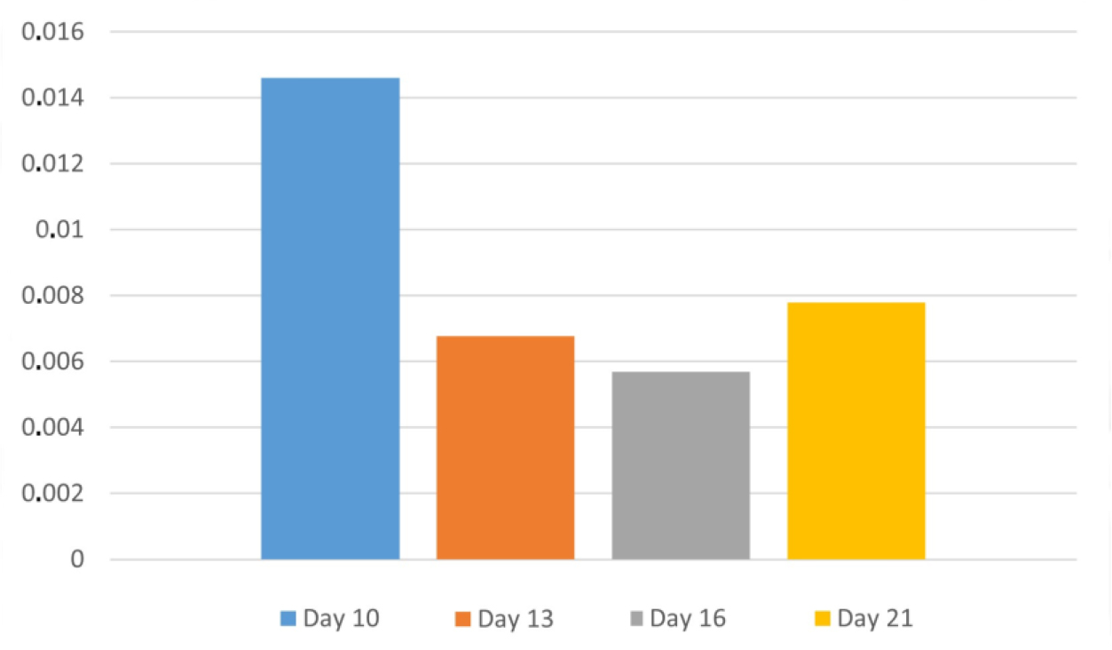
Fig. 4.
Coefficients of error in chick embryonic heart volumes
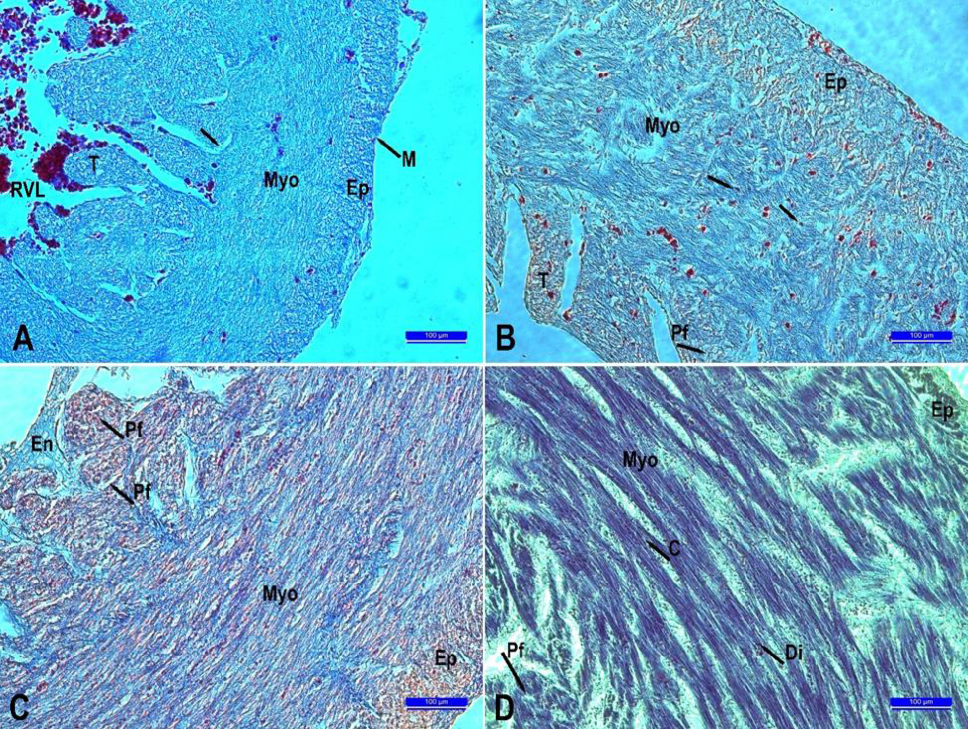
Fig. 5.
Histological development of the heart according to different periods. A. 10th day. B. 13th day. C.16th day. D. 21st day. C – Collateral; Di – Intercalary disc; En – Endocardium; Ep – Epicardium; M – Mesothelium; Myo – Myocardium; Pf – Purkinje fibers; T – Trabecula; RVL – right ventricular lumen; → – cardiomyocyte nucleus. Crossmon’s trichrome staining. Scale bars = 100 μm