
Table 1.
Air sac tissue lesion grading after experimental infection of goslings with Mycoplasma gallisepticum
| Grade | Pathological damage |
|---|---|
| 0 | Air sacs appear thin and translucent |
| 1 | Air sacs show slight thickening with sparse grey or a few yellow pinpoint exudates |
| 2 | Foamy fluid is visible within the air sacs; partial thickening and the presence of grey or yellow exudates are observed |
| 3 | Numerous yellow-white caseous exudates are visible, with considerable thickening in most air sacs |
| 4 | Nearly the entire air sac is filled with yellow-white caseous exudates and is severely thickened |
Table 2.
Clinical outcomes of experimental infection of goslings with a Mycoplasma gallisepticum strain previously isolated from geese (Group A, n = 10) or with a strain previously isolated from chickens (Group B, n = 10). Uninfected controls made up Group C (n = 10)
| Group | Number of morbidities | Number of deaths | qPCR positivity rate (%) |
|---|---|---|---|
| A | 9 | 2 | 100 |
| B | 8 | 0 | 80 |
| C | 0 | 0 | 0 |
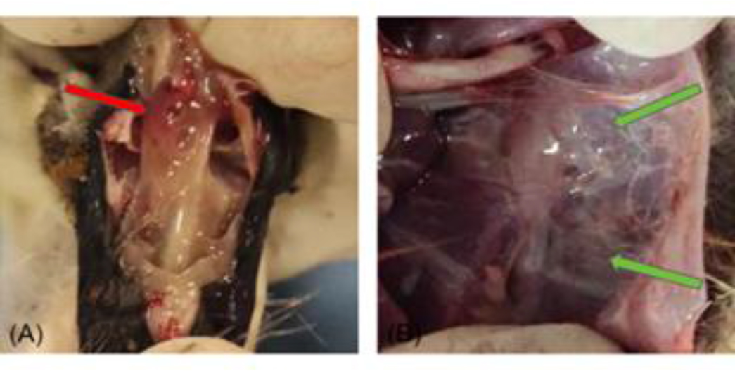
Fig. 1.
Dissection of gosling experimentally infected with Mycoplasma gallisepticum. (A)Numerous inflammatory and minor bloody secretions in the nasal cavity; (B) Yellow-white, cheese-like exudate in the air sacs and markedly thickening and grey discolouration of air sac walls
Table 3.
Distribution of pathological grades of air-sac tissue after experimental infection of goslings with a Mycoplasma gallisepticum strain previously isolated from geese (Group A, n = 10) or with a strain previously isolated from chickens (Group B, n = 10). Uninfected controls made up Group C (n = 10)
| Group | Gosling | Mean | |||||||||
|---|---|---|---|---|---|---|---|---|---|---|---|
| 1 | 2 | 3 | 4 | 5 | 6 | 7 | 8 | 9 | 10 | ||
| A | 1 | 3 | 2 | 3 | 2 | 2 | 1 | 3 | 3 | 3 | 2.3 |
| B | 2 | 1 | 3 | 3 | 2 | 2 | 1 | 1 | 2 | 1 | 1.9 |
| C | 0 | 0 | 0 | 0 | 0 | 0 | 0 | 0 | 0 | 0 | 0 |
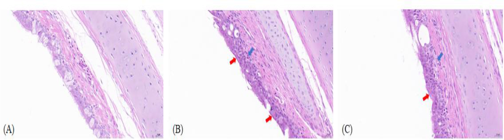
Fig. 2.
Histopathological lesions in tracheal tissue of uninfected goslings and goslings experimentally infected with Mycoplasma gallisepticum (MG). (A) Control goose tracheal tissue (400×); (B) – Goose-derived (MG)-GD01/22-infected goose tissue section (400×); (C) MG R(low)-infected goose tissue section (400×). Ciliated epithelial cell loss (red arrow); submucosal congestion and hemorrhage (blue arrow)
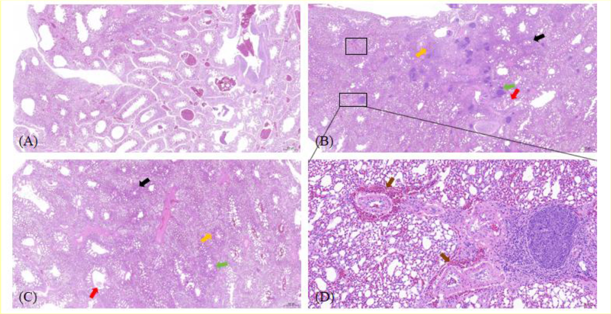
Fig. 3.
Histopathological lesions in lung tissue of uninfected goslings and goslings experimentally infected with Mycoplasma gallisepticum (MG). (A) Control goose lung tissue (20×); (B) MG-GD01/22-infected goose lung tissue (20×); (C) MG R(low)-infected goose lung tissue (20×); (D) MG-GD01/22-infected goose lung tissue (enlarged view of the lower outlined box in (B) 200×). Lymphocyte infiltration foci (green arrow); thickened capillary walls (black arrow); capillary congestion (yellow arrow); pink exudate in secondary and accessory bronchial lumens (red arrow); vascular hemorrhage in the pulmonary interstitium (brown arrow)
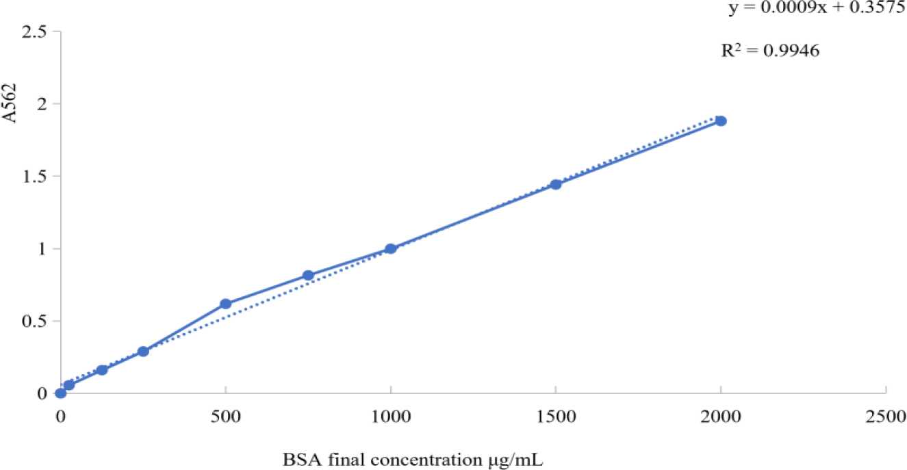
Fig. 4.
Bicinchoninic acid assay standard curve results after experimental infection of goslings with a Mycoplasma gallisepticum strain previously isolated from geese. A562 – absorbance at 562nm; R2 – coefficient of determination; BSA – bovine serum albumin
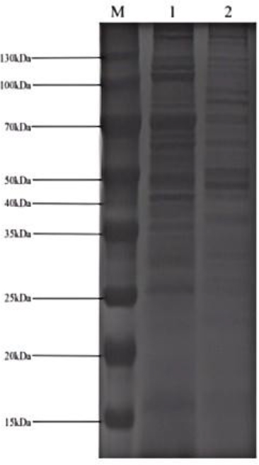
Fig. 5.
SDS-PAGE image after experimental infection of goslings with a Mycoplasma gallisepticum (MG) strain previously isolated from geese (MG-GD01/22) or with a strain previously isolated from chickens (MG R(low)). M – marker; 1 – MG-GD01/22; 2 – MG R(low)
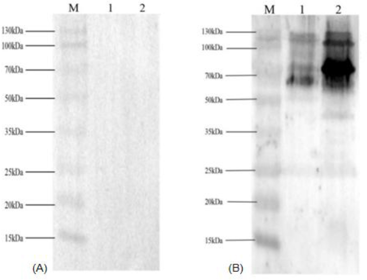
Fig. 6.
Western blot images of sera from uninfected goslings and goslings experimentally infected with Mycoplasma gallisepticum (MG). (A) Goose negative serum; M – marker; 1 – chicken-derived MG R(low); 2 – goose-derived MG-GD01/22. (B) Goose positive serum; M – marker; 1 – MG R(low); 2 – MG-GD01/22