
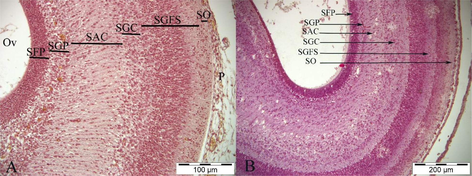
Fig 1.
Histological structure of a Japanese quail tectum opticum on embryonic days 7 (E7) and 9 (E9). A – E7; B – E9. SO – stratum opticum; SGFS – stratum griseum et fibrosum superficiale; SGC – stratum griseum centrale; SAC – stratum album centrale; SGP – stratum griseum periventriculare; SFP – stratum fibrosum periventriculare; Ov – optic ventricle; P – pia mater. Visualised with Crossmonn’s modified triple staining
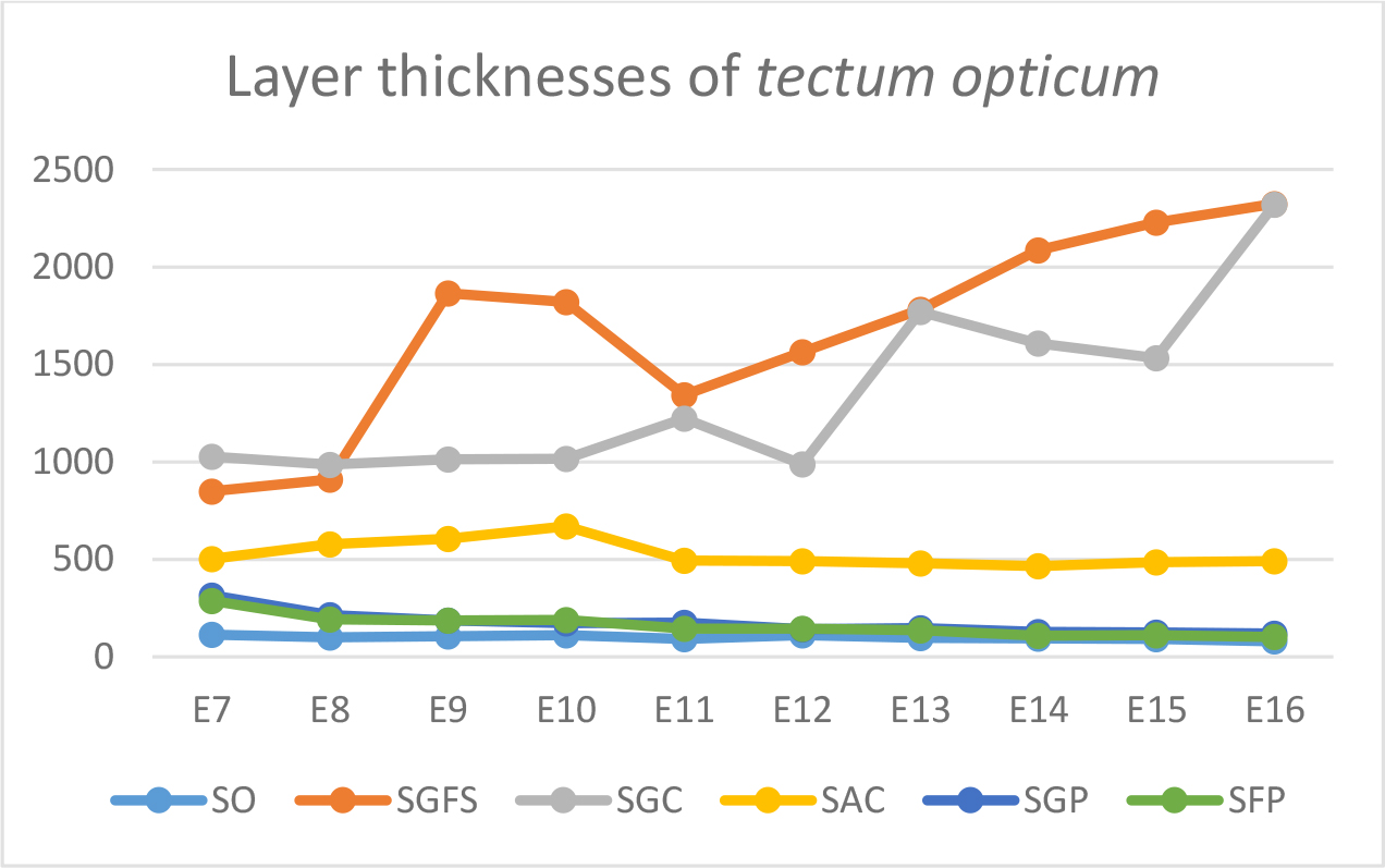
Fig 2.
Graphic representation of Japanese quail tectum opticum layer thicknesses. E7–16 – embryonic days 7–16; SO – stratum opticum; SGFS – stratum griseum et fibrosum superficiale; SGC – stratum griseum centrale; SAC – stratum album centrale; SGP – stratum griseum periventriculare; SFP – stratum fibrosum periventriculare
Table 1.
Tectum opticum layer thickness measurements in Japanese quail between embryonic days 7 (E7) and 16 (E16)
| SO | SGFS | SGC | SAC | SGP | SFP | |
|---|---|---|---|---|---|---|
| E7 | 113.7±12.7a | 849.2±41.8a | 1028.1±112.4a | 503.5±38.5a | 316.4±60.9a | 287.6±37.2a |
| E8 | 99.2±13.1b | 910.7±52.5a | 986.22±103.8a | 578.63±51.8b | 215.92±28.1b | 192.11±41.2b |
| E9 | 106.6±19.8a | 1865.4±65.5b | 1012.74±102.5a | 606.2±40.5b | 185.9±19.8b | 187.2±19.6b |
| E10 | 111.15±9.3a | 1820±189.8b | 1015.5±87.8a | 669.61±85.2c | 171.63±19.3b | 189.44±22.7b |
| E11 | 92.62±9.1b | 1343.9±55.9c | 1222.31±103.3a | 494.82±50.9a | 174.88±26.8c | 143.51±21.1c |
| E12 | 112.1±9.4a | 1562.2±39.9c | 987.3±93.7a | 490.32±39.2a | 140.7±16.9d | 145.3±33.2d |
| E13 | 97.4±10.2b | 1780.2±65.6b | 1770.9±88.6b | 478.9±68.2a | 148.3±21.1c | 137.23±26.7c |
| E14 | 93.4±14.4b | 2085.3±105.8d | 1607.82±186.1b | 465.4±48.2a | 128.44±23.5d | 109.88±11.9d |
| E15 | 91.7±11.9b | 2226.8±102.8d | 1532.8±161.4b | 486.4±52.3a | 125.63±15.5d | 111.17±16.8d |
| E16 | 79.4±13.2d | 2324.11±111.5d | 2318.73±120.1c | 490.71±3.1a | 119.75±14.9d | 101.64±15.4d |
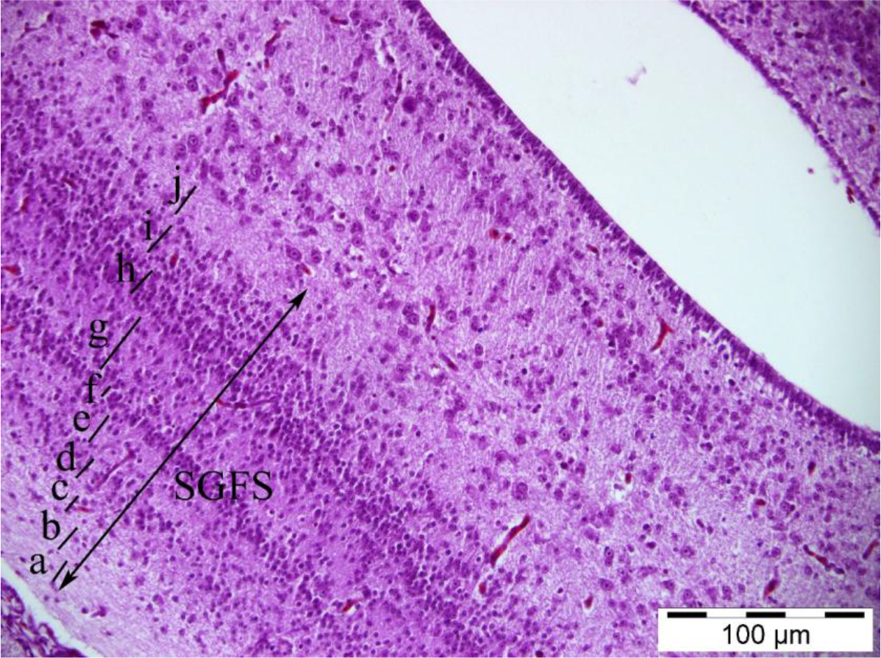
Fig 3.
Histological structure of the stratum griseum et fibrosum superficiale (SGFS) layer of a Japanese quail tectum opticum on embryonic day 12. a–j – sublayers from deep to superficial. Visualised with Crossmonn’s modified triple staining
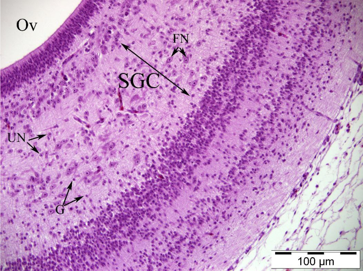
Fig 4.
Neurons in the stratum griseum centrale (SGC) layer of a Japanese quail tectum opticum on embryonic day 11. Ov – optic ventricle; UN – unipolar neuron; G – glial cells; FN – fusiform neuron. Crossmonn’s modified triple staining
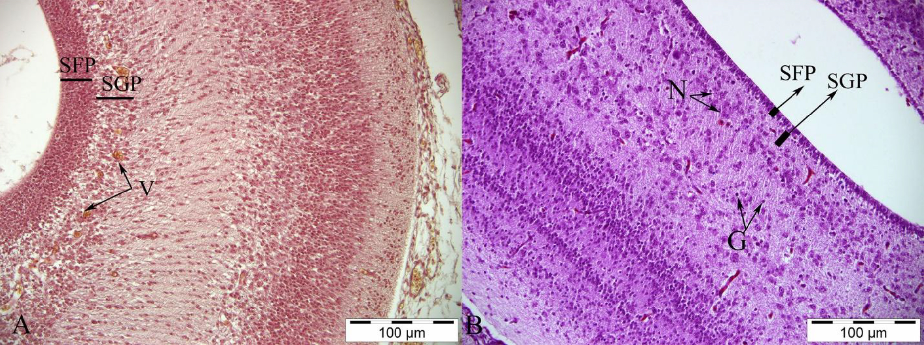
Fig 5.
Histological structure of the stratum griseum periventriculare (SGP) and stratum fibrosum periventriculare (SFP) layers of a Japanese quail tectum opticum on embryonic days 7 (E7) and 12 (E12). A – E7; B – E12. N – spherical neuron; V – blood vessel; G – glial cells. Visualised with Crossmonn’s modified triple staining
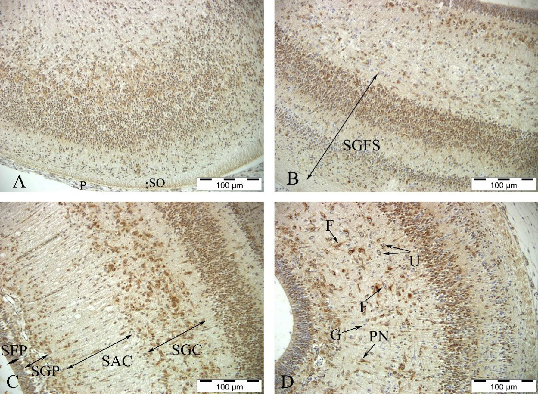
Fig 6.
Heat-shock protein 70 immunostaining in a Japanese quail tectum opticum. A – on embryonic (E) day 7; B – E9; C – E10; D – E11. SO – stratum opticum; SGFS – stratum griseum et fibrosum superfiiciale; SFP – stratum fibrosum perivenriculare; SGP – stratum griseum periventriculare; SAC – stratum album centrale; SGC – stratum griseum centrale; P – pia mater; F – fusiform neuron; G – glial cells; PN – pyramidal neuron; U – unipolar neuron
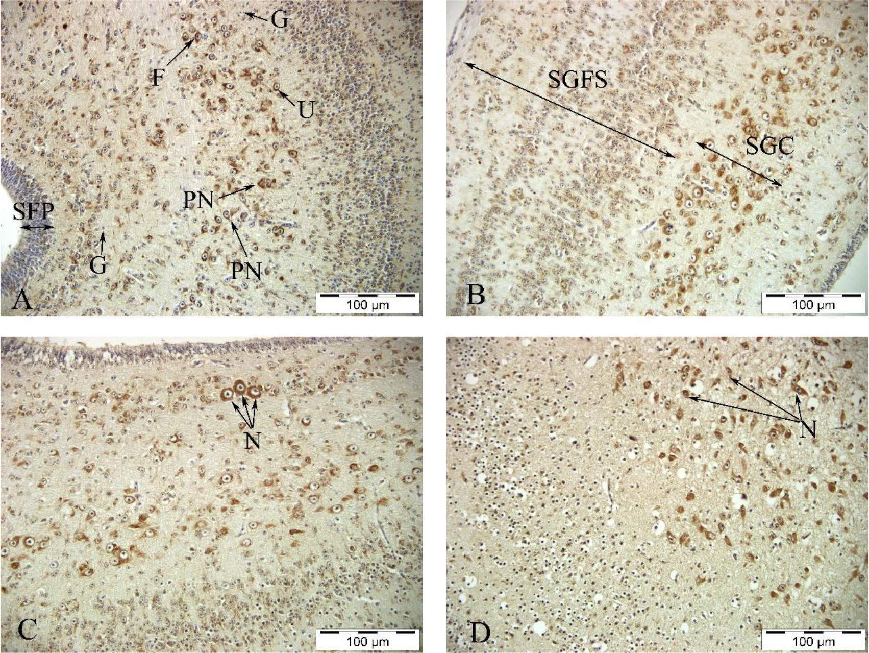
Fig 7.
Heat-shock protein 70 immunostaining in a Japanese quail tectum opticum. A – on embryonic (E) day 13; B – E14; C – E15; D – E16. SFP – stratum fibrosum perivenriculare; SGFS – stratum griseum et fibrosum superficiale; SGC – stratum griseum centrale; G – glial cells; F – fusiform neuron; PN – pyramidal neuron; U – unipolar neuron; N – neurons showing high immunoreactivity