
Table 1.
Primers used in the analysis of gene expression
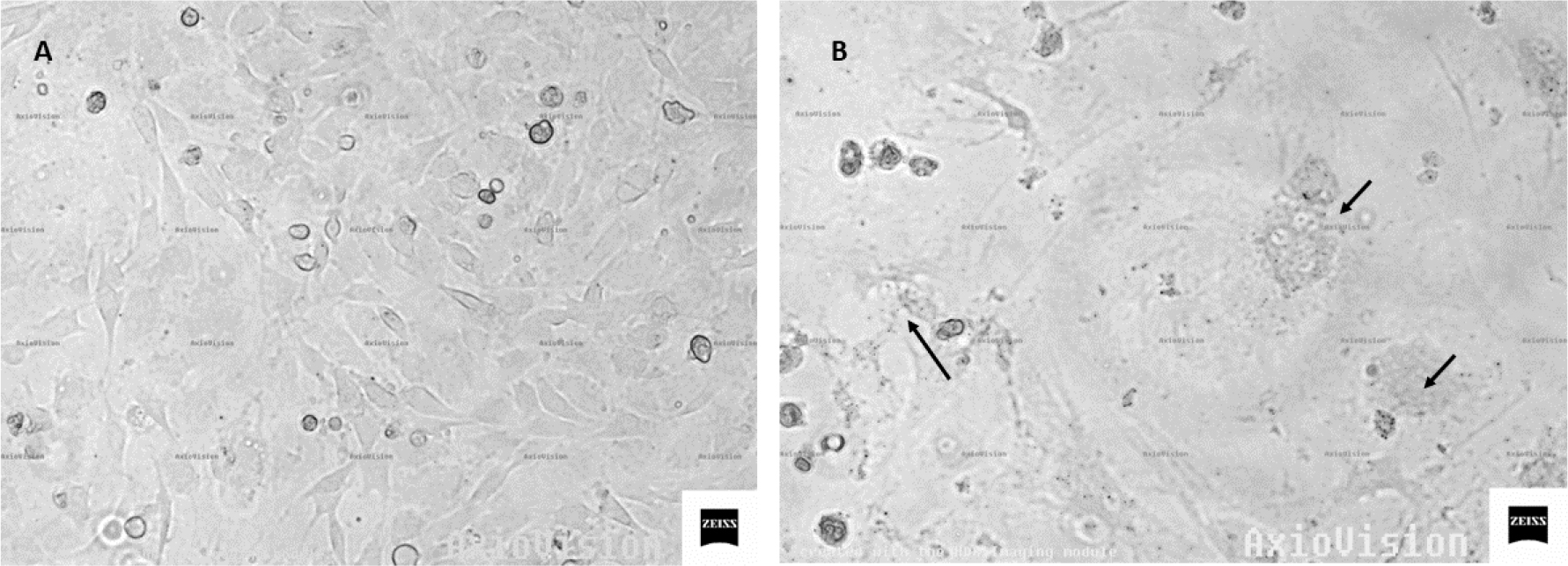
Fig. 1.
Photomicrographs of skin cells: A – uninfected cells; B – cultured cells at 7 days after SRLV infection (arrows indicate syncytia)
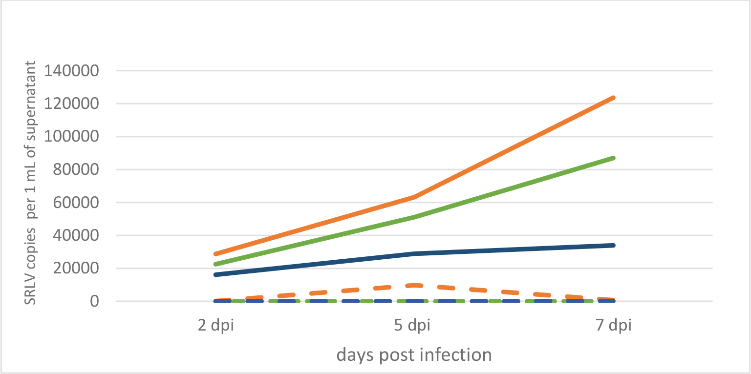
Fig. 2.
Kinetics of SRLV replication in experimentally infected skin cells from a high-proviral-load goat (solid orange line), a low-proviral-load goat (solid blue line) and an uninfected goat (solid green line) and these kinetics in cells left without experimental infection from the same goats (dashed lines)
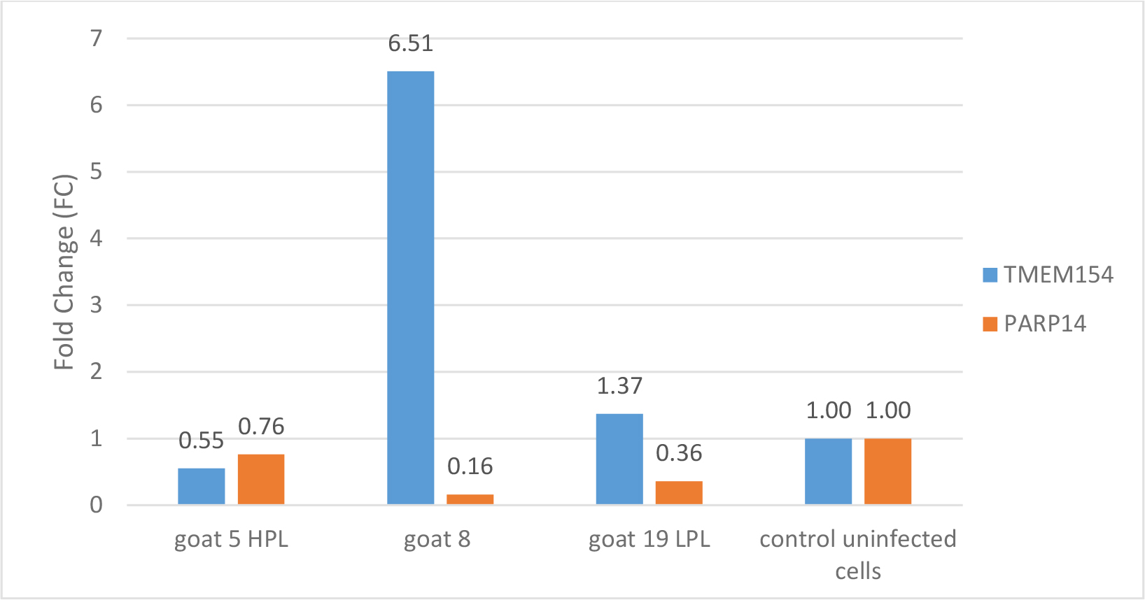
Fig. 3.
Relative gene expression in skin cells of goats Nos 5, 8 and 19 experimentally infected with SRLV and in these cells from all three animals left without experimental infection as controls. HPL – high proviral load; LPL – low proviral load; TMEM154 – transmembrane protein 154; PARP14 – poly ADP-ribose polymerase
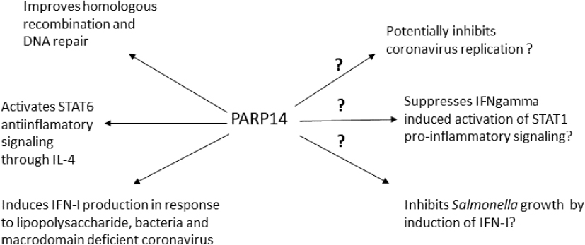
Fig. 4.
Known and probable functions of the PARP14 protein. STAT – signal transducer and activator of transcription; IFN – interferon; IL-4 – interleukin 4