
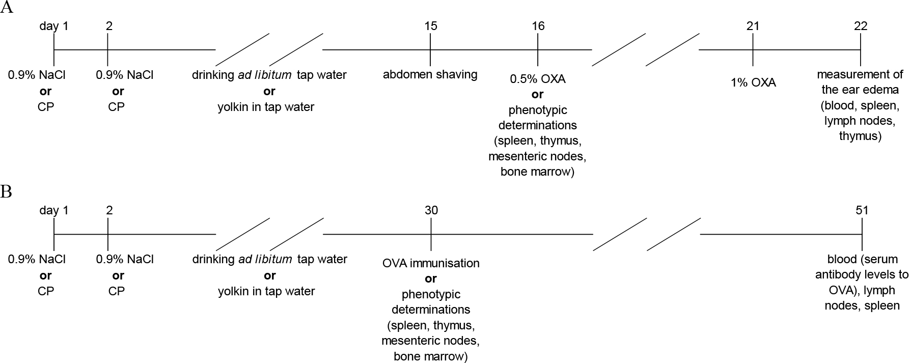
Fig. 1.
Experimental design. Immunological determinations in (A) the model of contact sensitivity to oxazolone (OXA) and (B) the model of humoral response to ovalbumin (OVA). CP – cyclophosphamide
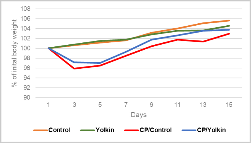
Fig. 2.
Changes in cyclophosphamide (CP)-immunocompromised mouse body weight during the experimental period. The results are presented as % of initial body weight (mean values) of mice from individual experimental groups from both research models: contact sensitivity to oxazolone and humoral response to ovalbumin. Control – control mice given only 0.9% NaCl; Yolkin – mice given 0.9% NaCl and yolkin; CP/Control – mice given only CP; CP/Yolkin – mice given CP and yolkin
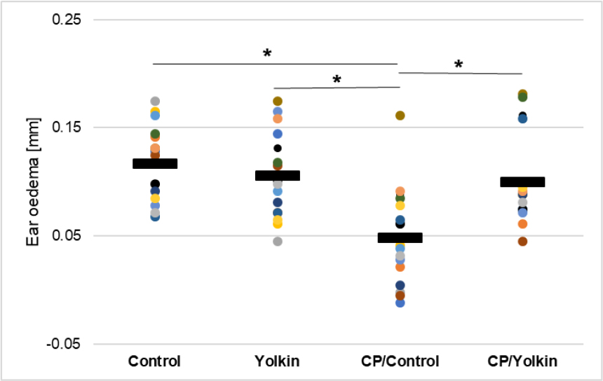
Fig. 3.
Antigen specific increase in ear oedema in cyclophosphamide (CP)-immunocompromised mice expressing contact sensitivity to oxazolone. The antigen-specific increase of ear thickness (the difference obtained by subtracting the baseline measurement from the measurement taken from sensitised mice) is presented as the mean value of ear thickness measured in eight mice (16 determinations) in mm ± standard error of the mean. * – statistically significant (P-value < 0.05); Control – control mice given only 0.9% NaCl; Yolkin – mice given 0.9% NaCl and yolkin; CP/Control – mice given only CP; CP/Yolkin – mice given CP and yolkin
Table 1.
Effects of treatment with yolkin for 13 d on total number of immune cells and the numbers of lymphocyte subpopulations in the lymphoid organs of cyclophosphamide (CP)-immunocompromised mice
| Total number of cells in the examined organs | ||||||
|---|---|---|---|---|---|---|
| Thymus | Spleen | Lymph nodes | Bone marrow | |||
| Control | 64.3 ± 4.9 ab | 82.1 ± 10.7 a | 36.1 ± 2.6 a | 21.5 ± 1.3 a | ||
| Yolkin | 68.8 ± 5.4 a | 87.4 ± 12.9 a | 39.1 ± 2.5 a | 24.3 ± 2.4 a | ||
| CP/Control | 48.7 ± 2.9 bc | 61.6 ± 7.7 a | 14.8 ± 1.7 b | 14.7 ± 1.4 b | ||
| CP/Yolkin | 60.0 ± 5.8 abc | 75.8 ± 11.6 a | 16.1 ± 0.8b | 14.9 ± 0.9 b | ||
| Thymocytes | ||||||
| CD8+ | CD4+CD8+ | CD4–CD8– | CD4+ | |||
| Control | 2.9 ± 0.6 a | 41.6 ± 3.4 a | 2.3 ± 0.2 ab | 17.5 ± 1.7 a | ||
| Yolkin | 2.9 ± 0.6 a | 46.1 ± 4.2 a | 2.7 ± 0.2 a | 17.2 ± 1.6 a | ||
| CP/Control | 1.5 ± 0.2 a | 30.1 ± 4.5 a | 1.4 ± 0.2 c | 10.0 ± 2.1 a | ||
| CP/Yolkin | 1.8 ±0.4 a | 36.7 ± 5.8 a | 1.5 ± 0.3 bc | 15.4 ± 2.7 a | ||
| Splenocytes | ||||||
| CD8+ | CD4+ | CD3+ | CD19+ | CD14+CD169+ | CD25+Foxp3+ | |
| Control | 7.7 ± 1.7 a | 15.25 ± 1.7 a | 28.4 ± 3.9 a | 44.6 ± 5.7 a | 1.9 ± 0.2 a | 6.9 ± 1.0 a |
| Yolkin | 6.7 ± 1.8 a | 15.41 ± 3.0 a | 29.8 ± 5.7 a | 48.0 ± 6.0 a | 2.01 ± 0.2 a | 7.7 ± 0.9a |
| CP/Control | 3.3 ± 0.6 a | 7.8 ± 1.3 a | 19.1 ± 1.4 a | 33.7 ± 5.2 a | 2.7 ± 0.5 a | 6.1 ± 0.7 a |
| CP/Yolkin | 5.9 ± 1.1 a | 13.6 ± 2.4 a | 25.5 ± 3.2 a | 38.7 ± 6.8 a | 3.1 ± 0.6 a | 6.2 ± 1.2 a |
| Mesenteric lymph node cells | ||||||
| CD8+ | CD4+ | CD3+ | CD19+ | |||
| Control | 5.4 ± 0.6 a | 21.1 ± 1.8 a | 25.3 ± 2.1 a | 10.2 ± 0.6 a | ||
| Yolkin | 4.6 ± 0.1a | 21.1 ± 1.6 a | 25. 9 ± 1.8 a | 9.2 ± 1.5a | ||
| CP/Control | 2.1 ± 0.2 b | 9.9 ± 1.1 b | 12.0 ± 1.4 b | 2.3 ± 0.3b | ||
| CP/Yolkin | 2.6 ± 0.1 b | 10.4 ± 0.5 b | 12.7 ± 0.5 b | 3.1 ± 0.3b | ||
| Bone marrow cells | ||||||
| CD19+ | ||||||
| Control | 12.8 ± 0.9 ab | |||||
| Yolkin | 14.5 ± 1.4 a | |||||
| CP/Control | 9.7 ± 0.8 bc | |||||
| CP/Yolkin | 9.0 ± 0.9 bc | |||||
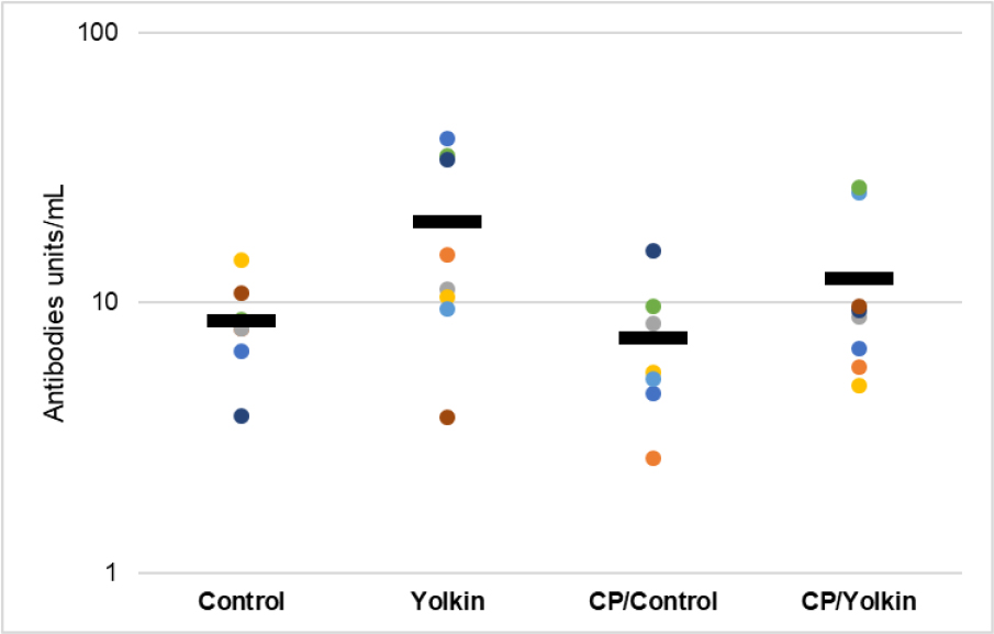
Fig. 4.
The levels of antibodies to ovalbumin in cyclophosphamide (CP)-immunocompromised mice followed by 26-d oral treatment with yolkin. The data are presented as mean and individual values of the antibody levels on a logarithmic scale. Control – control mice given only 0.9% NaCl; Yolkin – mice given 0.9% NaCl and yolkin; CP/Control – mice given only CP; CP/Yolkin – mice given CP and yolkin
Table 2.
Effects of treatment with yolkin for 26 d on the total number of cells and the numbers of lymphocyte subpopulations in the lymphoid organs of cyclophosphamide (CP)-immunocompromised mice
| Total number of cells in the examined organs | ||||||
|---|---|---|---|---|---|---|
| Thymus | Spleen | Lymph nodes | Bone marrow | |||
| Control | 97.3 ± 3.5 a | 141.2 ± 6.7 a | 34.05 ± 1.4 ab | 20.3 ± 1.0 ab | ||
| Yolkin | 94.3 ± 2.8 ab | 126.9 ± 3.7 a | 37.5 ± 2.9 a | 21.1 ± 1.2 a | ||
| CP/Control | 82.1 ± 3.1 bc | 82.0 ± 6.2 b | 24.7 ± 1.9 c | 11.6 ± 0.8 c | ||
| CP/Yolkin | 93.0 ± 4.5 abc | 92.9 ± 5.2 b | 30.6 ± 2.6 abc | 20.2 ± 2.0 ab | ||
| Thymocytes | ||||||
| CD8+ | CD4+CD8+ | CD4–CD8– | CD4+ | |||
| Control | 3.9 ± 0.4 a | 59.2 ± 4.2 a | 7.3 ± 1.4 a | 25.9 ± 1.8 a | ||
| Yolkin | 4.1 ± 0.4 a | 54.9 ± 2.7 ab | 6.0 ± 0.5 a | 28.6 ± 3.3 a | ||
| CP/Control | 4.1 ± 0.5 a | 43.0 ± 2.3 c | 5.1 ± 0.5 a | 30.1 ± 1.5 a | ||
| CP/Yolkin | 4.9 ± 0.7 a | 50.4 ± 1.8 abc | 5.1 ± 0.5 a | 32.6 ± 2.2 a | ||
| Splenocytes | ||||||
| CD8+ | CD4+ | CD3+ | CD19+ | CD14+CD169+ | CD25+Foxp3+ | |
| Control | 12.6 ± 0.6 a | 24.5 ± 1.0 a | 48.6 ± 2.8 a | 76.8 ± 3.3 a | 3.4 ± 0.4 a | 11.4 ± 0.8 a |
| Yolkin | 8.1 ± 0.4 b | 20.5 ± 1.1 ab | 38.7 ± 1.2 b | 75.3 ± 3.4 a | 4.5 ± 0.8 a | 10.4 ± 0.5 a |
| CP/Control | 4.4 ± 0.8 c | 14.2 ± 1.7 c | 25.4 ± 3.3 c | 44.4 ± 2.4 b | 3.1 ± 0.4 a | 6.6 ± 0.5 b |
| CP/Yolkin | 7.3 ± 0.9 b | 15.4 ± 1.1 bc | 31.1 ± 2.2 bc | 52.0 ± 3.4 b | 4.0 ± 0.8 a | 7.2 ± 0.7 b |
| Mesenteric lymph node cells | ||||||
| CD8+ | CD4+ | CD3+ | CD19+ | |||
| Control | 4.2 ± 0.4 a | 19.6 ± 0.7 a | 22.5 ± 1.6 a | 9.9 ± 1.3 a | ||
| Yolkin | 4.2 ± 0.3 a | 19.7 ± 1.3 a | 23.0 ± 1.2 a | 13.2 ± 2.1 a | ||
| CP/Control | 3.3 ± 0.4 a | 17.4 ± 1.5 a | 20.6 ±.1.8 a | 3.2 ± 0.4 b | ||
| CP/Yolkin | 4.0 ± 0.4 a | 21.6 ± 1.7 a | 24.9 ± 2.1 a | 4.0 ± 0.7 b | ||
| Bone marrow cells | ||||||
| CD19+ | ||||||
| Control | 13.8 ± 0.6 a | |||||
| Yolkin | 12.6 ± 0.8 ab | |||||
| CP/Control | 7.7 ± 0.5 c | |||||
| CP/Yolkin | 13.1 ± 0.6 ab | |||||