
Fig. 1.
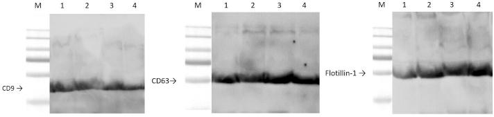
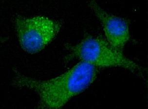
Fig. 2.
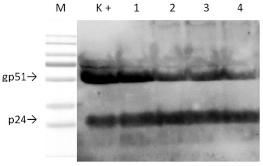
Fig. 3.
Fig. 5.
Fig. 6.
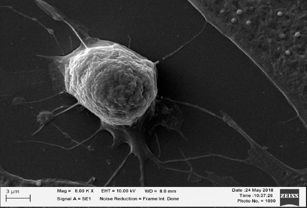
Fig. 7.
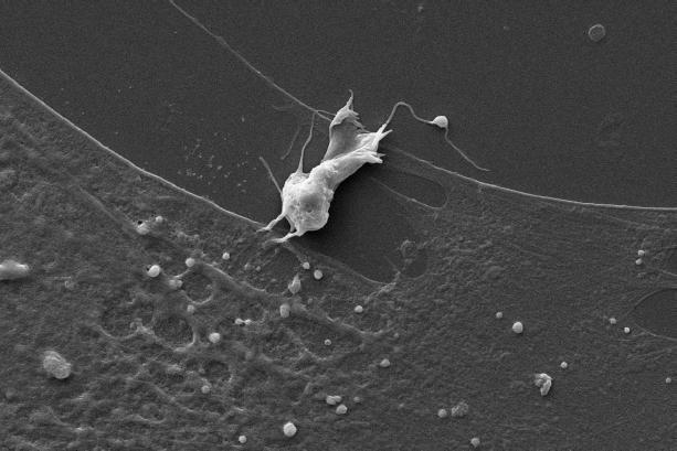
Fig. 9.
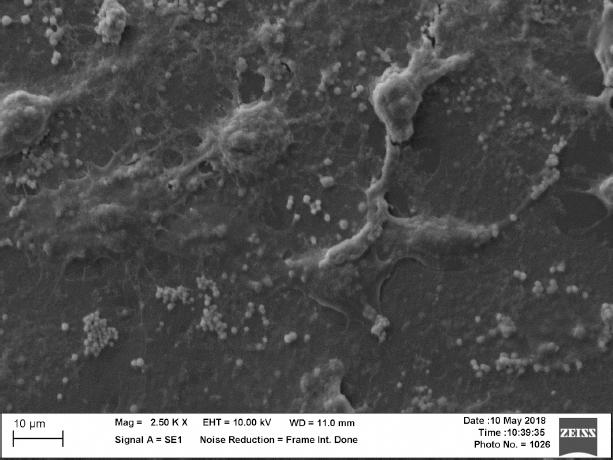
Fig. 8.
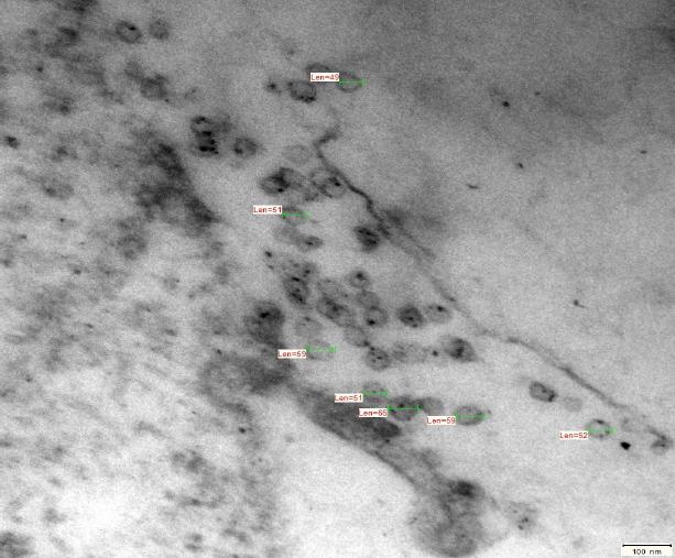
Fig. 10.
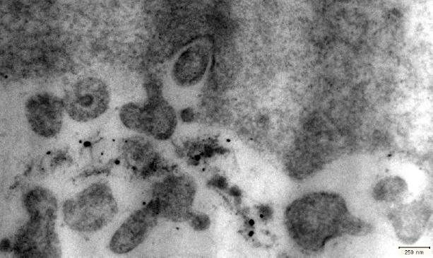
Fig. 4.
The viral and exosomal markers detected by Western blot in bovine leukaemia virus (BLV)-infected cows
| Origin of exosomes | BLV infection | BLV markers | Cellular markers | ||||
|---|---|---|---|---|---|---|---|
| gp51 | p24 | CD63 | CD9 | flotillin-1 | |||
| Positive control – supernatant of FLK-BLV culture | + | + | + | + | + | + | |
| Supernatant of cultured in vitro BLV-infected bovine DCs | 1 | + | + | + | + | + | + |
| 2 | + | + | + | + | + | + | |
| 3 | + | + | + | + | + | + | |
| 4 | + | + | + | + | + | + | |
| 5 | + | + | + | + | + | + | |
| Negative control – plasma BLV− | − | − | − | + | + | + | |
| Positive control – lysate of FLK–BLV cells | + | + | + | + | + | + | |
| Bovine BLV+ sera (1–7) and BLV− sera (8–11) | 1 | + | + | + | + | + | + |
| 2 | + | + | + | + | + | + | |
| 3 | + | + | + | + | + | + | |
| 4 | + | + | + | + | + | + | |
| 5 | + | + | + | + | + | + | |
| 6 | + | + | + | + | + | + | |
| 7 | + | + | + | + | + | + | |
| 8 | − | − | − | + | + | + | |
| 9 | − | − | − | + | + | + | |
| 10 | − | − | − | + | + | + | |
| 11 | − | − | − | + | + | + | |