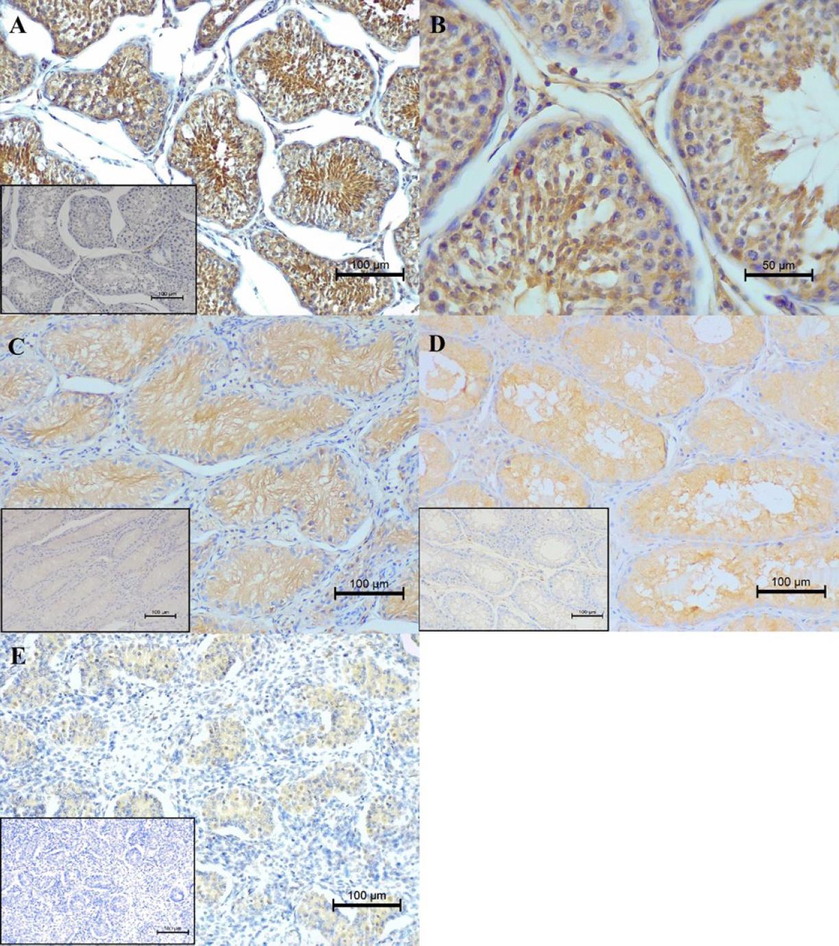
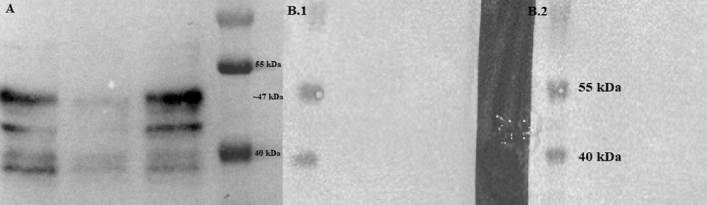
Have a personal or library account? Click to login
Study on the expression of testin in the testes of dogs Cover

Figures & Tables

Fig. 1.

Immunohistochemical examination of the expression of testin in the testes of dogs at various life stages. (A) Intense cytoplasmic testin expression in the Sertoli cells (SCs) and spermatids of an adult dog aged between 2 and 6 and weaker expression in the interstitium and interstitial endocrine Leydig cells. The lower left corner shows the negative control. (B) The same cells at higher magnification. (C) Very weak cytoplasmic reaction exclusively in the SCs of an older dog aged 9 or 10. The left corner shows the negative control. (D) Weaker expression of testin compared to normal adult canine testes in the SCs and interstitial endocrine Leydig cells of a puppy aged 2 months. The lower left corner shows the negative control. (E) Positive but weak cytoplasmic expression in the SCs of a miscarried canine foetus. The lower left corner shows the negative control. (A) and (C–E) scale bar 100 μm; (B) scale bar 50 μm

Fig. 2.

Representative Western blot results of TES protein expression in testes. (A) A marked dark band between 55 kDa and 40 kDa corresponds to a testin protein of ~47 kDa. (B.1) Negative control without primary antibody. (B.2) Negative control with rabbit serum

Immunohistochemical results

 Germ cells/SpermatocytesSertoli cellsLeydig cells
% of positive cellsReaction intensity% of positive cellsReaction intensity% of positive cellsReaction intensity
Normal mature testes434332
Atrophic areas of aged canine testes004100
Normal immature testes004131
Testes of miscarried foetuses002100

Immunoreactive scores on the semiquantitative scale established by Remmele and Stegner (28)

Percentage of positive cellsPointsImmunohistochemical reactionPoints
00Absent0
1–10%1Weak1
11–50%2Moderate2
5–80%3Intense3
>80%4  
Language: English
Page range: 627 - 633
Submitted on: May 17, 2023
Accepted on: Oct 3, 2023
Published on: Dec 19, 2023
Published by: National Veterinary Research Institute in Pulawy
In partnership with: Paradigm Publishing Services
Publication frequency: 4 issues per year

© 2023 Rafał Ciaputa, Marcin Nowak, Stanisław Dzimira, Eleonora Brambilla, Małgorzata Kandefer-Gola, Alicja Tomaszek, Aneta Popiel-Kopaczyk, Piotr Dzięgiel, Valeria Grieco, published by National Veterinary Research Institute in Pulawy
This work is licensed under the Creative Commons Attribution-NonCommercial-NoDerivatives 3.0 License.