
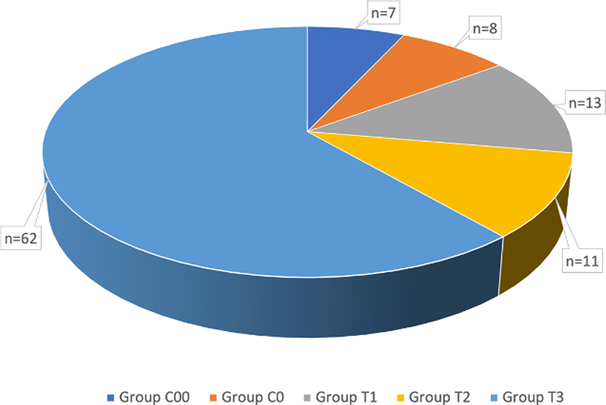
Fig. 1.
The division of animals into groups according to the length of pregnancy
Group C00 – control group of non-pregnant heifers; Group C0 – control group of non-pregnant cows; Group T1 – experimental heifers at day of pregnancy (dop) ≤ 28; Group T2 – experimental heifers at dop ≥ 29–dop < 45; Group T3 – experimental cows at dop ≥ 29–dop ≤ 50
Table 1.
Average values of haematological parameters of Polish black-and-white Holstein-Friesian cattle, comparing those of non-pregnant heifers to those of pregnant cows grouped by length of pregnancy
| Parameter | Group 1 | Group 2 | Group 3 |
|---|---|---|---|
| WBC (103/μL) | 8.34 ± 2.03** | 7.30 ± 2.38** | 9.66 ± 4.46 |
| GRA (103/μL) | 4.26 ± 1.04 | 3.72 ± 1.31** | 5.34 ± 3.86 |
| GRA% (%) | 51.76 ± 8.86** | 51.70 ± 11.04** | 50.86 ± 12.66 |
| LYM (103/μL) | 3.43 ± 1.38** | 3.07 ± 1.50* | 3.58 ± 1.08 |
| LYM% (%) | 40.63 ± 8.85** | 40.91 ± 11.02** | 41.34 ± 12.40 |
| MON (103/μL) | 0.64 ± 0.23 | 0.52 ± 0.15 | 0.75 ± 0.33 |
| MON% (%) | 7.61 ± 1.86 | 7.40 ± 1.42 | 7.80 ± 0.55 |
| RDW (103/μL) | 19.42 ± 1.06 | 19.88 ± 1.72 | 20.36 ± 1.75 |
| RBC (106/μL) | 7.63 ± 1.10** | 7.44 ± 1.46** | 8.40 ± 0.58 |
| HGB (g/dL) | 11.06 ± 1.58** | 11.40 ± 2.32* | 11.59 ± 0.54 |
| MCH (pg) | 14.51 ± 0.84 | 15.36 ± 1.12 | 13.85 ± 1.13 |
| MCHC (g/dL) | 34.34 ± 0.74* | 34.60 ± 1.05 | 35.45 ± 0.93 |
| MCV (μm3) | 42.28 ± 2.58 * | 44.44 ± 3.40 | 39.13 ± 3.56 |
| MPV (μm3) | 6.54 ± 0.81 | 6.50 ± 0.73 | 6.83 ± 0.66 |
| PCT (%) | 0.18 ± 0.11 | 0.19 ± 0.13 | 0.17 ± 0.08 |
| PDW (%) | 31.96 ± 13.08** | 29.53 ± 13.71** | 42.23 ± 11.63 |
| PLT (103/μL) | 290.17 ± 191.98** | 300.00 ± 219.29** | 248.88 ± 117.54 |
| HCT (%) | 32.15 ± 4.11** | 31.95 ± 7.52** | 32.73± 1.92 |
1 Group 1 – pregnant cows up to the 45th day of pregnancy (dop); Group 2 – pregnant cows between the 45th and 50th dop; Group 3 – non-pregnant heifers (group C00); - mean; SD - standard deviation;
1 WBC – white blood cell count; GRA – granulocytes; GRA% – percentage of granulocytes; LYM – lymphocytes; LYM% –percentage of lymphocytes; MON – monocytes; MON% – percentage of monocytes; RDW – red blood cell distribution width; RBC – red blood cell count; HGB – the haemoglobin concentration; MCH – mean corpuscular haemoglobin; MCHC – mean corpuscular haemoglobin concentration; MCV – mean corpuscular volume; MPV –mean platelet volume; PCT – thrombocrit; PDW – platelet distribution width; PLT – platelets; HCT – haematocrit
Table 2.
Comparison of average haematological parameters of Polish black-and-white Holsteina-Friesian cattle setting heifers against pregnant cows
| Parameter | H | C |
|---|---|---|
| WBC (103/μL) | 9.11a ± 2.85 | 6.74b ± 1.76 |
| GRA (103/μL) | 4.35 ± 2.17** | 4.02 ± 1.30 |
| GRA% (%) | 46.49A ± 9.57** | 58.80B ± 5.66* |
| LYM (103/μL) | 4.09A ± 1.27** | 2.20B ± 0.49 |
| LYM% (%) | 46.10A ± 9.60** | 33.45B ± 4.82 |
| MON (103/μL) | 0.67 ± 0.23 | 0.51 ± 0.21 |
| MON% (%) | 7.41 ± 1.14 | 7.75 ± 1.98 |
| RDW (103/μL) | 19.96 ± 1.30 | 19.44 ± 1.66 |
| RBC (106/μL) | 8.46a ± 0.94** | 6.56b ± 0.49* |
| HGB (g/dL) | 12.21A ± 1.67* | 9.95B ± 0.98* |
| MCH (pg) | 14.44 ± 1.01 | 15.19 ± 1.16 |
| MCHC (g/dL) | 35.08 ± 0.91* | 33.95 ± 0.57 |
| MCV (μm3) | 41.20A ± 3.09 | 44.72B ± 3.06* |
| MPV (μm3) | 6.76 ± 0.77 | 6.27 ± 0.62 |
| PCT (%) | 0.14 ± 0.07 | 0.26 ± 0.13 |
| PDW (%) | 40.59A ± 11.38* | 20.25B ± 4.03* |
| PLT (103/μL) | 206.52A ± 110.26** | 402.41B ± 224.99** |
| HCT (%) | 34.18A ± 6.05** | 29.31B ± 2.62 |
1A, B, C – values in columns with different uppercase letters differ highly significantly (P ≤ 0.01);
1 WBC – white blood cell count; GRA – granulocytes; GRA% – percentage of granulocytes; LYM – lymphocytes; LYM% – percentage of lymphocytes; MON – monocytes; MON% – percentage of monocytes; RDW – red blood cell distribution width; RBC – red blood cell count; HGB – haemoglobin concentration; MCH – mean corpuscular haemoglobin; MCHC – mean corpuscular haemoglobin concentration; MCV – mean corpuscular volume; MPV -mean platelet volume; PCT – thrombocrit; PDW – platelet distribution width; PLT – platelets; HCT – haematocrit
Table 3.
Reference values for haematological parameters of dairy cattle
| Reference value | |
|---|---|
| WBC | 4.0–12.0 (103/μL) |
| GRA | 0.6–4.0 (103/μL) |
| GRA% | 15.0–50.0 (%) |
| LYM | 2.5–7.5 (103/μL) |
| LYM% | 45.0–75.0 (%) |
| MON | 0.1–0.8 (103/μL) |
| MON% | 2.0–7.0 (%) |
| RDW | 16.0–24.0 (103/μL) |
| RBC | 5.0–10.0 (106/μL) |
| HGB | 8.0–15.0 (g/dL) |
| MCH | 11.0–18.0 (pg) |
| MCHC | 30.0–37.0 (g/dL) |
| MCV | 37.0–60.0 (μm3) |
| MPV | 5.5–7.5 (μm3) |
| PCT | 0.250–0.650 (%) |
| PDW | 10.0–20.0 (%) |
| PLT | 150–730 (103/μL) |
| HCT | 24.0–46.0 (%) |
1 WBC – white blood cell count; GRA – granulocytes; GRA% – percentage of granulocytes; LYM – lymphocytes; LYM% – percentage of lymphocytes; MON – monocytes; MON% – percentage of monocytes; RDW – red blood cell distribution width; RBC – red blood cell count; HGB – haemoglobin concentration; MCH – mean corpuscular haemoglobin; MCHC – mean corpuscular haemoglobin concentration; MCV – mean corpuscular volume; MPV – mean platelet volume; PCT – thrombocrit; PDW – platelet distribution width; PLT – platelets; HCT – haematocrit
Table 4.
Pearson’s correlation values for haematological parameters of cows’ blood
| WBC | RBC | HGB | HCT | PLT | LYM | MON | GRA | LYM% | |
|---|---|---|---|---|---|---|---|---|---|
| WBC | 1 | 0.64145** | 0.58184** | 0.55768** | 0.12123ns | 0.71413** | 0.83838** | 0.87833ns | 0.03639ns |
| RBC | 1 | 0.94734** | 0.90814** | 0.12114ns | 0.68151** | 0.44398** | 0.35080** | 0.44345** | |
| HGB | 1 | 0.95365** | 0.03649ns | 0.66429** | 0.32751* | 0.26443* | 0.48052** | ||
| HCT | 1 | 0.06239ns | 0.58727** | 0.28291* | 0.28316* | 0.37719* | |||
| PLT | 1 | 0.04063ns | 0.09542ns | 0.13425ns | 0.15948ns | ||||
| LYM | 1 | 0.60442** | 0.29617* | 0.64392** | |||||
| MON | 1 | 0.69739** | 0.00841ns | ||||||
| GRA | 1 | 0.48615** | |||||||
| LYM% | 1 | ||||||||
| MON% | GRA% | MCV | MCH | MCHC | RDW | MPV | PCT | PDW | |
| WBC | 0.23363* | 0.07747ns | - | - | - | 0.36385* | 0.02595ns | 0.12563ns | 0.41233** |
| RBC | 0.30012* | 0.37933* | 0.45821** | 0.20889* | 0.64494** | 0.25965* | 0.01210ns | 0.12181ns | 0.56941** |
| HGB | 0.41980** | 0.39406** | 0.19525* | 0.08335ns | 0.73003** | 0.25594* | 0.01719ns | 0.03862ns | 0.45478** |
| HCT | 0.44251** | 0.28904* | 0.17111ns | 0.05845ns | 0.59783** | 0.23689* | 0.07609ns | 0.06153ns | 0.36950* |
| PLT | 0.04885ns | 0.14702ns | 0.17235ns | 0.17466ns | 0.07183ns | 0.03623ns | 0.15619ns | 0.99610** | 0.30944* |
| LYM | 0.15027ns | 0.60204** | 0.27350* | 0.09556ns | 0.45777** | 0.28579* | 0.06002ns | 0.03566ns | 0.40430** |
| MON | 0.27067* | 0.04035ns | 0.38371* | 0.26092* | 0.28212* | 0.36855* | 0.09669ns | 0.09607ns | 0.31521* |
| GRA | 0.27286* | 0.52385** | 0.24411* | 0.12630ns | 0.30521* | 0.29278* | 0.01533ns | 0.13912ns | 0.21823* |
| LYM% | 0.03879ns | 0.98379** | 0.13328ns | 0.03809ns | 0.23664* | 0.07820ns | 0.11351ns | 0.15558ns | 0.30458* |
| MON% | GRA% | MCV | MCH | MCHC | RDW | MPV | PCT | PDW | |
| MON% | 1 | 0.21733* | 0.30243* | 0.43501** | 0.46124** | 0.06582ns | 0.10003ns | 0.03840ns | 0.07662ns |
| GRA% | 1 | 0.18446ns | 0.11527ns | 0.14839ns | 0.08820ns | 0.12886ns | 0.14511ns | 0.28382* | |
| MCV | 1 | 0.93347** | 0.02221ns | 0.23940* | 0.05386ns | 0.16434ns | 0.45209** | ||
| MCH | 1 | 0.37543* | 0.16699ns | 0.03119ns | 0.16664ns | 0.28139* | |||
| MCHC | 1 | 0.16761ns | 0.05376ns | 0.07050ns | 0.36066* | ||||
| RDW | 1 | 0.05475ns | 0.03287ns | 0.20184* | |||||
| MPV | 1 | 0.09250ns | 0.36006* | ||||||
| PCT | 1 | 0.28095* | |||||||
| PDW | 1 |
1 WBC – white blood cell count; RBC – red blood cell count; HGB – haemoglobin concentration; HCT – haematocrit; PLT – platelets; LYM – lymphocytes; MON – monocytes; GRA – granulocytes; LYM% – percentage of lymphocytes; MON% – percentage of monocytes; GRA% – percentage of granulocytes; MCV – mean corpuscular volume; MCH – mean corpuscular haemoglobin; MCHC – mean corpuscular haemoglobin concentration; RDW – red blood cell distribution width; MPV – mean platelet volume; PCT – thrombocrit; PDW – platelet distribution width
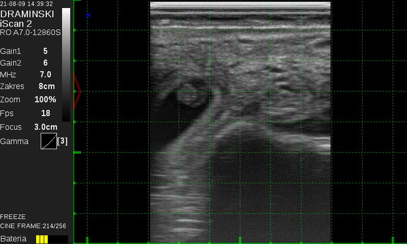
Fig. 2.
Transrectal ultrasound image showing late embryo development at around the 29th day of pregnancy
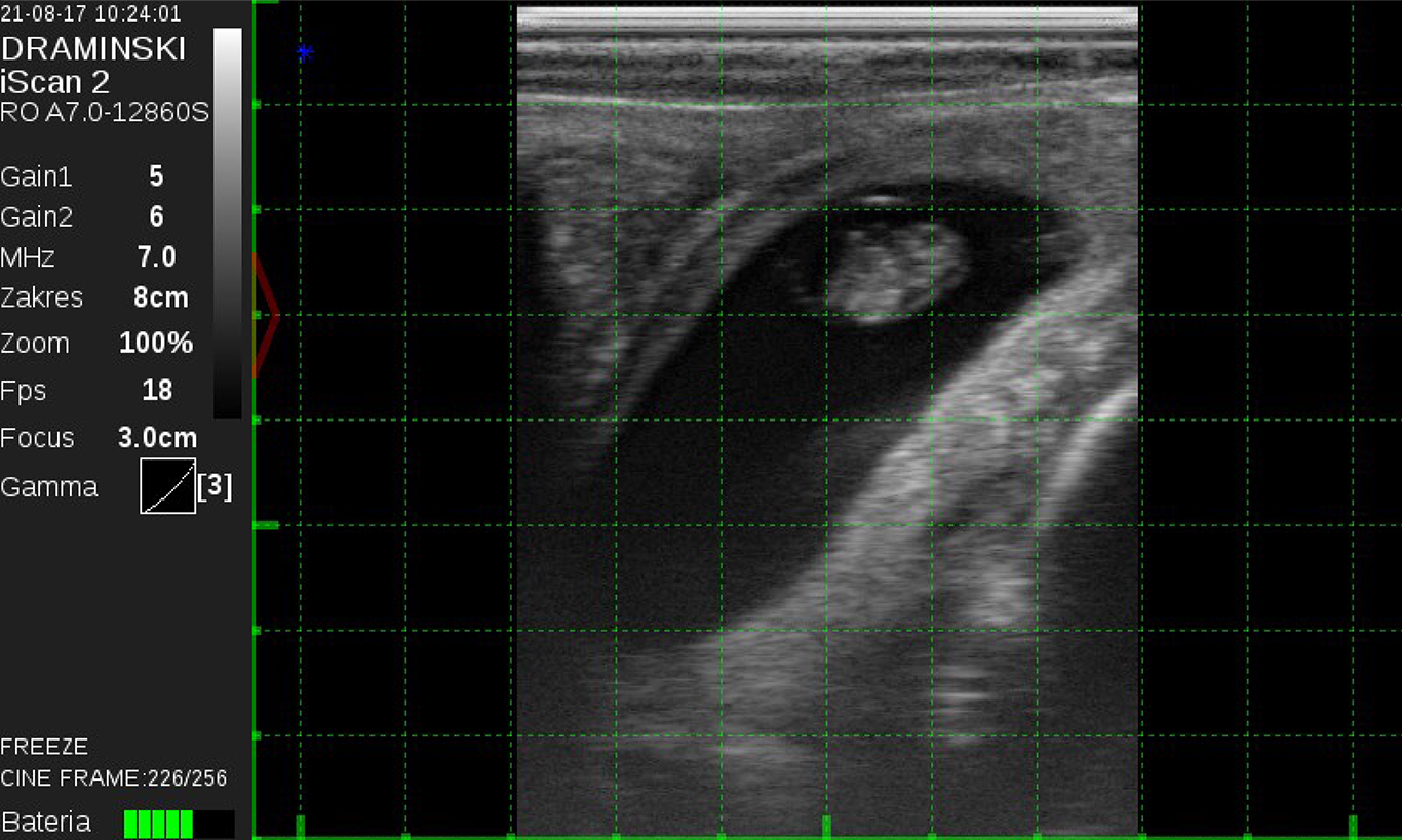
Fig. 3.
Transrectal ultrasound image showing late embryo development at around the 30th day of pregnancy
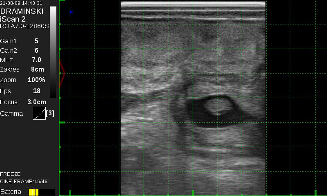
Fig. 4.
Transrectal ultrasound image showing late embryo development at around the 31st day of pregnancy
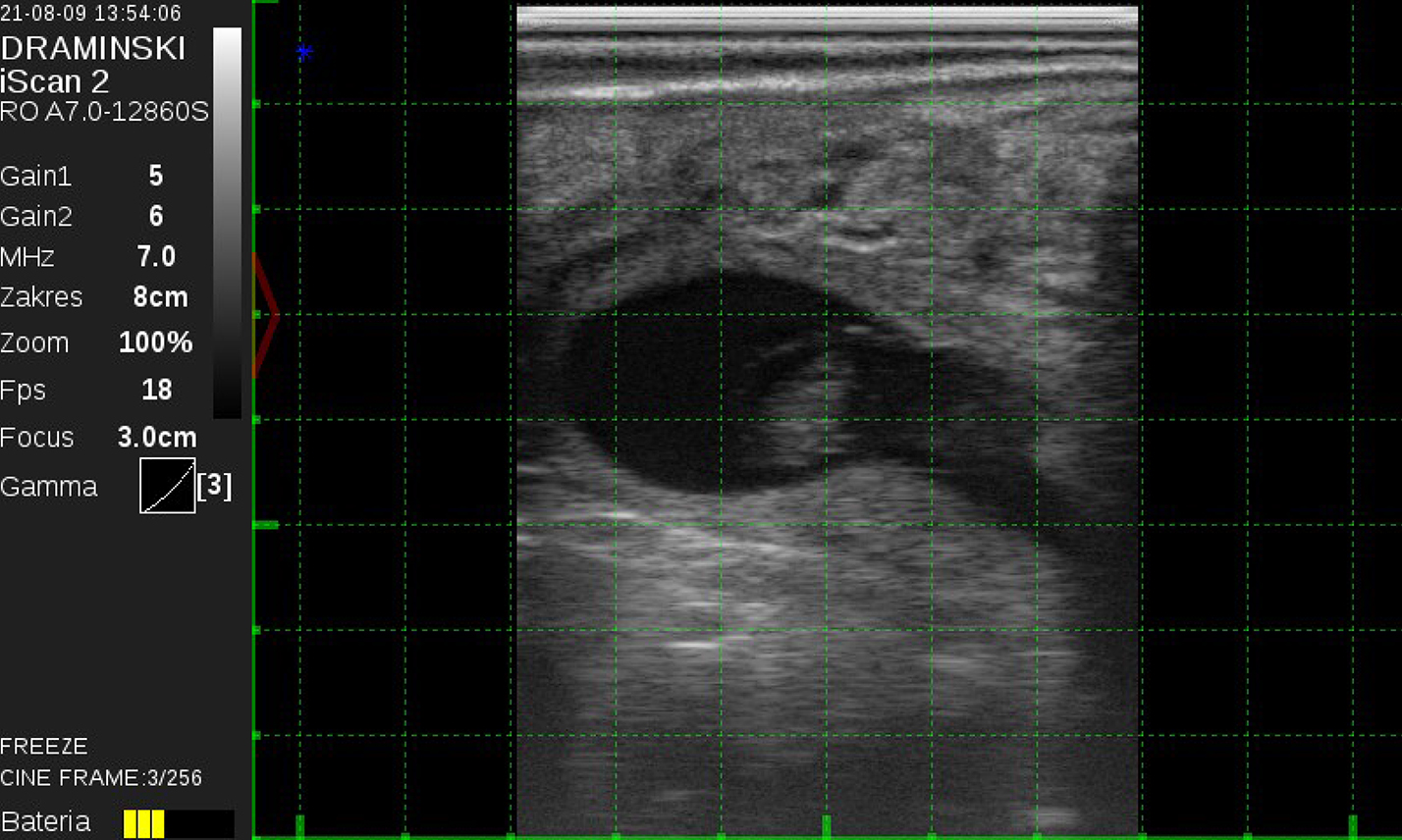
Fig. 5.
Transrectal ultrasound image showing late embryo development at around the 32nd day of pregnancy
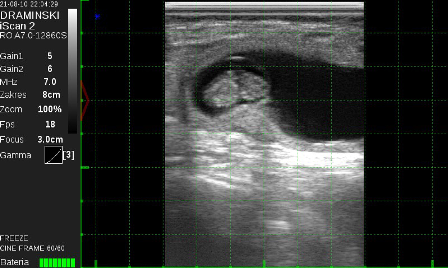
Fig. 6.
Transrectal ultrasound image showing late embryo development at around the 35th day of pregnancy
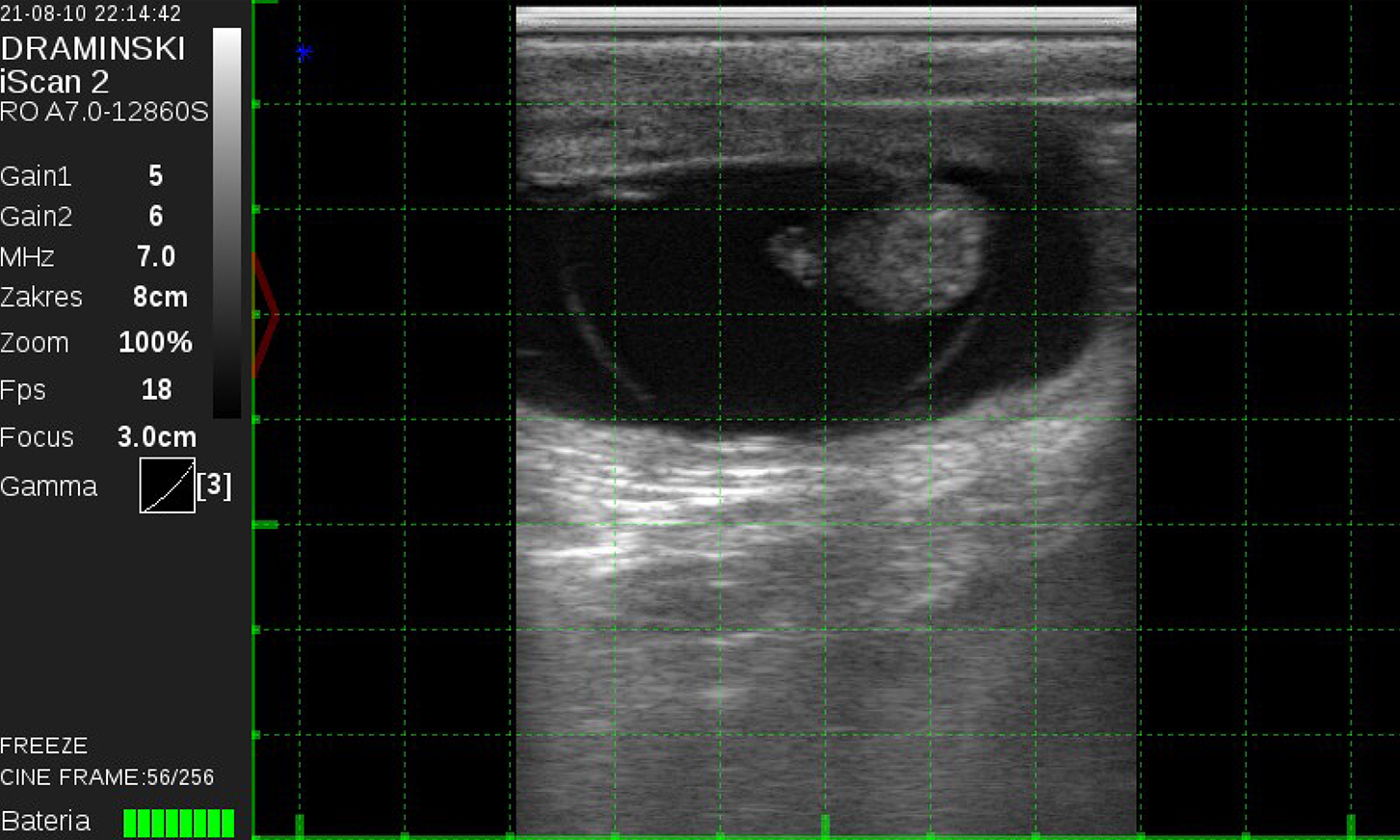
Fig. 7.
Transrectal ultrasound image showing late embryo development at around the 37th day of pregnancy
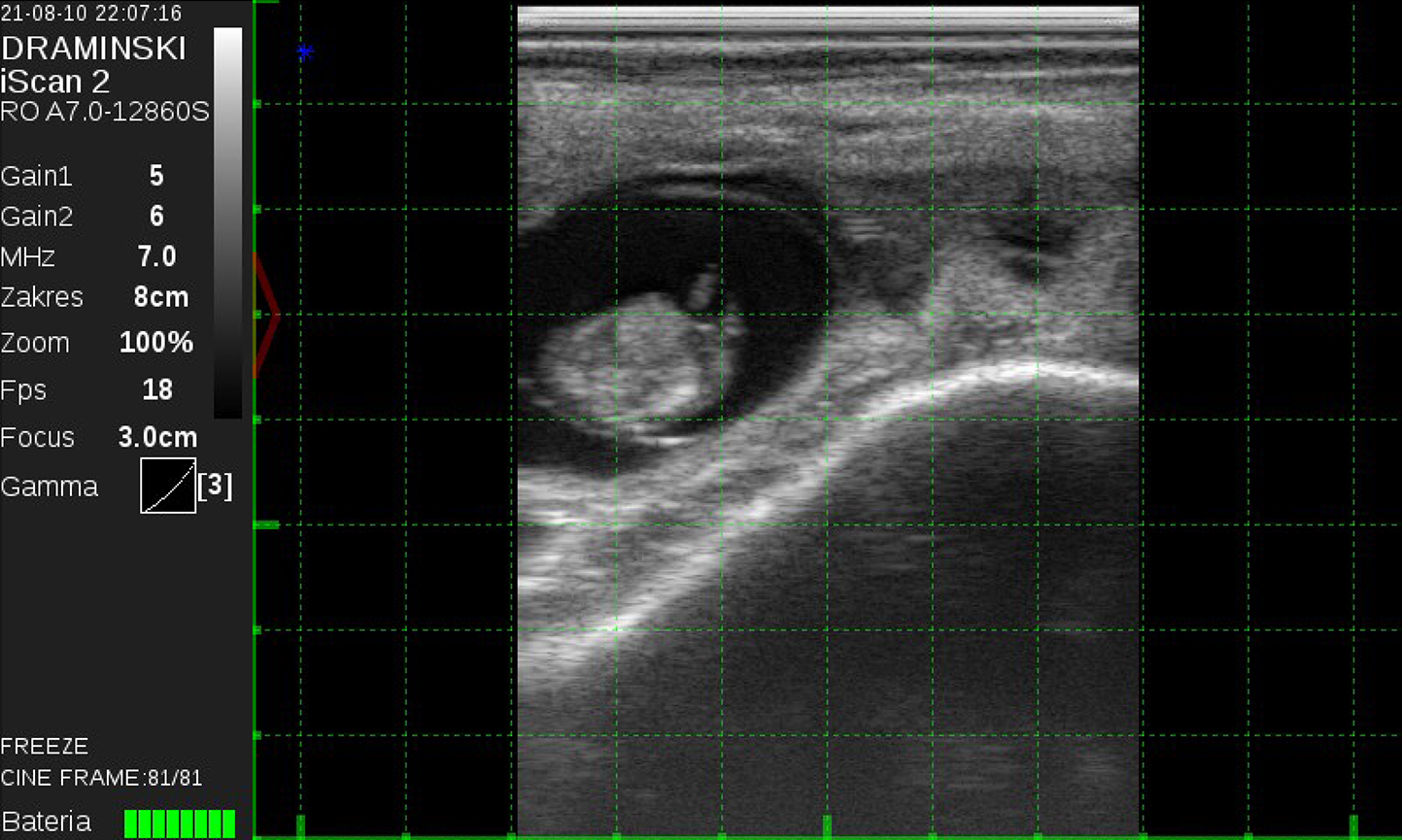
Fig. 8.
Transrectal ultrasound image showing late embryo development at around the 39th day of pregnancy