
Table 1
Bovine papillomatosis prevalence in Nuevo León, Chiapas, Veracruz and Tabasco, Mexico
| State: | Papillomatosis | n |
|---|---|---|
| Nuevo León | ||
| Farm 1 (Linares) | 3.77% | 450 |
| Farm 2 (Linares) | 18% | 250 |
| Farm 3 (Cadereyta) | 11.43% | 700 |
| Chiapas | ||
| Farm 4 (Palenque) | 12.83% | 1200 |
| Farm 5 (Libertad) | 11.6% | 500 |
| Farm 6 (Salto de Agua) | 32.8% | 125 |
| Farm 7 (Agua Fria) | 20% | 50 |
| Veracruz | ||
| Farm 8 (Temapache) | 10.67% | 150 |
| Farm 9 (Tihuatlán) | 10% | 160 |
| Tabasco | ||
| Farm 10 (Emiliano Zapata) | 9.2% | 1500 |
| Farm 11 (Balancan) | 16.25% | 80 |
| Farm 12 (Tenosique) | 23.44% | 320 |
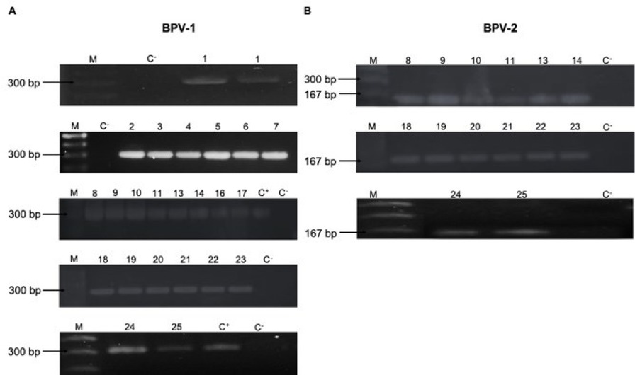
Fig. 1
Bovine papillomavirus BPV-1 and BPV-2 shown in representative samples which were positive for viral genetic material when electrophoresed in 1.5% agarose gel
M – 100 base pair molecular weight marker; C+ – BPV-1 positive sample from Nuevo León; C− – DNA-free reaction mix; A – BPV-1 presence in cutaneous/genital warts: lanes 1–11, 13–14 and 16–25; B – BPV-2 presence in cutaneous/genital warts: lanes 8–11, 13–14 and 18–25 (Nuevo León, lanes 1–5 and 24–25; Veracruz, 18–23; Chiapas, 6–7 and 14–17; and Tabasco, 8–13)
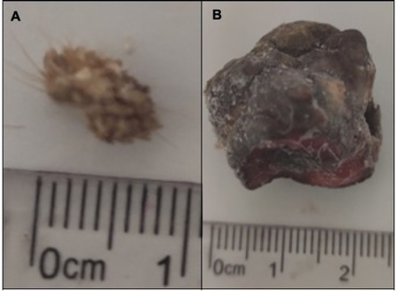
Fig. 2
Samples negative for bovine papillomavirus (BPV)-1/2/4 and therefore with genotype unknown A – sample from Emiliano Zapata, Tabasco; B – sample from Libertad, Chiapas. Samples had cauliflower morphology suggesting that they be considered BPV positive
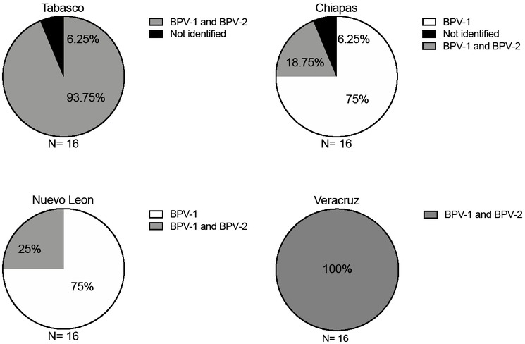
Fig. 3
Bovine papillomavirus (BPV)-1 and BPV-2 genotype distribution per state from representative samples. BPV-1 was found as a unique genotype or as a co-infection with BPV-2, but BPV-2 was found only as a co-infection
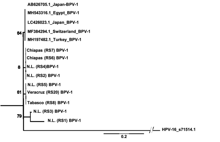
Fig. 4
Bovine papillomavirus (BPV)-1 phylogenetic tree of Mexican isolates (partial L1 sequence). Sequences were compared with sequences from different nationalities recorded in the NCBI database, of which the accession numbers are indicated. The comparison used a neighbour-joining method in MEGA 10.1. RS – representative sample
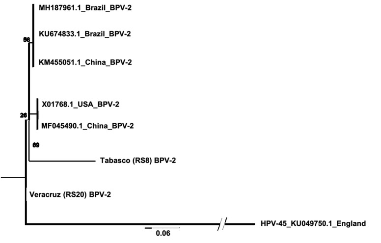
Fig. 5
Bovine papillomavirus (BPV)-2 phylogenetic tree of Mexican isolates (partial L2 sequence). Sequences were compared with sequences from different nationalities recorded in the NCBI database, of which the accession numbers are indicated. The comparison used a neighbour-joining method in MEGA 10.1. RS – representative sample
Table 2
Nucleotide sequence identity analysis of bovine papillomavirus 1 L1 fragments isolated from samples positive for the virus
| Sequence | Expect (E) value | Identity % (alignment with KC595244.2) | Gaps |
|---|---|---|---|
| Chiapas (RS7) | 7e−129 | 99% | 2/265 |
| Chiapas (RS6) | 4e−118 | 99% | 0/242 |
| Nuevo León (RS4) | 2e−129 | 99% | 3/267 |
| Nuevo León (RS2) | 2e−101 | 99% | 0/209 |
| Nuevo León (RS5) | 3e-131 | 99% | 2/266 |
| Veracruz (RS20) | 1e−125 | 99% | 1/257 |
| Tabasco (RS8) | 1e−125 | 99% | 1/257 |
| Nuevo León (RS3) | 2e−126 | 99% | 2/261 |
| Nuevo León (RS1) | 6e−128 | 99% | 4/267 |
Table 3
Nucleotide sequence identity analysis of bovine papillomavirus 2 L2 fragments isolated from samples positive for the virus
| Sequence | Expect (E) value | Identity % (alignment with MH187961.1) | Gaps |
|---|---|---|---|
| Veracruz (RS20) | 7e−25 | 97% | 0/71 |
| Tabasco (RS8) | 6e−45 | 91% | 3/132 |
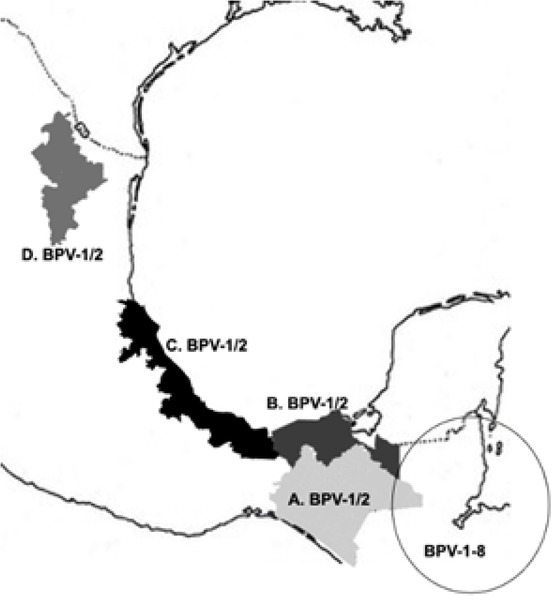
Fig. 6
Geographical bovine papillomavirus (BPV) genotype localisation in Mexico. Tabasco, Chiapas, and Veracruz states are considered breeder states and cattle collection centres for Central America. Nuevo León is a final feedlot destination state for national consumption or international exportation. The intensity of grey indicates the percentage of co-infection
A – Chiapas; B – Tabasco; C – Veracruz; D – Nuevo León
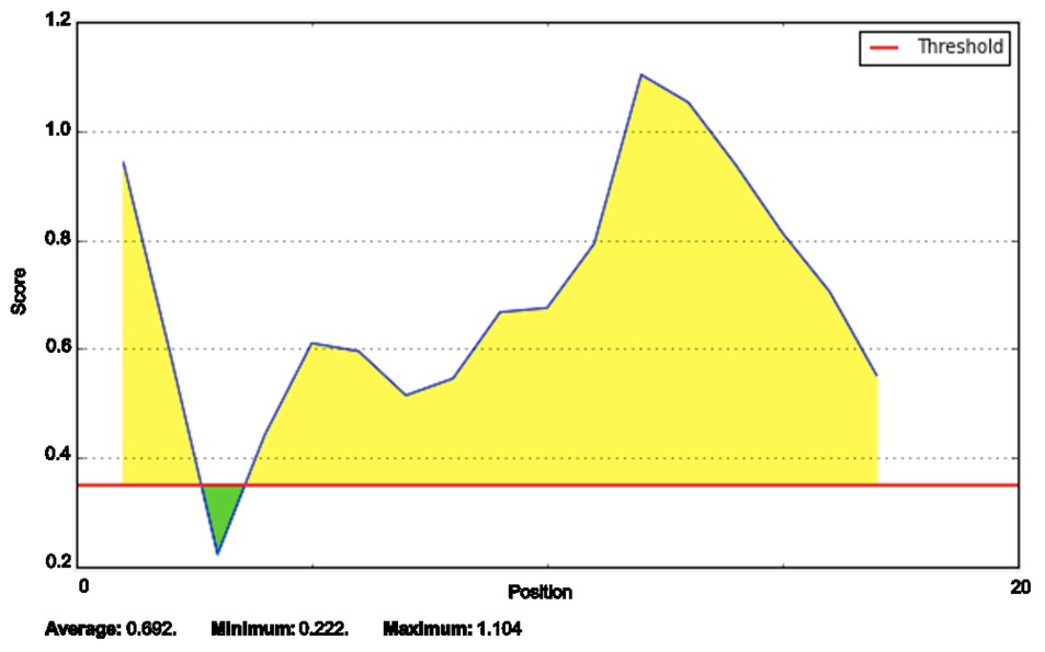
Fig. 7
Candidate peptide prediction by online server. The amino acid sequence of the selected peptide possesses antigenicity. Prediction with default values was performed in Bepipred Linear Epitope Predictor
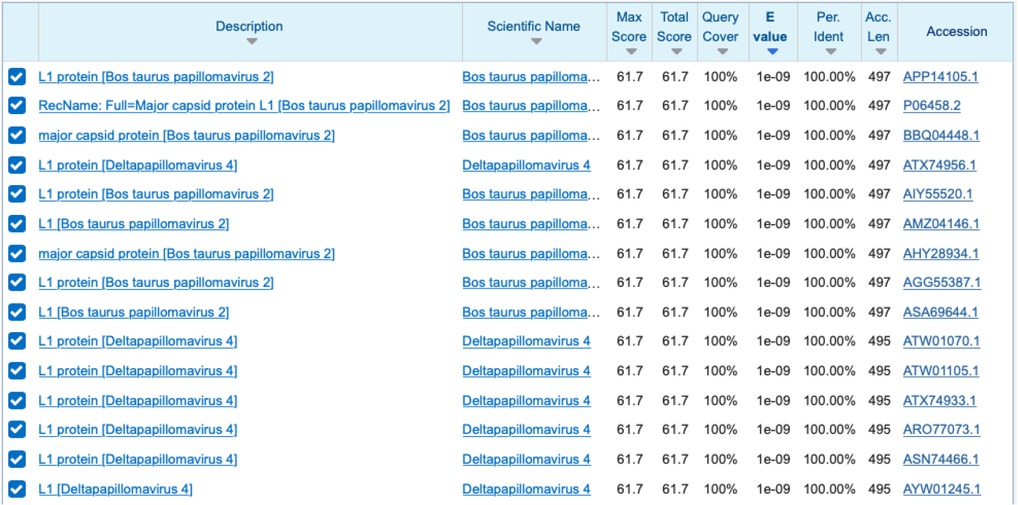
Fig. 8
Specificity of the candidate peptide for BPV-1. BLASTp showed it to be specific to BPV-1 and BPV-2 with 100% homology
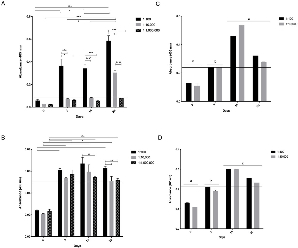
Fig. 9
Sera reactivity against WWL/synthetic peptide and antibody specificity against BPV
A – Anti-wart antibody reactivity against WWL at 1: 100 dilution on day 7 and 1:10,000 on days 14 and 35; B – Anti-peptide serum reactivity against synthetic peptide at a titre of 1:1,000,000 on days 7, 14 and 30. Cut-off points (horizontal solid line) were calculated at 0.087 and 0.053, respectively. **** – extremely significant difference (Tukey test P < 0.0001); *** – highly significant difference (Tukey test P < 0.0002) (n = 2); ** – significant difference (Tukey test P < 0.0021); * - significant difference (Tukey test P < 0.0332) (n = 2); C – Anti-wart serum reactivity against peptide at titres of 1 : 10,000–1 : 1,000,000 on day 14 and at a titre of 1 : 10,000 on day 35; D – Anti-peptide serum reactivity against WWL at a titre of 1 : 10,000 on days 7, 14 and 35, and at a titre of 1 : 1,000,000 on days 14 and 35. Letters a, b, and c represent significant difference based on the cut point (horizontal line) of 0.219 in both C and D (Tukey’s test P < 0.05) (n = 2)
Table 4
Amino acid matches between epitope predictions in different software
| Online server | L1 epitope | Position | Threshold | Score |
|---|---|---|---|---|
| Bepitope | SPATKCASNVIPAK | 421 | 0.75 | Yes |
| ABCpred | SILEDTYRYESPATK | 410 | 0.75 | 0.87 |
| Bepipred | PSVLQNWEIGVQPPTSSILEDTYRYIESPATKCASNVIPAKEDPYAGFKF | 394 | 0.75 | Yes |
| LBtope | VQPPTSSILEDTYRYIES | 404 | 0.75 | 85.2 |
| BCEpred | DTYRYIESPATKCASNV | 414 | 1.9 | Yes |
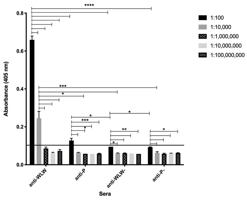
Fig. 10
Anti-wart serum reactivity against WWL at titres of 1 : 100–1 : 10,000 and at a titre of 1 : 100 on day 42 in anti-peptide serum against WWL. The solid line indicates the cut-off point at 0.08; **** – extremely significant difference (Tukey test P < 0.0001); *** – highly significant difference (Tukey test P < 0.0002) (n = 2); ** – significant difference (Tukey test P < 0.0021); * – significant difference (Tukey test P < 0.0332) (n = 2)