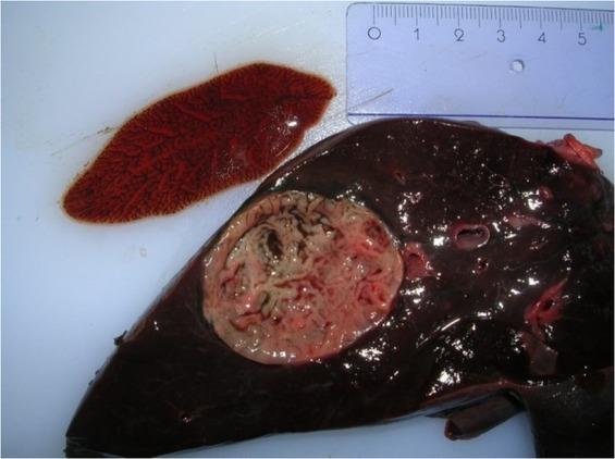
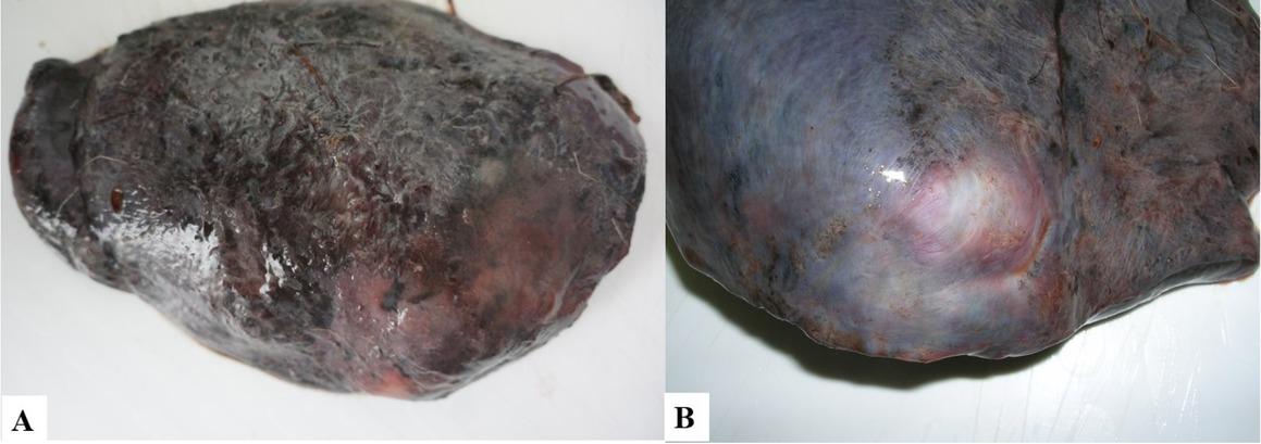
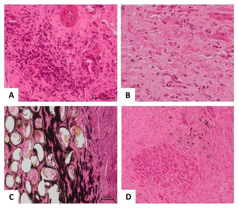
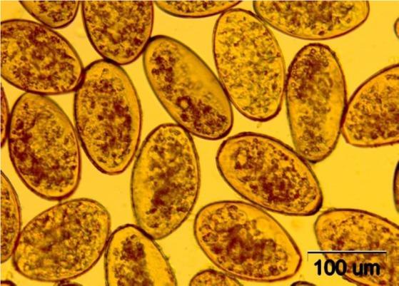
Have a personal or library account? Click to login
The occurrence of Fascioloides magna (Bassi, 1875) in the wild cervid population in the Lower Silesian Wilderness – epidemiological and pathological aspects Cover

The occurrence of Fascioloides magna (Bassi, 1875) in the wild cervid population in the Lower Silesian Wilderness – epidemiological and pathological aspects

Open Access
|Aug 2022

Figures & Tables

Fig. 1

Fascioloides magna isolated from a pseudocyst in the liver parenchyma of an infected red deer

Fig. 2

A – Surface of infected red deer liver covered with fibrin; B – A pseudocyst visible through the liver capsule

Fig. 3

Histopathological picture of the liver parenchyma in the course of trematode infection. A – Focus of lymphoplasmacytic inflammatory infiltrates in the liver parenchyma; B – Coagulative necrosis of individual hepatocytes mixed with inflammatory cells; C – Oval structures with an opaque sheath embedded in the liver parenchyma suggestive of parasite eggs; D – Extensive areas of fibrosis of the liver parenchyma with a central focus of the necrotic area

Fig. 4

Eggs of Fascioloides magna isolated from the liver of an infected red deer

Prevalence and intensity of infection of Fascioloides magna in the livers of red deer from the Lower Silesian Wilderness during the three hunting seasons of the study

YearNumber of examined red deerNumber of infected red deerPrevalence (CI 95%)Intensity
20143026.7 (1.8–21.3)2, 9

201634926.5 (14.6–43.1)2–19 a

2017351028.6 (16.3–45.1)9, 3–24 b

Prevalence and number of Fascioloides magna eggs per gram of faeces (EPG) in roe deer, red deer and fallow deer in 2015 and 2017

Year of sampling
20152017P value
Roe deer

n = 35n = 17
Prevalence a16 (45.7; 30.5–61.8)5 (29.4; 13.3–53.1)0.256
FEC (EPG) b18, 4–35 (1–84)2, 1–6 (1–83)0.160

Red deer

n = 61n = 12
Prevalence a32 (52.5; 40.2–64.5)7 (58.3; 32.0–80.7)0.709
FEC (EPG) b25, 11–43 (1–97)31, 11–219 (3–395)0.419

Fallow deer

n = 33n = 12
Prevalence a16 (48.5; 32.5–64.8)7 (58.3; 32.0–80.7)0.558
FEC (EPG) b12, 5–26 (1–76)33, 14–57 (11–63)0.181
Language: English
Page range: 381 - 387
Submitted on: Apr 14, 2022
Accepted on: Jul 26, 2022
Published on: Aug 12, 2022
Published by: National Veterinary Research Institute in Pulawy
In partnership with: Paradigm Publishing Services
Publication frequency: 4 issues per year

© 2022 Katarzyna Filip-Hutsch, Anna M. Pyziel-Serafin, Tomasz Hutsch, Kamila Bulak, Michał Czopowicz, Dorota Merta, Janusz Kobielski, Aleksander W. Demiaszkiewicz, published by National Veterinary Research Institute in Pulawy
This work is licensed under the Creative Commons Attribution-NonCommercial-NoDerivatives 3.0 License.