
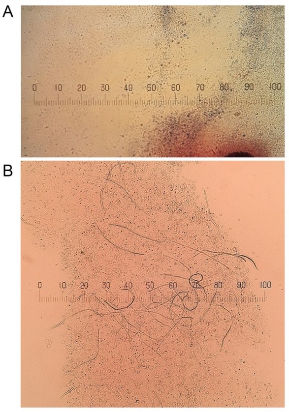
Fig. 1
Microfilariae detected by modified Knott’s test. One sample contained few larvae (A), while the other presented many (B). The parasites were photographed using a Nikon microscope and Easy Share C 613 camera (Kodak, Rochester, NY, USA). Stage micrometer: 1 mm
Table 1
Dogs that tested positive in at least one assay
| Dog number | Gender | Age (years) | Breed | History of travelling | Ownership (Region) | Knott’s test | Dirofilaria species by real-time PCR |
|---|---|---|---|---|---|---|---|
| 76 | male | 12 | mixed | none | private (Mura river region) | positive | D. repens |
| 389 | male | 2 | GSP | DE | State police (Ljubljana) | negative | D. repens |
| 390 | female | 1 | GSP | none | State police (Ljubljana) | positive | D. repens |
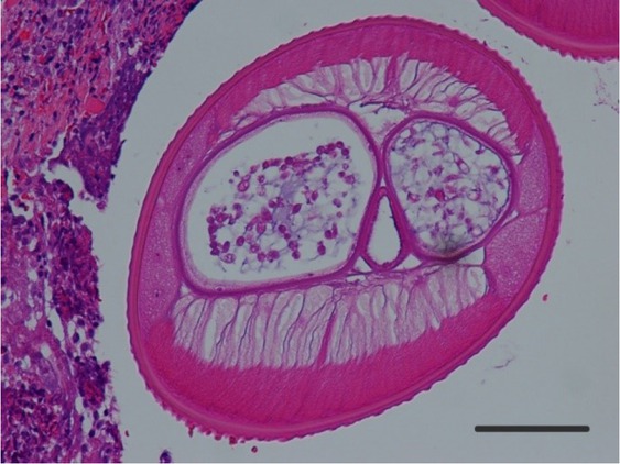
Fig. 2
Haematoxylin and eosin-stained cross-section of D. repens; external cuticular ridges are well visible; case 1. The parasite was photographed using a microscope and DS-Fi1 camera (Nikon). Scale bar: 100 μm
Table 2
Demographic and clinical data of the human cases described in this study
| Case | Gender | Age (years) | Region | Year of established diagnosis | Site of infection | Isolate | Species identified |
|---|---|---|---|---|---|---|---|
| 1 | Female | 20 | Central Slovenia (Ljubljana) | 2010 | N/A | Subcut. nodule | D. repens |
| 2 | Female | 45 | North (Mura East river Slovenia region) | 2013 | Breast | Subcut. nodule | D. repens |
| 3 | Male | 49 | North (Mura East river Slovenia region) | 2014 | Right upper arm | Subcut. nodule | D. repens |
| 4 | Male | 63 | North (Drava East river Slovenia region) | 2017 | Scalp | Subcut. nodule | D. repens |