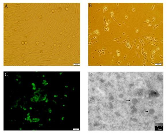
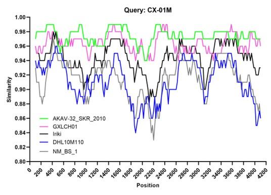
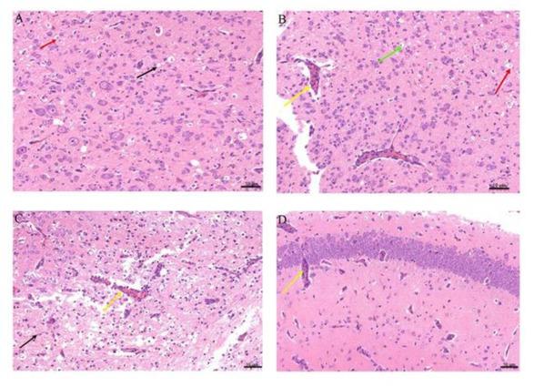
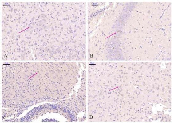
Have a personal or library account? Click to login
Genetic and pathogenic characterisation of a virulent Akabane virus isolated from goats in Yunnan, China Cover

Genetic and pathogenic characterisation of a virulent Akabane virus isolated from goats in Yunnan, China

Open Access
|Mar 2022

Figures & Tables

Fig. 1

Histopathological images of baby hamster kidney (BHK)-21 cells infected with Akabane virus strain CX01. A – Control BHK-21 cells; B – Cytopathic effect of CX-01 in BHK-21cells; C – Immunofluorescence results of BHK-21 cells infected with CX-01 after 36 h; D – Negatively stained Akabane virus particles 50–100 nm in diameter. Scale bar - 100 nm

Fig. 3

Recombination analysis of the CX-01 strain using a 200-bpsliding window and a 20-bp step. The y-axis indicates the percentage similarity between the query sequence and the reference sequences. Comparison of genome scale similarity of CX-01 (query) with DHL10M110 (green), GXLCH01(pink), Iriki (black), AKAV-32/SKR/2010 (blue) and NM/BS/1 (grey)

Fig. 4

Histopathological characteristics in the central nervous tissues of mice with AKAV CX-01 strain. A – brain of a 7-day-old mouse inoculated intraperitoneally (IP) with the CX-01 virus. Neuronal degeneration, necrosis (red arrow) and cavities are visible in the brain stem tissue (black arrow); B–brain of a 7-day-old mouse inoculated intracerebrally (IC) with the CX-01 virus. Enlarged vascular endothelial cells and perivascular infiltration of mononuclear cells (yellow arrow), gaps in the neuron cells (green arrow) and neuronal degeneration and necrosis are visible (red arrow); C – brain of a 7-day-old mouse inoculated IP with the CX-01 virus. The cortex, thalamus and hypothalamus on one side of the tissue have extensive necrosis, and fragmented nuclei are visible (black arrows); D – brain of a 7-day-old mouse inoculated IC with the CX-01 virus. Hippocampal pyramidal cells are regularly arranged, with clear demarcation, and round nuclei with white blood cells in the vascular cavity (yellow arrow). Haematoxylin and eosin staining. Scale bar– 50 μm

Fig. 5

Immunohistochemical characteristics in the central nervous tissues of mice with AKAV CX01 strain. A– brain stem of a mouse inoculated intraperitoneally (IP). Cells detected as antigen positive were observed as dark brown (red arrow); B– brain of a mouse inoculated intracerebrally (IC). Viral antigen is present mainly in neurons of the hippocampus, of which some cells were detected as antigen positive (red arrow); C – brain of a mouse inoculated IP. The hippocampal pyramidal cells were virus antigen–positive (red arrow); D– brain of a mouse inoculated IC. The hippocampal pyramidal cells were virus antigen positive (red arrow). Haematoxylin and eosin staining. Scale bar– 50 μm

Comparison of the three segments S, M and L (ORF) and the coding region of the M segment among AKAVs

GenotypeStrainGeographic origin and hostPairwise % identity (nt/aa)
SMML
GnGcNSm
Genogroup IaDHL10M110ChinaAnopheles vagus94.7/99.591.7/95.692.7/80.791.3/78.490.5/75.797.5/99.4
NM/BS/1ChinaBovine98.1/99.191.8/96.292.6/80.791.5/78.091.9/79.6-
GXLCH01ChinaRhizomys pruinosus97.6/99.696.6/97.996.8/91.496.4/91.196.7/91.794.5/98.4
GXLCH70NChinaRhizomys pruinosus97.7/99.696.8/97.996.4/90.797.2/93.295.4/89.094.6/98.7
HN10174ChinaCulex quinquefasciatus-91.7/96.392.0/78.991.5/78.091.9/79.695.1/98.8
KM-1/Br/06JapanBovine98.4/10097.3/97.997.4/93.697.2/93.597.1/92.396.2/98.9
AKAV-32/SKR/2010KoreaBovine94.9/98.797.7/98.498.1/95.497.7/94.397.2/92.8-
IRIKIJapanBovine95.0/98.794.7/97.094.9/81.694.5/86.093.4/82.9-
Genogroup IbFO-90-3JapanCulicoides spp.95.3/98.391.7/95.691.0/71.091.8/79.692.3/81.2-
Genogroup IIOBE-1JapanBovine95.3/98.787.5/93.889.3/66.886.4/66.789.4/71.891.8/97.6
Genogroup IIIB8935AustraliaBovine92.7/97.484.4/91.985.7/57.684.1/63.283.9/58.696.4/95.8
Genogroup IVMP496KenyaAnopheles funestus83.5/90.670.3/74.475.2/-69.9/38.365.9/30.4-

The primers used for the amplification of the small (S), medium (M) and large (L) segments of the Akabane virus genome

GenePrimerSequence (5´–3´)PositionProduct size (bp)
SAKVS1CTCCACTATTAACTACGCAT9–26804
AKVS2GGTGTGCACCACATAGACAT793–812
MAKVM1AGTAGTGAACTACCACAACAAAATG1–254,308
AKVM2AGTAGTGTTCTACCACAACAAATAATTAT4,280–4,308
LAKVL1AGTAGTGTACCCCTAAATACAACATACA1–284,151
AKVL2GTCAGCTTGCTTAAATCCC4,133–4,151
AKVL3GTGATTGTGCATTCCTTGG3,764–3,7823,106
AKVL4AGTAGTGTGCCCCTAAATGCAATAATAT6,842–6,869
Language: English
Page range: 35 - 42
Submitted on: Jun 2, 2021
Accepted on: Feb 1, 2022
Published on: Mar 10, 2022
Published by: National Veterinary Research Institute in Pulawy
In partnership with: Paradigm Publishing Services
Publication frequency: 4 issues per year

© 2022 Hua-Feng Gao, Jin-Ping Wang, Zhen-Xing Yang, Jia-Rui Xie, Yu-Wen He, Qiong-Hua Hong, Ai-Guo Xin, published by National Veterinary Research Institute in Pulawy
This work is licensed under the Creative Commons Attribution-NonCommercial-NoDerivatives 3.0 License.