
Of the many avian parasites, the Histomonas meleagridis protozoan flagellate and causative agent of histomonosis, or blackhead disease, is a particularly dangerous one. Histomonosis is most common among 2–16-week-old turkeys; however, it is also diagnosed in older birds (26). Even though chickens and other bird species are considered less susceptible to infection and despite the milder course of the disease in them, many clinical cases of histomonosis in poultry in different production systems have recently been described in gallines (3, 8, 27, 28, 33).
Turkey infection with H. meleagridis occurs per os with Heterakis gallinarum eggs. Infection with these flagellates may also spread without the participation of these nematodes, and the penetration route of the protozoa is then through the cloaca (14, 18, 19). The pathogenesis of histomonosis starts with the colonisation of the caecum by the parasite, leading to severe inflammation and necrosis. Following the destruction of intestinal tissue, the parasite may infiltrate blood vessels and reach the liver via the portal veins. As a consequence, areas of inflammation and destruction can be created in the liver. In the final stage, the disease may become systemic when the parasite spreads to other organs of the host (13). The incidence and mortality of turkeys infected with this protozoan can reach 100%. Histomonosis manifests with clinical signs such as reduced appetite, sulphur-yellow diarrhoea, dehydration, and emaciation (19, 20, 26).
Cases of histomonosis in poultry flocks are frequent in many countries of the European Union and across the world. This re-emerging endemic disease poses a serious threat to the breeding of birds, mainly turkeys, and confronts the industry with major clinical, epidemiological, and economic problems (9, 17, 25). This unfavourable situation is caused by a ban on the prophylactic and therapeutic use of effective chemotherapeutics in poultry, notable preparations affected by the ban being those containing derivatives of nitroimidazoles (metronidazole, ronidazole, and dimetridazole) (6) and nitrofurans (nifursol) (7). International guidelines for food safety, including those on veterinary drug residues in food, are outlined in the Codex Alimentarius of the Food and Agriculture Organization of the United Nations and the World Health Organization, and regarding histomonostats, they strongly recommend not to use nitroimidazoles such as metronidazole or dimetridazole in food-producing birds (5). The ban on administering these substances to domestic birds addresses the risk of their residues being present in foodstuffs and posing a threat to consumer health (2).
This situation and the fact that biosecurity measures do not always prevent H. meleagridis introduction into a flock, infection (4), or ultimate disease outbreak, have forced a new approach to the problem of histomonosis prevention and treatment. That is why recent research focuses on various areas of disease control from the use of phytocompounds (1, 12, 31) to administration of vaccines (25, 29). The presented study aims to assess the therapeutic and preventive efficacy of phytoncides contained in adiCoxSOLPF (AdiFeed, Warsaw, Poland), a feed supplement for poultry, in a clinical case of histomonosis in a parent turkey flock, as well as to determine their effect on selected parameters of bird cell-mediated and humoral immunity.
Dietary supplement. The adiCoxSOLPF dietary supplement for turkeys contains 52.0% herbal extracts including sweet flag (Acorus calamus L.), saponaria (Saponaria officinalis L.), mustard (Sinapis alba L.), and pepper (Piper nigrum L.); 9.2% flavouring additives including thymol; and preservatives including acetic acid (4.6%), calcium lactate (0.6%), and sodium diacetate (0.2%).
Case presentation, clinical history, and treatment. The turkey farm where the studied birds were reared operated on the all-in/all-out system. There were two turkey houses (J-1 and J-2) on the farm and 9,339 one-day-old turkey poults were housed in them in total. In the J-2 turkey house, a clinical form of histomonosis occurred when the turkeys were aged 11 weeks in a flock designated as breeders. Within the next seven days, 48 turkey hens out of 3,870 placed in J-2 were found dead. In the same poultry house, 1,019 turkey toms were reared in a sector separated by a solid wall. The turkeys were administered adiCoxSOLPF in a dose of 2.5 mL/L water in 3 seven-day cycles, with a two-day break after each.
In J-1, the 4,450 turkey hens kept there were administered the preparation for seven days in a dose of 2.5 mL/L water and then in two subsequent cycles in a dose of 1 mL/L. Next, the depressed and emaciated turkey hens from J-2 were culled. Afterwards, the floor in J-2 was bedded with a thick straw layer, and all birds were dewormed by two-day administration of Levamol 10% (Vetoquinol Biowet Sp. z o.o., Gorzów Wielkopolski, Poland) in a dose of 0.3 mL/kg body weight in drinking water. For two consecutive days, the gastrointestinal tract of turkeys was colonised by physiological bacterial flora (Protexin, Probiotics Int. Ltd., Lopen Head, UK) and then the birds were administered multivitamin preparations for another two days. After these treatments, adiCoxSOLPF was re-administered for seven days in a dose of 2.5 mL/L water in J-2, and 1 mL/L water in J-1.
Until the end of the growing-out stage, i.e., until week 28 of life, the birds received adiCoxSOLPF in a dose of 1 mL/L water in seven-day cycles with a three-day break between them. In week 29 of life, the birds were transferred to a laying farm. The turkey hens that had been reared in J-2 were placed in a separate poultry house (P-1) and were administered adiCoxSOLPF in a dose of 1 mL/L water in seven-day cycles with a seven-day break between them until the end of laying production. In turn, the hens reared in J-1 were transferred to poultry houses P-2 and P-3. Along with toms which were also transferred to poultry houses P-2 and P-3, these hens received the supplement in a dose of 1 mL/L water in three-day cycles with an eleven-day break between them until the end of the laying period.
Eggs/hen, fertility, hatchability, and mortality and culling rates were monitored in all poultry houses.
Necropsy, parasitological and microbiological examination. During anatomopathological examination, tissue sections (liver, spleen, heart, ileum, and caecum) were collected for parasitological and microbiological analyses and detection of H. meleagridis genetic material. Caecal contents were washed with water and viewed under a stereoscopic microscope for the presence of H. gallinarum. Additionally, faeces samples from sick birds were tested for the presence of this nematode by the flotation technique. Three grams of faeces from each turkey were mixed with 15 mL of Fecasol (Vetoquinol Biowet) solution and strained through a 0.5 mm mesh sieve. The suspension was transferred to a 15 mL tube and centrifuged at 1,500 × g for 5 min. The test sample was removed from each tube by touching the surface of the supernatant with a microscopic slide and then it was examined under 100 × magnification.
Microbiological examination was performed on samples from the liver, spleen, heart, and ileum, which were cultured in standard and selective media (Columbia agar with 5% sheep blood and MacConkey agar, Oxoid Deutschland GmbH, Wesel, Germany).
DNA isolation and PCR amplification. Liver and caecum samples (0.2 g each) were individually cut into fragments 1–2 mm in diameter. The fragments were placed in an Eppendorf tube, 700 μL of sterile PBS was added, and the samples were homogenised using a TissueLyser II automatic device (Qiagen, Hilden, Germany). The resulting homogenate was centrifuged at 8,000 × g for 30 s. Next, a 200 μL cell pellet was collected for DNA isolation, which was performed using a DNeasy Blood & Tissue Kit (Qiagen), following the manufacturer’s instructions. The concentration and quality of the isolated DNA were evaluated with a NanoDrop 2000 spectrophotometer (Thermo Fisher Scientific, Waltham, MA, USA).
The PCR method described by Grabensteiner and Hess (11) was employed to detect H. meleagridis genetic material in the liver and caecum samples. The eluted DNA concentration was optimised to 150 ng/reaction and amplified with the use of a HotStarTaq Plus Master Mix Kit (Qiagen) in a Vapo Protect thermal cycler (Eppendorf, Hamburg, Germany).
The PCR amplicon size was analysed by electrophoresis in 2% agarose gel (Prona Agarose; ABO, Gdańsk, Poland) in 1X concentrated TAE buffer (Fermentas, Vilnius, Lithuania) with ethidium bromide addition at 0.5 μL/mL (Sigma-Aldrich, St. Louis, MO, USA) using 5V/cm gel voltage for 60 min. DNA Marker (A&A Biotechnology, Gdynia, Poland) was used as a molecular size marker. The results of the electrophoretic separation were read via GelDoc XR+ visualisation (Bio-Rad, Hercules, CA, USA) and saved as digital images.
Turkeys, experimental groups and handling. Experiment I was conducted on thirty 46-day-old Hybrid Converter turkeys (Grelavi S.A., Olsztyn, Poland) randomly divided into three groups (A1–A3) of 10 birds each. Group A1 was the control, while birds from groups A2 and A3 were administered adiCoxSOLPF for three days in drinking water at a final dose of 1 mL or 3 mL/L, respectively.
Experiment II was conducted on sixty-nine 46-day-old Hybrid Converter broiler turkeys (Grelavi S.A., Olsztyn, Poland) randomly divided into three groups (B1–B3) of 23 birds each. The birds from group B1 were given adiCoxSOLPF for three days in drinking water at a final dose of 1 mL/L. All birds from groups B1, B2, and B3 were vaccinated against Ornithobacterium rhinotracheale (ORT) at 49 days of life. After vaccination, birds from group B2 were administered adiCoxSOLPF for three days in drinking water at a final dose of 1 mL/L, while birds from group B3 did not receive the phytoncidal preparation at any time during the experiment.
Turkeys were housed in isolated pens in a biosafety level-3 facility. Water and feed were provided ad libitum. The experimental procedures and animal handling procedures were approved by the Local Ethics Committee for Animal Experiments in Olsztyn, Poland.
Vaccine and vaccination. In experiment II, turkeys from groups B1, B2, and B3 were vaccinated against ORT with Ornitin (ABIC Biological Laboratories Ltd., Airport City, Israel) at 49 days of life. The vaccine was administered subcutaneously following the manufacturer’s instructions.
Sample collection. In experiment I, blood samples were collected from turkeys of groups A1–A3 by venipuncture of the wing into sterile tubes without anticoagulant for biochemical analyses (n = 10) and into sterile tubes containing EDTA K anticoagulant for flow cytometry analysis (n = 7). Samples were collected 10 h after the last adiCoxSOLPF supplement was given. Seven birds from each group were euthanised, samples of their spleens were collected, and together with blood samples these were used to isolate mononuclear cells and determine the percentages of CD4+, CD8+, and CD4+CD8+ T cell subpopulations and the IgM+ B cell subpopulation by flow cytometry. Humane euthanasia of the birds was performed using a professional UNO Euthanasia CO2 unit (UNO, Zevenaar, the Netherlands).
In experiment II, blood samples for serological analyses were collected from 23 birds from groups B1–B3 on the day of ORT vaccination and three weeks post vaccination.
Serological analysis. Anti-ORT IgY titres were determined using a commercial immunoenzymatic ELISA kit (IDEXX Laboratories, Westbrook, MN, USA) according to the manufacturer’s recommendations. The ELISA assay was performed using an epMotion 5075 LH automated pipetting system (Eppendorf), an Elx405 washer, and an Elx800 absorbance microplate reader (BioTek, Winooski, VT, USA).
Biochemical analyses. The levels of total protein (TP) and the activities of the alanine aminotransferase (ALT), aspartate aminotransferase (AST), alkaline phosphatase (ALP), lactate dehydrogenase (LDH), and creatine kinase (CK) enzymes were determined in blood serum samples. The tests were performed using a VetTest Chemistry Analyzer (IDEXX) according to the manufacturer’s instructions.
Isolation of mononuclear cells and flow cytometry. Peripheral blood mononuclear cells (PBMCs) were isolated according to a previously described procedure (22). The isolation of mononuclear cells from the spleen and the determination of the percentages of CD4+ and CD8+ T cell and IgM+B cell subpopulations in PBMCs and spleen were carried out as described by Kubińska et al. (24).
Statistical analysis. The significance of differences in values of the measured parameters between the investigated groups was analysed with Student’s t-test for independent variables. All calculations were made using Statistica 13.1 software (StatSoft, Tulsa, OK, USA). Differences were considered statistically significant at P ≤ 0.05.
Clinical observations. Sick turkeys displayed apathy, were emaciated with their wings down, and had dull plumage and sulphur-yellow faeces adhering to the area around the cloaca. All dead turkeys were dissected, and the following pathological changes were noticed during necropsy. The livers were enlarged and ulcerated lesions with circular depressions up to 1 cm in diameter had formed on the surface, which were light yellow in the centre and raised at their peripheries. The caeca were bloated and contained caseous matter; after the removal of the contents, necrosis and ulcers of the caecal wall were seen. No adult H. gallinarum nematodes were found in the caecal contents, and no eggs of these parasites were found in faeces during parasitological examination with the flotation method. Microbiological analyses demonstrated numerous E. coli in the internal organs of dead birds. Histomonosis was additionally diagnosed based on the presence of the parasite’s genetic material in the liver and caecum samples (Fig. 1).
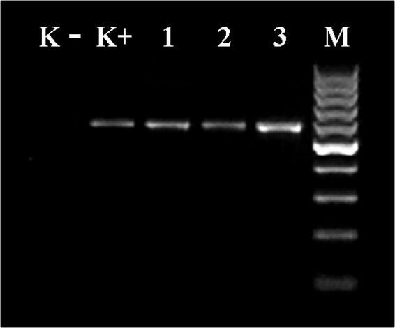
Detection of H. meleagridis by PCR assay
K− – negative control; K+ – positive control (574 bp); 1, 2 – DNA samples isolated from liver; 3 – DNA sample isolated from caecum; M – molecular size marker (100–1000 bp)
During the administration of the tested preparation, no disease cases were found among toms from J-2 or hens from J-1. Within four weeks of the onset of the disease, 229 (5.92%) turkey hens died and 630 (16.28%) were euthanised, representing 22.20% of the total. Over the entire 28-week growing-out period, the mortality and culling rate was 28.3% in J-2 and 4.53% in J-1.
After transfer to the production facilities, turkey hens began laying in week 32 of life. Production parameters (eggs/hen, fertility, and hatchability) and the mortality and culling rate determined over the laying production period are summarised in Table 1. On average, 112 eggs were produced by each turkey hen from P-1, whereas in P-2 and P-3 the production rate was higher by 5 and 7 eggs, respectively. Irrespective of the turkey house, fertility and hatchability were at 97% and 85%, respectively. In the studied period, 6.1% of birds died or were culled in P-1, whereas the mortality and culling rate in P-2 and P-3 was 2.7% and 3.4%, respectively. No cases of histomonosis were recorded in any of the birds which were found dead or were euthanised.
Mean values of production parameters and the mortality and culling rate in the laying production period (week 32 to week 56 of life) in a reproductive flock of turkeys treated for histomonosis diagnosed in the rearing period
| Parameter | Turkey house | ||
|---|---|---|---|
| P-1 | P-2 | P-3 | |
| Eggs/hen | 112 | 117 | 119 |
| Mortality and culling (%) | 6.1 | 2.7 | 3.4 |
| P-1, P-2, and P-3 | |||
| Fertility (%) | 97 | ||
| Hatchability (%) | 85 |
P-1 – the poultry house used to hold hens from J-2 where there was an outbreak of clinical histomonosis during the rearing period
P-2, P-3 – the poultry houses used to hold hens from J-1 and toms from J-2
Experiment I. The dose of the preparation administered to turkeys had no effect on water and feed intake. The results of biochemical analyses are summarised in Table 2. All presented values fell within the ranges of standard physiological values. Graded levels of supplementation with adiCoxSOLPF had no significant effect on the analysed parameters.
Mean value ± SD of biochemical markers in blood serum of turkeys from experiment I (n = 10)
| Group | ALT (U/L) | AST (U/L) | ALP (U/L) | CK (U/L) | LDH (U/L) | TP (g/L) |
|---|---|---|---|---|---|---|
| A1 | 15.83 ± 1.26 | 261.67 ± 14.39 | 2056.67 ± 110.32 | 1287.90 ± 260.08 | 942.00 ± 91.42 | 35.45 ± 2.26 |
| A2 | 16.00 ± 2.10 | 250.50 ± 18.97 | 1768.83 ± 360.05 | 1450.92 ± 255.59 | 1002.83 ± 88.23 | 35.61 ± 1.65 |
| A3 | 17.33 ± 3.39 | 275.33 ± 39.26 | 2311.33 ± 542.02 | 1934.52 ± 399.42 | 1034.17 ± 67.31 | 35.63 ± 4.25 |
A1 – control group
A2 – group administered adiCoxSOLPF for three days in drinking water at a final dose of 1 mL/L
A3 – group administered adiCoxSOLPF for three days in drinking water at a final dose of 3 mL/L
ALT – alanine aminotransferase; AST – aspartate aminotransferase; ALP – alkaline phosphatase; CK – creatine kinase; LDH – lactate dehydrogenase; TP – total protein
The results of the selected cell-mediated immunity markers are presented in Table 3. They indicate that the therapeutic dose of the preparation (3 mL/L water) administered to birds in group A3 for three days caused a statistically significant increase in the percentage of IgM+ B lymphocytes and a considerable (although statistically insignificant) increase in the percentage of CD8+ T lymphocytes in the spleen. The prophylactic dose of adiCoxSOLPF (1 mL/L water) brought about a statistically significant multiplication in the CD4+ T lymphocyte subpopulation in blood from Group A2 turkeys.
Mean percentage of CD4+, CD8+, and CD4+CD8+ T lymphocytes and IgM+ B cells ± SD in blood and spleen of turkeys from experiment I (n = 7)
| Blood | Spleen | |||||||
|---|---|---|---|---|---|---|---|---|
| Group | CD4+ | CD8+ | CD4+CD8+ | IgM+ | CD4+ | CD8+ | CD4+CD8+ | IgM+ |
| A1 | 16.95a ± 2.44 | 2.20 ± 0.38 | 0.57 ± 0.08 | 10.86 ± 0.99 | 34.80 ± 6.80 | 21.35 ± 6.14 | 1.32 ± 0.36 | 17.47a ± 2.21 |
| A2 | 23.68b ± 4.88 | 2.19 ± 0.70 | 0.71 ± 0.17 | 11.55 ± 1.15 | 37.27 ± 7.38 | 25.15 ± 10.00 | 1.46 ± 0.37 | 20.85ab ± 2.10 |
| A3 | 18.53ab ± 6.05 | 2.17 ± 0.42 | 0.57 ± 0.11 | 11.54 ± 0.98 | 33.15 ± 3.19 | 28.40 ± 8.49 | 1.55 ± 0.54 | 22.10b ± 3.32 |
A1 – control group
A2 – group administered adiCoxSOLPF for three days in drinking water at a final dose of 1 mL/L
A3 – group administered adiCoxSOLPF for three days in drinking water at a final dose of 3 mL/L
a, b – means with different superscripts in the same column differ significantly (t-test, P < 0.05)
Experiment II. The results of serological analyses are provided in Table 4. They show that both before (Group B1) and after (Group B2) vaccination, the turkeys receiving the prophylactic dose of the test preparation (1 mL/L water) produced higher titres of anti-ORT antibodies than control birds (Group B3). However, the differences observed were statistically insignificant. A markedly higher titre of post-vaccination antibodies was determined in birds receiving adiCoxSOLPF in drinking water for three days post vaccination.
Mean geometric titre of post-vaccination anti-ORT IgY antibodies in blood serum and %CV of turkeys from experiment II (n = 23)
| Day of vaccination | 3 weeks post vaccination | |||
|---|---|---|---|---|
| Group | ORT (titre) | ORT (%CV) | ORT (titre) | ORT (%CV) |
| B1 | 128 | 137 | 2,312 | 64 |
| B2 | 123 | 129 | 2,778 | 75 |
| B3 | 125 | 134 | 1,567 | 54 |
B1 – group administered adiCoxSOLPF in drinking water at a final dose of 1 mL/L for three days prior to ORT vaccination
B2 – group administered adiCoxSOLPF in drinking water at a final dose of 1 mL/L for three days after ORT vaccination
B3 – control group
CV – coefficient of variation
Turkey histomonosis, which was first described in 1983, has caused considerable losses in the turkey industry (25, 26). This situation prompted research aimed at developing effective preparations for the prevention and treatment of this dangerous disease. The widespread use of the preparations consequently developed minimised the problem of histomonosis in turkeys and other poultry species for many years (17). However, health problems in flocks of turkeys and chickens caused by H. meleagridis infection have reemerged in recent years since the imposition of a ban in the EU countries and the US on the use of histomonostats in poultry production based on international guidelines on food safety (5, 6, 7).
The resurgence of turkey histomonosis was the stimulus for investigations to intensify again after a several-year break in research to develop effective preparations to combat this disease. It is now known that biosecurity measures alone are not effective enough (4) and that the main risk factors that predispose a domestic flock to histomonosis include the presence of wild birds within 1 km of the farm, water acidification, and failure on the part of workers and visitors to change shoes before entering the poultry house (9). This is also indicated by the results of a recently published study by Sulejmanovic et al. (30), who demonstrated the presence of H. meleagridis DNA in dust samples from a building housing clinically healthy turkeys.
The action strategy aimed at countering H. meleagridis infection in poultry flocks mainly entails searching for effective preparations of plant origin and developing a vaccine. As indicated by research findings (29), experimental vaccination using in vitro attenuated H. meleagridis effectively protects turkeys from histomonosis and is safe to use, which recommends it as the most promising means of prevention of histomonosis in poultry for the future. However, numerous publications show that, despite satisfactory in vitro results, phytocompounds fail to produce promising effects in vivo in the fight against this flagellate (1, 12, 14, 32). The likely reason for this failure is that in vivo, the H. meleagridis protozoan interacts with other pathogens and E. coli in particular (10, 16). This was not the case in our study, despite the isolation of E. coli from the internal organs of turkeys which died of histomonosis. This could be due to the immunomodulatory effect of the adiCoxSOLPF preparation, because our study results showed a tangible effect of its substances on the size of T and B lymphocyte subpopulations in both the blood and spleen of turkeys. The tested preparation also stimulated humoral immunity, which resulted in higher levels of anti-ORT antibodies in the groups of birds vaccinated against ornithobacteriosis and receiving adiCoxSOLPF. The results obtained correspond with those of previous studies, which demonstrated the immunomodulatory effect of phytoncides on humoral and cell-mediated immunity in chickens and turkeys (21). Our research also showed that, regardless of the dose used, adiCoxSOLPF had no adverse effects on internal organs, as confirmed by the results of biochemical analyses. The values of all biochemical parameters analysed in our study did not exceed the standard values of these parameters described by Krasnodębska-Depta and Koncicki (23).
The results of the present research confirm the high therapeutic and prophylactic effectiveness of the adiCoxSOLPF preparation used in a turkey breeder flock in which the clinical form of histomonosis was diagnosed during autopsy and confirmed by PCR. Over the entire 28-week rearing period, the mortality and culling rate was 28.3% in the flock of turkeys that underwent treatment and 4.53% in the flock of turkeys that did not get sick but received the prophylactic dose of adiCoxSOLPF. It is worth noting that no cases of histomonosis were recorded among toms receiving the prophylactic dose even though they are considered more susceptible to infection by H. meleagridis than hens (9, 15). However, many publications show that histomonosis has recently been the cause of very high losses in flocks of broiler and breeder turkeys. The mortality rates in untreated turkey flocks ranged from 23% to over 83%. With the use of preparations based on plant extracts or antibiotics (e.g. paromomycin), they were slightly lower but still ranged from 9 to 42% (9, 15, 30). The birds were emaciated and very often culled before reaching slaughter age out of concern for their welfare. In addition to numerous deaths, laying declined or even completely ceased in affected breeding turkey flocks (9).
The very high effectiveness of the adiCoxSOLPF preparation tested is evidenced by the fact that no relapses occurred in the turkey breeder flock under therapeutic and prophylactic treatment due to histomonosis developed at 11 weeks of life. In week 28, these turkeys were transferred to laying facilities and kept there until their 56th week. Laying began in week 32. Production parameters (eggs/hen, fertility, and hatchability) as well as the mortality and culling rate determined over the 24-week production period did not deviate from the standard values for this type of turkey. An average of 112 eggs were obtained from a turkey hen from the facility where histomonosis was diagnosed in the rearing period. On the other hand, hens each produced five to seven eggs more as birds administered the prophylactic dose of adiCoxSOLPF during the rearing period, and no histomonosis was diagnosed in them even though they were kept in the turkey house adjacent to the one with the sick birds. Irrespective of the turkey house, the fertilisation and hatching rates were 97% and 85%, respectively. Not a single case of histomonosis was detected in birds which were found dead or were euthanised, for which the respective rates ranged from 2.7% to 6.1% over the laying period.
The results of the present study allow it to be concluded that the active ingredients of the adiCoxSOLPF preparation are effective in the prevention and therapy of turkey histomonosis. The disease did not spread to turkey hens kept in an adjacent building and breeder turkeys were cured during the rearing period; these were both outcomes which demonstrated the efficacy of the preparation. That no disease recurred by the end of the rearing period nor within 24 weeks of laying production also confirms its high efficacy in eradicating turkey histomonosis as do the production results obtained (eggs/hen, fertility, and hatchability). It was also shown that, besides acting on pathogenic H. meleagridis flagellates, the plant extracts contained in the evaluated preparation stimulated both the humoral and cell-mediated defence mechanisms of the birds without suppressing the functions of their internal organs. This was regardless of the dose being to prevent or to treat, as evidenced by the biochemical markers studied matching the physiological norms in value. Such therapeutic and prophylactic effects against histomonosis after the use of plant extracts have never been described before.